
Paper:
Glass Bonding Method Utilizing Relatively Mildly Acidic Treatment and Fluorinated Plasma for Stable Micro/Nano Device Fabrication
Masafumi Watanabe*,**
 , Yuki Sakurai*, and Yutaka Kazoe*
*Department of System Design Engineering, Keio University
3-14-1 Hiyoshi, Kohoku-ku, Yokohama, Kanagawa 223-8522, Japan
**Research and Development Department, Kanagawa Institute of Industrial Science and Technology
705-1 Shimoimaizumi, Ebina, Kanagawa 243-0435, Japan

The bonding process is one of the most critical processes in the fabrication of stable, long-term, glass-based micro/nano devices for various applications, including biosensors. However, the current bonding process causes delamination and topographical damage to the detection unit of a biosensor, such as electrodes patterned on a glass substrate, owing to the strong acid treatment used for surface cleaning and activation. Herein, we propose a low-temperature glass bonding method that utilizes a relatively mildly acidic treatment without sacrificing bonding strength. To explore this method, we examined the effects of various acids, such as piranha solution, fuming nitric acid, and hydrochloric acid, on the bonding strength. To measure bonding strength, we treated fused-silica glass substrates with an acid solution, activated with oxygen plasma, bonded to each other, and annealed at 110°C. A crack-opening test using bonded glass substrates revealed that acid treatment is necessary to achieve reliable bonding strength. Relatively mildly acidic treatment, such as with hydrochloric acid, exhibited comparable bonding strength to strong acid treatment, such as piranha solution. Furthermore, we found that the selection of the bonding surface (top or bottom) and the use of fluorinated plasma with polytetrafluoroethylene pieces during oxygen plasma significantly enhance the bonding strength, e.g., 0.76 J/m2 with hydrochloric acid. We believe that our findings will contribute to the development of reliable long-term glass-based biosensors.
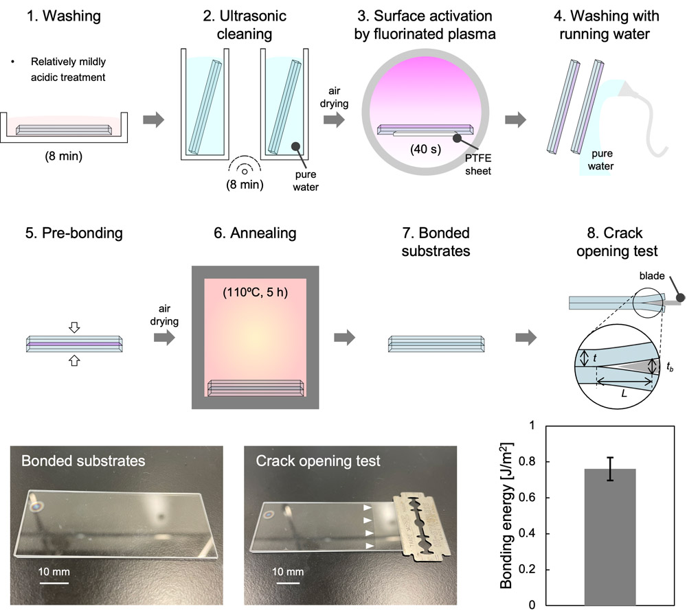
Glass bonding using mildly acidic treatment
- [1] S. Aralekallu, R. Boddula, and V. Singh, “Development of glass-based microfluidic devices: A review on its fabrication and biologic applications,” Mater. Des., Vol.225, Article No.111517, 2023. https://doi.org/10.1016/j.matdes.2022.111517
- [2] G. M. Whitesides, “The origins and the future of microfluidics,” Nature, Vol.442, No.7101, pp. 368-373, 2006. https://doi.org/10.1038/nature05058
- [3] D. Huh, G. A. Hamilton, and D. E. Ingber, “From 3D cell culture to organs-on-chips,” Trends Cell Biol., Vol.21, No.12, pp. 745-754, 2011. https://doi.org/10.1016/j.tcb.2011.09.005
- [4] Y. Liu et al., “Recent progress in microfluidic biosensors with different driving forces,” TrAC Trends Anal. Chem., Vol.158, Article No.116894, 2023. https://doi.org/10.1016/j.trac.2022.116894
- [5] T. Tang et al., “Glass based micro total analysis systems: Materials, fabrication methods, and applications,” Sens. Actuators B Chem., Vol.339, Article No.129859, 2021. https://doi.org/10.1016/j.snb.2021.129859
- [6] D. Han, Y.-R. Kim, C. M. Kang, and T. D. Chung, “Electrochemical signal amplification for immunosensor based on 3D interdigitated array electrodes,” Anal. Chem., Vol.86, No.12, pp. 5991-5998, 2014. https://doi.org/10.1021/ac501120y
- [7] D.-S. Shin et al., “Micropatterned surfaces functionalized with electroactive peptides for detecting protease release from cells,” Anal. Chem., Vol.85, No.1, pp. 220-227, 2013. https://doi.org/10.1021/ac302547p
- [8] Z. Matharu et al., “Detecting transforming growth factor- release from liver cells using an aptasensor integrated with microfluidics,” Anal. Chem., Vol.86, No.17, pp. 8865-8872, 2014. https://doi.org/10.1021/ac502383e
- [9] T. Nakao et al., “Cytokine analysis on a countable number of molecules from living single cells on nanofluidic devices,” Analyst, Vol.144, No.24, pp. 7200-7208, 2019. https://doi.org/10.1039/c9an01702j
- [10] K. Eyer, S. Stratz, P. Kuhn, S. K. Küster, and P. S. Dittrich, “Implementing enzyme-linked immunosorbent assays on a microfluidic chip to quantify intracellular molecules in single cells,” Anal. Chem., Vol.85, No.6, pp. 3280-3287, 2013. https://doi.org/10.1021/ac303628j
- [11] M. Mirasoli et al., “Portable chemiluminescence multiplex biosensor for quantitative detection of three B19 DNA genotypes,” Anal. Bioanal. Chem., Vol.405, Nos.2-3, pp. 1139-1143, 2013. https://doi.org/10.1007/s00216-012-6573-7
- [12] M. Malik, S. A. Steele, D. Mitra, C. J. Long, and J. J. Hickman, “Trans-epithelial/endothelial electrical resistance (TEER): Current state of integrated TEER measurements in organ-on-a-chip devices,” Curr. Opin. Biomed. Eng., Vol.34, Article No.100588, 2025. https://doi.org/10.1016/j.cobme.2025.100588
- [13] J. Yeste et al., “A compartmentalized microfluidic chip with crisscross microgrooves and electrophysiological electrodes for modeling the blood-retinal barrier,” Lab Chip, Vol.18, No.1, pp. 95-105, 2018. https://doi.org/10.1039/c7lc00795g
- [14] S. Palma-Florez et al., “BBB-on-a-chip with integrated micro-TEER for permeability evaluation of multi-functionalized gold nanorods against Alzheimer’s disease,” J. Nanobiotechnol., Vol.21, No.1, Article No.115, 2023. https://doi.org/10.1186/s12951-023-01798-2
- [15] M. A. Holzreuter and L. I. Segerink, “Innovative electrode and chip designs for transendothelial electrical resistance measurements in organs-on-chips,” Lab Chip, Vol.24, No.5, pp. 1121-1134, 2024. https://doi.org/10.1039/d3lc00901g
- [16] W. Wei, F. Cardes, A. Hierlemann, and M. M. Modena, “3D in vitro blood-brain-barrier model for investigating barrier insults,” Adv. Sci., Vol.10, No.11, Article No.2205752, 2023. https://doi.org/10.1002/advs.202205752
- [17] R. Van den Eeckhoudt et al., “Full-electric microfluidic platform to capture, analyze and selectively release single cells,” Lab Chip, Vol.23, No.19, pp. 4276-4286, 2023. https://doi.org/10.1039/d3lc00645j
- [18] Z. Zhang, T. Zheng, and R. Zhu, “Single-cell individualized electroporation with real-time impedance monitoring using a microelectrode array chip,” Microsyst. Nanoeng., Vol.6, Article No.81, 2020. https://doi.org/10.1038/s41378-020-00196-0
- [19] M. H. Fakhr et al., “Cleaning of LTCC, PEN, and PCB Au electrodes towards reliable electrochemical measurements,” Sci. Rep., Vol.12, Article No.20431, 2022. https://doi.org/10.1038/s41598-022-23395-3
- [20] V. Bhalla, S. Carrara, C. Stagni, and B. Samorì, “Chip cleaning and regeneration for electrochemical sensor arrays,” Thin Solid Films, Vol.518, No.12, pp. 3360-3366, 2010. https://doi.org/10.1016/j.tsf.2009.10.022
- [21] P. Mao and J. Han, “Fabrication and characterization of 20 nm planar nanofluidic channels by glass-glass and glass-silicon bonding,” Lab Chip, Vol.5, No.8, pp. 837-844, 2005. https://doi.org/10.1039/b502809d
- [22] B. R. Fonslow and M. T. Bowser, “Free-flow electrophoresis on an anodic bonded glass microchip,” Anal. Chem., Vol.77, No.17, pp. 5706-5710, 2005. https://doi.org/10.1021/ac050766n
- [23] S. Queste, R. Salut, S. Clatot, J.-Y. Rauch, and C. G. Khan Malek, “Manufacture of microfluidic glass chips by deep plasma etching, femtosecond laser ablation, and anodic bonding,” Microsyst. Technol., Vol.16, No.8, pp. 1485-1493, 2010. https://doi.org/10.1007/s00542-010-1020-1
- [24] J. S. Mellors, V. Gorbounov, R. S. Ramsey, and J. M. Ramsey, “Fully integrated glass microfluidic device for performing high-efficiency capillary electrophoresis and electrospray ionization mass spectrometry,” Anal. Chem., Vol.80, No.18, pp. 6881-6887, 2008. https://doi.org/10.1021/ac800428w
- [25] B. Renberg, K. Sato, T. Tsukahara, K. Mawatari, and T. Kitamori, “Hands on: Thermal bonding of nano- and microfluidic chips,” Microchim. Acta, Vol.166, Nos.1-2, pp. 177-181, 2009. https://doi.org/10.1007/s00604-009-0166-y
- [26] T. Mayer, A. N. Marianov, and D. W. Inglis, “Comparing fusion bonding methods for glass substrates,” Mater. Res. Express, Vol.5, No.8, Article No.085201, 2018. https://doi.org/10.1088/2053-1591/aad166
- [27] C. Wang et al., “Recycled low-temperature direct bonding of Si/glass and glass/glass chips for detachable micro/nanofluidic devices,” J. Mater. Sci. Technol., Vol.46, pp. 156-167, 2020. https://doi.org/10.1016/j.jmst.2019.11.034
- [28] K. Shoda, M. Tanaka, K. Mino, and Y. Kazoe, “A simple low-temperature glass bonding process with surface activation by oxygen plasma for micro/nanofluidic devices,” Micromachines, Vol.11, No.9, Article No.804, 2020. https://doi.org/10.3390/mi11090804
- [29] C. Mai, M. Li, and S. Yang, “Low temperature direct bonding of silica glass via wet chemical surface activation,” RSC Adv., Vol.5, No.53, pp. 42721-42727, 2015. https://doi.org/10.1039/c5ra06705g
- [30] J. Zhou, N. Mei, Z. Leonenko, N. Zhou, and M. Mayer, “Direct glass-to-glass bonding obtained via simplified ammonia-based low-temperature procedure resists high shear stress and powerful CW fiber laser irradiation,” RSC Adv., Vol.12, No.48, pp. 31016-31023, 2022. https://doi.org/10.1039/d2ra04234g
- [31] P. B. Allen and D. T. Chiu, “Calcium-assisted glass-to-glass bonding for fabrication of glass microfluidic devices,” Anal. Chem., Vol.80, No.18, pp. 7153-7157, 2008. https://doi.org/10.1021/ac801059h
- [32] Z.-J. Jia, Q. Fang, and Z.-L. Fang, “Bonding of glass microfluidic chips at room temperatures,” Anal. Chem., Vol.76, No.18, pp. 5597-5602, 2004. https://doi.org/10.1021/ac0494477
- [33] S. Funano, N. Ota, and Y. Tanaka, “A simple and reversible glass-glass bonding method to construct a microfluidic device and its application for cell recovery,” Lab Chip, Vol.21, No.11, pp. 2244-2254, 2021. https://doi.org/10.1039/d1lc00058f
- [34] J. Xu et al., “Mechanisms for low-temperature direct bonding of Si/Si and quartz/quartz via VUV/O activation,” RSC Adv., Vol.8, No.21, pp. 11528-11535, 2018. https://doi.org/10.1039/c7ra13095c
- [35] E. C. Onyiriuka et al., “Effect of RCA cleaning on the surface chemistry of glass and polysilicon films as studied by ToF-SIMS and XPS,” Surf. Interface Anal., Vol.26, No.4, pp. 270-277, 1998. https://doi.org/10.1002/(SICI)1096-9918(199804)26:4<270::AID-SIA371>3.0.CO;2-H
- [36] Q. Kang, C. Wang, K. Liu, and T. Kitamori, “Room-temperature bonding of glass chips via PTFE-assisted plasma modification for nanofluidic applications,” Lab Chip, Vol.23, No.12, pp. 2710-2719, 2023. https://doi.org/10.1039/d3lc00169e
- [37] J. Kang and P. A. Rowntree, “Gold film surface preparation for self-assembled monolayer studies,” Langmuir, Vol.23, No.2, pp. 509-516, 2007. https://doi.org/10.1021/la0518804
- [38] S. Massey, E. Alizadeh, P. A. Rowntree, and L. Sanche, “On the possibility of using low-energy electron stimulated desorption of ions as a surface probe: Analysis of Au substrates,” Int. J. Mass Spectrom., Vol.394, pp. 33-41, 2016. https://doi.org/10.1016/j.ijms.2015.11.005
- [39] S. D’Elia et al., “Ellipsometry investigation of the effects of annealing temperature on the optical properties of indium tin oxide thin films studied by Drude–Lorentz model,” Appl. Surf. Sci., Vol.255, No.16, pp. 7203-7211, 2009. https://doi.org/10.1016/j.apsusc.2009.03.064
- [40] C. Wang, X. Gao, K. Mawatari, and T. Kitamori, “Clogging-free irreversible bonding of polycarbonate membranes to glass microfluidic devices,” J. Electrochem. Soc., Vol.164, No.5, pp. B3087-B3090, 2017. https://doi.org/10.1149/2.0141705jes
- [41] H. Sano, Y. Kazoe, and T. Kitamori, “Stable formation of aqueous/organic parallel two-phase flow in nanochannels with partial surface modification,” Anal. Sci., Vol.37, No.11, pp. 1611-1616, 2021. https://doi.org/10.2116/analsci.21P138
- [42] K. Shirai, K. Mawatari, and T. Kitamori, “Extended nanofluidic immunochemical reaction with femtoliter sample volumes,” Small, Vol.10, No.8, pp. 1514-1522, 2014. https://doi.org/10.1002/smll.201302709
- [43] W. P. Maszara, G. Goetz, A. Caviglia, and J. B. McKitterick, “Bonding of silicon wafers for silicon-on-insulator,” J. Appl. Phys., Vol.64, No.10, pp. 4943-4950, 1988. https://doi.org/10.1063/1.342443
- [44] A. Plößl and G. Kräuter, “Wafer direct bonding: Tailoring adhesion between brittle materials,” Mater. Sci. Eng. R Rep., Vol.25, Nos.1-2, pp. 1-88, 1999. https://doi.org/10.1016/S0927-796X(98)00017-5
- [45] Y. Liao et al., “Piranha solution treatment: A facile method for improving the antithrombotic ability and regulating smooth muscle cell growth on blood contact materials,” Front. Bioeng. Biotechnol., Vol.11, Article No.1166334, 2023. https://doi.org/10.3389/fbioe.2023.1166334
- [46] J. Yang, H. Kamai, and Y. Xu, “A simple chemical method to nondestructively regenerate functional nanochannels for single-molecule studies,” Sens. Actuators B Chem., Vol.393, Article No.134106, 2023. https://doi.org/10.1016/j.snb.2023.134106
- [47] M. Holmes, J. Keeley, K. Hurd, H. Schmidt, and A. Hawkins, “Optimized piranha etching process for SU8-based MEMS and MOEMS construction,” J. Micromech. Microeng., Vol.20, No.11, Article No.115008, 2010. https://doi.org/10.1088/0960-1317/20/11/115008
- [48] R. Ohta, K. Morikawa, Y. Tsuyama, and T. Kitamori, “Relationship between bonding strength and surface roughness in low-temperature bonding of glass for micro/nanofluidic device,” J. Micromech. Microeng., Vol.34, No.1, Article No.017002, 2024. https://doi.org/10.1088/1361-6439/ad104c
- [49] H. G. Schmidt, “Safe piranhas: A review of methods and protocols,” ACS Chem. Health Saf., Vol.29, No.1, pp. 54-61, 2022. https://doi.org/10.1021/acs.chas.1c00094
- [50] H. K. Jang et al., “Effects of chemical etching with hydrochloric acid on a glass surface,” J. Vac. Sci. Technol. A, Vol.18, No.5, pp. 2563-2567, 2000. https://doi.org/10.1116/1.1287445
- [51] M. L. Hobbs et al., “The role of nitric acid, kitty litter, and confinement in the 2014 WIPP accident,” Sci. Technol. Energ. Mater., Vol.86, No.1, pp. 11-18, 2025. https://doi.org/10.34571/stem.86.1_11
- [52] H. K. Jang et al., “Effects of chemical etching with nitric acid on glass surfaces,” J. Vac. Sci. Technol. A, Vol.19, No.1, pp. 267-274, 2001. https://doi.org/10.1116/1.1333087
- [53] N. V. Reichhardt, T. Nylander, B. Klösgen, V. Alfredsson, and V. Kocherbitov, “Porosity and surface properites of SBA-15 with grafted PNIPAAM: A water sorption calorimetry study,” Langmuir, Vol.27, No.22, pp. 13838-13846, 2011. https://doi.org/10.1021/la203093u
- [54] Z. Wang et al., “Glass surface treatment and bonding methods for microsystem packaging,” Mater. Today Chem., Vol.46, Article No.102698, 2025. https://doi.org/10.1016/j.mtchem.2025.102698
- [55] Y. Xu et al., “Low-temperature direct bonding of glass nanofluidic chips using a two-step plasma surface activation process,” Anal. Bioanal. Chem., Vol.402, No.3, pp. 1011-1018, 2012. https://doi.org/10.1007/s00216-011-5574-2
- [56] Y. Xu et al., “Bonding of glass nanofluidic chips at room temperature by a one-step surface activation using an O/CF plasma treatment,” Lab Chip, Vol.13, No.6, pp. 1048-1052, 2013. https://doi.org/10.1039/c3lc41345d
- [57] R. Ohta, K. Mawatari, T. Takeuchi, K. Morikawa, and T. Kitamori, “Detachable glass micro/nanofluidic device,” Biomicrofluidics, Vol.13, No.2, Article No.024104, 2019. https://doi.org/10.1063/1.5087003
This article is published under a Creative Commons Attribution-NoDerivatives 4.0 Internationa License.