
Review:
Sensor Introduction into Cells and Tissues for Information Transmission
Yoko Yamanishi

Department of Mechanical Engineering, Kyushu University
744 Motooka, Nishi-ku, Fukuoka, Fukuoka 819-0395, Japan

Accurate transmission of biological information from within cells and living tissues often requires direct sensing of intracellular states. This review discusses recent advances, challenges, and future perspectives in intracellular sensing technologies and bio-integrated information systems. Embedding sensors or artificial constructs inside cells, rather than relying solely on surface-based approaches, are particularly important for acquiring intracellular biochemical and molecular information. In contrast, certain physical parameters, such as temperature, can in some cases be monitored from the cell surface or extracellular environment. However, intracellular sensing systems face significant challenges, including weak signal output, low signal-to-noise ratios, and limited biocompatibility that restrict their long-term functionality. Addressing these limitations is critical for advancing bio-integrated information technologies.
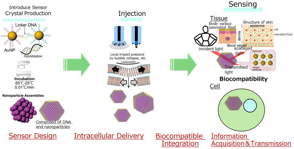
Concept of sensor introduction into cells and tissues
- [1] A. Roth et al., “Quantification of polystyrene uptake by different cell lines using fluorescence microscopy and label-free visualization of intracellular polystyrene particles by raman microspectroscopic imaging,” Cells, Vol.13, No.5, Article No.454, 2024. https://doi.org/10.3390/cells13050454
- [2] H. Hirose et al., “A noncanonical endocytic pathway is involved in the internalization of 3 μm polystyrene beads into HeLa cells,” Biomater. Sci., Vol.10, No.24, pp. 7093-7102, 2022. https://doi.org/10.1039/d2bm01353c
- [3] K. McNear et al., “Understanding cellular internalization pathways of silicon nanowires,” J. Nanobiotechnol., Vol.15 No.1, Article No.17, 2017. https://doi.org/10.1186/s12951-017-0250-0
- [4] J. F. Zimmerman et al., “Cellular uptake and dynamics of unlabeled freestanding silicon nanowires,” Sci. Adv., Vol.2, No.12, Article No.e1601039, 2016. https://doi.org/10.1126/sciadv.1601039
- [5] S. Durán et al., “Internalization and viability studies of suspended nanowire silicon chips in HeLa cells,” Nanomaterials, Vol.10, No.5, Article No.893, 2020. https://doi.org/10.3390/nano10050893
- [6] R. A. J. F. Oerlemans et al., “Artificial organelles: Towards adding or restoring intracellular activity,” ChemBioChem, Vol.22, No.12, pp. 2051-2078, 2021. https://doi.org/10.1002/cbic.202000850
- [7] C. Yao et al., “Dynamic assembly of DNA-ceria nanocomplex in living cells generates artificial peroxisome,” Nat. Commun., Vol.13, Article No.7739, 2022. https://doi.org/10.1038/s41467-022-35472-2
- [8] A. C. Saito et al., “Introducing micrometer-sized artificial objects into live cells: A method for cell–giant unilamellar vesicle electrofusion,” Plos One, Vol.9, No.9, Article No.e106853, 2014. https://doi.org/10.1371/journal.pone.0106853
- [9] D. Singh et al., “Smart nanofluidic systems powered by DNA origami for targeted intracellular delivery: A newer approach,” Explor. Target. Antitumor Ther., Vol.6, Article No.1002349, 2025. https://doi.org/10.37349/etat.2025.1002349
- [10] K. Kansara et al., “Lipid modification of DNA nanocages enhances cellular uptake, migration, and in vivo uptake,” bioRxiv, 2023. https://doi.org/10.1101/2023.05.06.539685
- [11] M. Lee et al., “Stimuli-responsive DNA hydrogel design strategies for biomedical applications,” Biosensors, Vol.15, No.6, Article No.355, 2025. https://doi.org/10.3390/bios15060355
- [12] H. Al-Zarah et al., “A versatile approach for geometry-based self-assembly of DNA-protein hybrid nanostructures using histone-DNA interactions,” Chem. Commun., Vol.61, No.3, pp. 532-535, 2025. https://doi.org/10.1039/d4cc05253f
- [13] X. Ma et al., “DNA-functionalized gold nanoparticles: Modification, characterization, and biomedical applications,” Front. Chem., Vol.10, Article No.1095488, 2022. https://doi.org/10.3389/fchem.2022.1095488
- [14] Z. Peng et al., “Unraveling the time course of interaction between DNA nanopores and lipid bilayers using QCM-D: Role of cholesterol anchors and bilayer supporting substrates,” Nanoscale, Vol.17, No.18, pp. 11668-11678, 2025. https://doi.org/10.1039/d5nr01299f
- [15] A. Ismail and S.-F. Chou, “Polyethylenimine Carriers for Drug and Gene Delivery,” Polymers, Vol.17, No.15, Article No.2150, 2025. https://doi.org/10.3390/polym17152150
- [16] P. Roy et al., “Quinine-Based Polymers Are Versatile and Effective Vehicles for Intracellular pDNA, mRNA, and Cas9 Protein Delivery,” Biomacromolecules, Vol.25, No.10, pp. 6693-6707, 2024. https://doi.org/10.1021/acs.biomac.4c00925
- [17] D. Tang et al., “Autonomous nanorobots as miniaturized surgeons for intracellular applications,” Nanomaterials, Vol.14, No.7, Article No.595, 2024. https://doi.org/10.3390/nano14070595
- [18] S. Mukherjee et al., “Anisotropy, topography and non-newtonian properties of cellular interiors probed by helical magnetic nanobots,” J. Microbio Robot., Vol.21, No.1, Article No.6, 2025. https://doi.org/10.1007/s12213-024-00176-x
- [19] J. Behera et al., “Stable and highly steerable magnetic nanorobots demonstrate therapeutical functionality in crowded biological media,” ACS Nano, Vol.19, No.18, pp. 17785-17795, 2025. https://doi.org/10.1021/acsnano.5c03144
- [20] M. I. Arjona et al., “Intracellular mechanical drugs induce cell-cycle altering and cell death,” Adv. Mater., Vol.34, No.17, Article No.2109581, 2022. https://doi.org/10.1002/adma.202109581
- [21] M. I. Arjona et al., “Integrating magnetic capabilities to intracellular chips for cell trapping,” Sci. Rep., Vol.11, Article No.18495, 2021. https://doi.org/10.1038/s41598-021-98095-5
- [22] N. Torras et al., “Suspended planar-array chips for molecular multiplexing at the microscale,” Adv. Mater., Vol.28, No.7, pp. 1449-1454, 2016.
- [23] J.-C. Lin et al., “Intracellular hydrogelation preserves fluid and functional cell membrane interfaces for biological interactions,” Nat. Commun., Vol.10, Article No.1057, 2019. https://doi.org/10.1038/s41467-019-09049-5
- [24] X. Han et al., “Exosome-coated oxygen nanobubble-laden hydrogel augments intracellular delivery of exosomes for enhanced wound healing,” Nat. Commun., Vol.15, Article No.3435, 2024. https://doi.org/10.1038/s41467-024-47696-5
- [25] C. Zhong et al., “Development of fluorescence lifetime biosensors for ATP, cAMP, citrate, and glucose using the mTurquoise2-based platform,” Cell Rep. Methods, Vol.4, No.11, Article No.100902, 2024. https://doi.org/10.1016/j.crmeth.2024.100902
- [26] R. Gómez-Martínez et al., “Intracellular silicon chips in living cells,” Small, Vol.6, No.4, pp. 499-502, 2010. https://doi.org/10.1002/smll.200901041
- [27] N. Kimura et al., “Cytotransducers for Visualization of Spatiotemporal Intercellular Communication,” Small, Vol.21, No.35, Article No.e2503749, 2025. https://doi.org/10.1002/smll.202503749
- [28] CRDS Research and Development Overview Report (2024), CRDS-FY2024-FR-04 (in Japanese). https://www.jst.go.jp/crds/pdf/2024/FR/CRDS-FY2024-FR-04/CRDS-FY2024-FR-04_20203.pdf [Accessed January 10, 2026]
This article is published under a Creative Commons Attribution-NoDerivatives 4.0 Internationa License.