
Paper:
Glass Bonding Method Utilizing Relatively Mildly Acidic Treatment and Fluorinated Plasma for Stable Micro/Nano Device Fabrication
Masafumi Watanabe*,**
 , Yuki Sakurai*, and Yutaka Kazoe*
*Department of System Design Engineering, Keio University
3-14-1 Hiyoshi, Kohoku-ku, Yokohama, Kanagawa 223-8522, Japan
**Research and Development Department, Kanagawa Institute of Industrial Science and Technology
705-1 Shimoimaizumi, Ebina, Kanagawa 243-0435, Japan

The bonding process is one of the most critical processes in the fabrication of stable, long-term, glass-based micro/nano devices for various applications, including biosensors. However, the current bonding process causes delamination and topographical damage to the detection unit of a biosensor, such as electrodes patterned on a glass substrate, owing to the strong acid treatment used for surface cleaning and activation. Herein, we propose a low-temperature glass bonding method that utilizes a relatively mildly acidic treatment without sacrificing bonding strength. To explore this method, we examined the effects of various acids, such as piranha solution, fuming nitric acid, and hydrochloric acid, on the bonding strength. To measure bonding strength, we treated fused-silica glass substrates with an acid solution, activated with oxygen plasma, bonded to each other, and annealed at 110°C. A crack-opening test using bonded glass substrates revealed that acid treatment is necessary to achieve reliable bonding strength. Relatively mildly acidic treatment, such as with hydrochloric acid, exhibited comparable bonding strength to strong acid treatment, such as piranha solution. Furthermore, we found that the selection of the bonding surface (top or bottom) and the use of fluorinated plasma with polytetrafluoroethylene pieces during oxygen plasma significantly enhance the bonding strength, e.g., 0.76 J/m2 with hydrochloric acid. We believe that our findings will contribute to the development of reliable long-term glass-based biosensors.
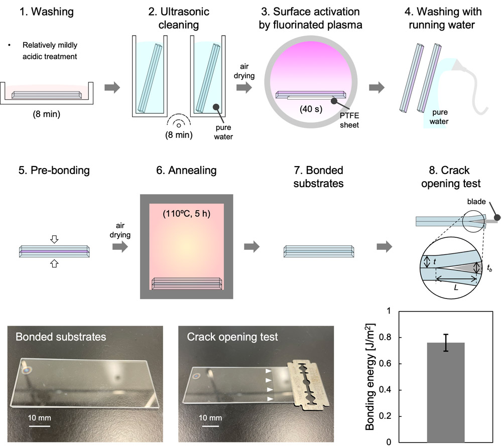
Glass bonding using mildly acidic treatment
1. Introduction
Recent advances in microfluidics have revolutionized the convergence of many scientific and technological fields, including engineering, chemistry, physics, biology, and medicine 1. Microfluidics has also contributed to several fields, including biological analysis, biomedical diagnostics, drug delivery, and biosensors, by manipulating small volumes of fluid in microfluidic devices 2,3,4. Over the past few decades, various materials, including silicon, glass, polydimethylsiloxane, and paper, have been used to fabricate microfluidic devices. Glass is an attractive material for microfluidic device fabrication because of its chemical, thermal, and mechanical stability; compatibility with biological samples; and transparency for observation and detection 5.
Glass-based microfluidic devices have been widely used in biological applications in combination with detection units, such as electrodes 6,7,8, antibodies 9,10, and oligonucleotides 11, to monitor single or multiple cells, proteins, and enzymes. Trans-epithelial/endothelial electrical resistance (TEER) measurement is one of the most common noninvasive methods for monitoring the physiological homeostasis and disease states of cells and tissues. It has been applied to organ-on-a-chip systems using microfluidic devices 12. Glass substrates with patterned electrodes, such as gold and platinum, enable the real-time resistance measurement of cells between electrodes in microfluidic devices based on Ohm’s law 13,14. Recently, indium tin oxide (ITO) has emerged as a promising alternative owing to its transparency and high electrical conductivity 15. For instance, a glass substrate with patterned ITO electrodes was developed for measuring the electrical resistance of a blood–brain barrier model 16. Moreover, other electrode-based models enable the real-time monitoring of biological activities at the single-cell level 17,18. However, these sensitive detection units can deteriorate under adverse conditions during device assembly, such as the surface treatment of substrates with strong acidic solutions 19,20.
Bonding, through which micro/nanochannels on one substrate are sealed with another, is one of the most critical steps in assembling a glass-based microfluidic device. This step is essential for achieving a stable device with reliable bonding strength for long-term use. The commonly used methods include anodic bonding, surface activation, fusion bonding, and adhesive bonding 5. However, anodic and fusion bonding require high temperatures, such as 300 °C–500 °C 21,22,23 and 500 °C–1100 °C 24,25, respectively, which suggests that detection units, such as electrodes, may not tolerate the necessary temperatures for bonding. Additionally, although adhesive bonding is recognized as the simplest method, it is not suitable for microfluidic applications because of its limited resistance to harsh environments over time 26. Conversely, surface-activated bonding using surface activation, e.g., using oxygen plasma, enables low-temperature bonding 27. The activated glass substrates covalently bond to each other via robust Si–O–Si bonds, achieving reliable bonding strength.
Surface activation bonding includes various approaches, such as acidic cleaning 28, basic cleaning 29,30, low-concentration ionic solutions 31, surfactant solutions 32,33, oxygen plasma 9,28, and vacuum ultraviolet (VUV) 34. For basic cleaning, an RCA solution (e.g., a mixture of ammonia and hydrogen peroxide) is one of the most commonly used cleaning methods. However, RCA cleaning is known to leach glass materials 35, potentially leading to the delamination of hybrid materials, such as electrodes on glass. Additionally, bonding using ionic solutions is occasionally combined with the use of surfactant solutions. Owing to the non-negligible size of surfactant micelles (10–20 nm in diameter), nanostructures fabricated on substrates can become clogged. For a stable bonding strength to be achieved using VUV, a high annealing temperature (\(>\)200 °C) is required. This may cause substrate cracking owing to the difference in the thermal expansion coefficients of the hybrid materials, as demonstrated in a previous study 28. Therefore, a combination of acidic cleaning and oxygen plasma appears to be the most appropriate approach for biosensors.
We have previously developed a simple, low-temperature glass bonding process using surface-activated bonding in standard cleanrooms 28. We achieved a reliable bonding strength through strong acid treatment, activation by oxygen plasma, and annealing at \(<\)200 °C. Furthermore, a recent study demonstrated that polytetrafluoroethylene (PTFE)-assisted plasma modification remarkably improves the bonding strength of glass devices by generating fluorine radicals via oxygen plasma 36. These studies required the use of strong acids, such as a piranha solution, to remove contaminants and activate the glass surface. However, compounding evidence shows that piranha solution can cause delamination and topographical damage to thin gold electrodes 37,38. Another study found that ITO thin films can be removed using piranha solution 39. These findings suggest the risk of deteriorating the detection sensitivity and accuracy of biosensors owing to damaged electrodes. Therefore, a new method should be developed for glass bonding using a relatively mildly acidic treatment without sacrificing the bonding strength.
In this study, we developed a low-temperature bonding method that uses fused-silica glass substrates and relatively mildly acidic treatment. First, we investigated the importance of acid treatment in glass bonding and found that this step is necessary to achieve a reliable bonding strength. Second, we compared various acidic solutions, such as piranha solution, fuming nitric acid, and hydrochloric acid. Notably, mildly acidic treatments, such as using hydrochloric acid, exhibited bonding strengths comparable to those of the piranha treatment. We then examined the effect of oxygen plasma treatment and found that prolonged treatment slightly enhanced the bonding strength. Finally, we found that selecting the bonding surface (top or bottom) and adding PTFE pieces during the oxygen plasma treatment significantly increased the bonding strength. Thus, we achieved reliable bonding strength using a mildly acidic treatment. These results will contribute to the development of a cutting-edge biosensor device with reliable bonding strength for long-term use, as well as reliable performance for the high-sensitivity monitoring of cells and tissues.
2. Materials and Methods
2.1. Materials
Fused-silica glass substrates with a thickness of 0.70 mm (70 mm \(\times\) 30 mm, VIO-SILSX, Shin-Etsu Quartz Co., Ltd., Tokyo, Japan) were prepared. The surface roughness of the glass substrates was less than 0.3 nm. PTFE sheets were purchased from MonotaRO Co., Ltd. (Osaka, Japan).
Sulfuric acid (H\(_2\)SO\(_4\), 95% m/m), hydrogen peroxide (H\(_2\)O\(_2\), 30.0–35.5% m/m), fuming nitric acid (HNO\(_3\), 97% m/m), diluted hydrochloric acid (HCl, 0.5 mol/L, equivalent to \({\approx 1.8}\)% m/m), isopropanol (IPA), and acetone were purchased from FUJIFILM Wako Pure Chemical Corporation (Osaka, Japan). Concentrated HCl (35.0–37.0% m/m) was purchased from Kanto Chemical Co., Inc. (Tokyo, Japan).
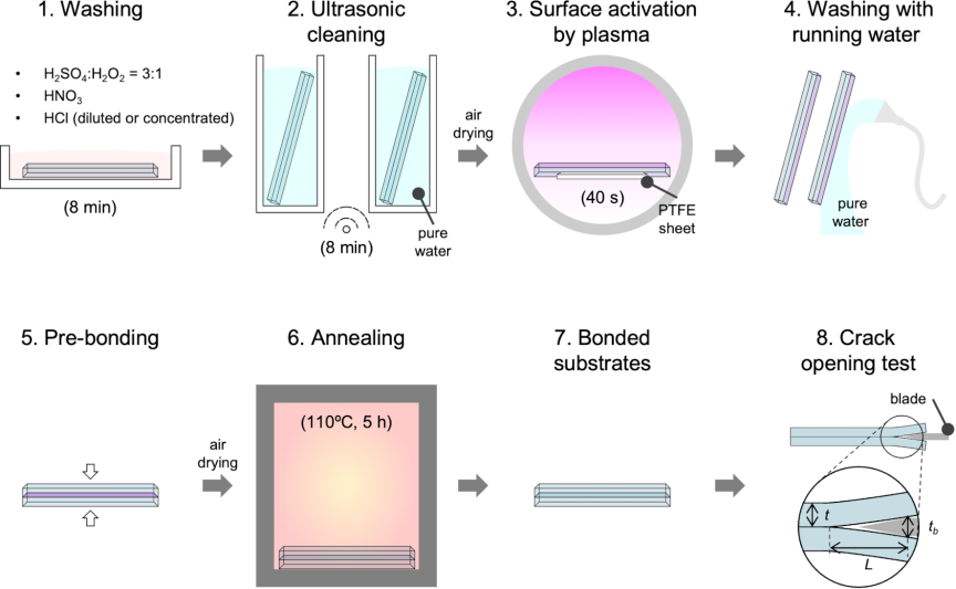
Fig. 1. Schematic of glass bonding process using relatively mildly acidic treatment and fluorinated plasma, as well as measurement of bonding strength of bonded substrates by crack-opening test.
2.2. Low-Temperature Bonding of Glass Substrates
We propose a new method for bonding fused silica glass in cleanrooms. Fig. 1 illustrates the proposed method. To remove organic contaminants and activate the surfaces of the glass substrates, we immersed them in a strong acidic solution, such as piranha solution (\({\textrm{H$_2$SO$_4$} : \textrm{H$_2$O$_2$}} = {3:1}\)) for 8 min, fuming HNO\(_3\) for 8 min, or diluted or concentrated HCl for 10 min in a class ISO 7 cleanroom. The substrates were cleaned in pure water using an ultrasonic cleaner for 8 min, and dried using an air gun. After cleaning, the substrate surfaces were activated with an oxygen plasma exposure of 250 W, with oxygen supplied at a flow rate of 70 mL/min for 40 s, using a gas plasma reactor (PR510, Yamato Scientific Co., Ltd., Tokyo, Japan). To generate fluorine radicals (\(\textrm{F*}\)) via the oxygen plasma, we placed pieces of PTFE in the gas plasma reactor during plasma exposure with the same settings mentioned above. Specifically, two pieces of PTFE (50 mm \(\times\) 20 mm \(\times\) 1 mm) per substrate (“\(\textrm{F*} \times 1\)”) or six pieces per substrate (“\(\textrm{F*} \times 3\)”) were placed close to the substrates in the plasma reactor.
The substrates were immersed in pure water and transported to a class ISO 5 cleanroom to prevent contamination by atmospheric small particles. After the substrates were washed under running pure water and dried with an air gun, they were brought into contact for pre-bonding. The top or bottom surfaces of the substrates were bonded to each other. The “top” surface was defined as the glass surface that was directly exposed to the plasma, whereas the “bottom” was defined as the surface that was indirectly exposed to the plasma. Finally, the pre-bonded substrates were annealed in an electric furnace (FO200, Yamato Scientific Co., Ltd.) at 110 °C for 5 h with an initial heating rate of 100 °C/h. For glass bonding, the treatment conditions for each step were determined based on previous studies 36,40,41,42. In particular, the flow rate of oxygen, power of the plasma treatment, and annealing temperature were optimized in our recent study 28.
2.3. Evaluation of Bonding Strength
The bonding strength of the glass substrates was measured using a crack-opening test 43, which is a standard method for evaluating the stability of bonded substrates. A stainless-steel blade with a thickness of 0.1 mm (Hi-Stainless, Feather Safety Razor Co., Ltd., Osaka, Japan) was used. As shown in Fig. 1, the stainless-steel blade was inserted at the bonding interface, and the crack opening length, \(L\), was measured. The bonding energy, \(\gamma\), between two glass substrates is given by
2.4. Statistical Analysis
A statistical analysis was performed using the statistical software R (version 4.2.1; R Core Team, Vienna, Austria). Data were presented as means \(\pm\) standard deviation (SD). The Shapiro–Wilk test was used to verify data normality. A one-way analysis of variance (ANOVA) and Tukey’s post-hoc test were used to compare differences between groups. A \(p\)-value of \(<0.05\) was considered statistically significant.
3. Results
3.1. Effects of Acid Treatment and Fluorinated Plasma on Bonding Strength
To understand the importance of acid treatment for fused-silica glass substrates, we first investigated its effect on bonding strength. Fig. 2(a) shows photographs of the bonded glass substrates that underwent different types of treatments, either non-acidic (e.g., IPA + acetone) or acidic (e.g., piranha or diluted HCl). Because diluted HCl ranging from 0.1 to 0.5 mol/L is commonly used to clean glass-based probes used for various applications, such as measuring pH or DNA content, we used diluted HCl at 0.5 mol/L. The IPA + acetone treatment produced a larger crack opening length (\(L\)) than piranha and HCl treatments. Fig. 2(b) confirms that the HCl treatment had a bonding energy approximately 4.2 times higher than that of the IPA + acetone treatment, whereas the piranha treatment had a bonding energy approximately 2.6 times higher than that of the IPA + acetone treatment. These results indicate that the acid treatment itself, rather than highly aggressive or concentrated acid conditions, is critical for achieving reliable bonding strength and stable micro/nanofluidic devices.
Furthermore, we investigated the effect of fluorinated plasma on the bonding strength. The addition of PTFE pieces during plasma exposure resulted in a significantly higher bonding energy than that without PTFE pieces when HCl treatment was used, whereas fluorinated plasma treatment without acid cleaning was not explored in the present study. This suggests that fluorinated plasma contributes to the bonding strength in low-temperature bonding with acid cleaning.
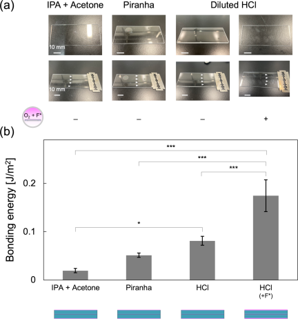
Fig. 2. Comparison of bonding strengths using non-acid and acid treatments. (a) Photographs of bonded glass substrates under different conditions. After acid cleaning, the glass surfaces were activated with an oxygen plasma, followed by fluorinated plasma using PTFE pieces (“\(\textrm{F*} \times 1\)”). The “bottom” surfaces, i.e., surfaces indirectly exposed to the plasma, of the glass substrates were bonded. The pre-bonded substrates were then annealed at 110 °C for 5 h. (b) Bonding energies of bonded substrates. Data represent mean \(\pm\) SD (\(n = 3\)/condition). \({}^{***}\)\({p<0.001}\), \(^{*}\)\({p<0.05}\) (one-way ANOVA followed by Tukey’s honestly significant difference test).
3.2. Comparison of Various Types of Acid Treatments
We conducted a comprehensive study to evaluate the effect of various acids on the bonding strength using piranha, fuming HNO\(_{3}\), and HCl. To achieve a relatively mildly acidic treatment, we used piranha solution 5 min after mixing H\(_{2}\)SO\(_{4}\) and H\(_{2}\)O\(_{2}\) at a \({3:1}\) ratio (“piranha (delayed)”) because piranha solution causes a sudden increase in temperature and rapid boiling. Fig. 3(a) shows photographs of the bonded glass substrates treated with different types of acids. The glass substrates were cleaned and activated using different types of acid treatments, followed by oxygen plasma treatment for 2 min and fluorinated plasma treatment for 40 s. The bottom sides of the substrates were bonded to each other under all conditions. The piranha (delayed) condition did not substantially change the bonding energy compared with the piranha condition, whereas the bonding energy decreased slightly. Diluted HCl exhibited a stronger bonding energy. Fuming HNO\(_3\) exhibited a similar level of bonding energy as the piranha and HCl conditions. Importantly, the relatively mildly acidic treatments, such as piranha (delayed), fuming HNO\(_3\), and diluted HCl, exhibited comparable levels of bonding energy to the strong acid treatment using piranha solution. The respective bonding energies were 0.16 J/m\(^{2}\) (piranha), 0.13 J/m\(^{2}\) (piranha (delayed)), 0.18 J/m\(^{2}\) (fuming HNO\(_3\)), 0.17 J/m\(^{2}\) (diluted HCl), and 0.09 J/m\(^{2}\) (concentrated HCl), as shown in Fig. 3(b).
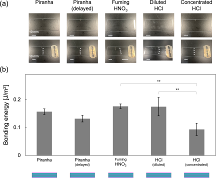
Fig. 3. Comparison of bonding strengths using various types of acid treatments. (a) Photographs of bonded glass substrates under different conditions. After acid cleaning, the glass surfaces were activated with an oxygen plasma for 2 min, followed by fluorinated plasma using PTFE pieces (“\({\textrm{F*} \times 1}\)”) for 40 s. The “bottom” surfaces of the glass substrates were bonded. The pre-bonded substrates were then annealed at 110 °C for 5 h. (b) Bonding energies of bonded substrates. Data represent mean \(\pm\) SD (\(n = 3\)/condition). \({}^{**}\)\({p<0.01}\) (one-way ANOVA followed by Tukey’s honestly significant difference test).
3.3. Effects of Plasma Exposure and Load Condition on Bonding Strength
We examined the effect of treatment time on plasma exposure by varying the duration of oxygen plasma exposure from 0 to 5 min, followed by 40 s of fluorinated plasma exposure. The bottom sides of the substrates were bonded to each other under all conditions. Fig. 4(a) shows photographs of the bonded glass substrates with oxygen plasma treatment times of 0, 2, and 5 min. Fig. 4(b) shows that the bonding energy increased slightly with increasing oxygen plasma treatment time, although the difference was not significant. Furthermore, we investigated the effect of load conditions during the annealing process in an electric furnace. Annealing with a 40 N load produced a bonding energy comparable to that achieved without a load. These results indicate that a relatively light load does not substantially affect the bonding strength, suggesting that a heavier load may be necessary to achieve a higher bonding energy.
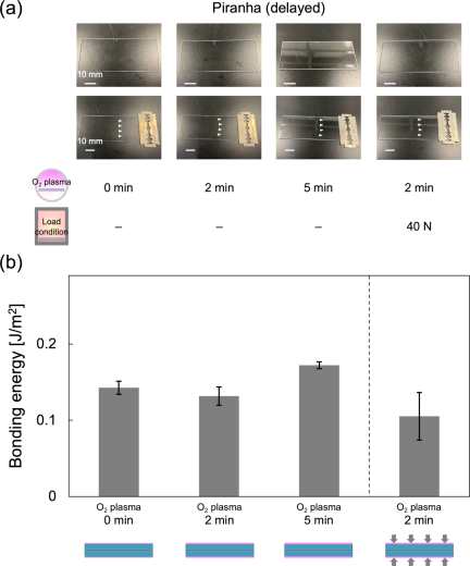
Fig. 4. Comparison of bonding strengths with different treatment times of oxygen plasma and load during the annealing process. (a) Photographs of bonded glass substrates under different conditions. After acid cleaning using piranha (delayed), the glass surfaces were activated with an oxygen plasma for 0, 2, or 5 min, followed by fluorinated plasma using PTFE pieces (“\(\textrm{F*} \times 1\)”) for 40 s. The “bottom” surfaces of the glass substrates were bonded. The pre-bonded substrates were then annealed at 110 °C for 5 h with 0 or 40 N load. (b) Bonding energies of bonded substrates. Data represent mean \(\pm\) SD (\({n = 3}\)/condition).
3.4. Effects of Bonding Surface and Amount of PTFE Pieces on Bonding Strength
We investigated whether the top and bottom surfaces of the substrate were activated by plasma in different ways because the plasma was irradiated onto the substrate from above. The top surface was directly exposed to the plasma, whereas the bottom surface was indirectly exposed. To address this, we compared two conditions: the top surfaces of the substrates were bonded to each other (top), and the bottom surfaces were bonded to each other (bottom). Furthermore, we investigated the effect of number of PTFE pieces on bonding strength by varying 2 pieces/substrate (“\({\textrm{F*} \times 1}\)”) and 6 pieces/substrate (“\({\textrm{F*} \times 3}\)”) of PTFE.
Figure 5(a) shows photographs of the bonded glass substrates under different conditions. The bonded substrates exhibited larger crack-opening lengths (\(L\)) when the bottom surfaces were bonded to each other than when the top surfaces were bonded. This suggested that the top surface was more efficiently activated and cleaned by the oxygen plasma than the bottom surface, resulting in a higher bonding strength. Furthermore, increasing the number of PTFE pieces yielded positive results when the top surfaces of the substrates were bonded. Fig. 5(b) shows that these conditions using PTFE pieces exhibited significantly higher bonding energies: 0.71 J/m\(^{2}\) (“\({\textrm{F*} \times 1}\)”) and 0.99 J/m\(^{2}\) (“\({\textrm{F*} \times 3}\)”), respectively, compared with the condition without PTFE. Collectively, we finalized our low-temperature bonding protocol utilizing a relatively mildly acidic treatment as follows: (i) acid treatment, (ii) ultrasonic cleaning in pure water for 8 min, (iii) oxygen plasma treatment for 2 min and fluorinated plasma treatment with 6 pieces/substrate of PTFE for 40 s, (iv) washing with running water, (v) pre-bonding, where the top surfaces of the substrates were bonded to each other, and (vi) annealing at 110 °C for 5 h without a load. Finally, we prepared bonded substrates using our finalized protocol with another relatively mildly acidic treatment, i.e., diluted HCl. This condition exhibited a high bonding energy of 0.76 J/m\(^2\), similar to that with piranha (delayed) treatment.
In summary, we developed a new method that considers the type of acid treatment, plasma conditions, and bonding surfaces. This method achieves high bonding strength even with relatively mildly acidic treatments, such as piranha (delayed) and diluted HCl treatments.
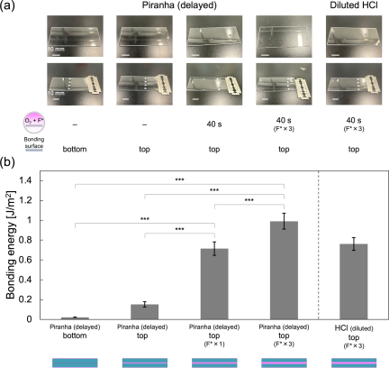
Fig. 5. Comparison of bonding strengths with different bonding surfaces (top and bottom) and amounts of polytetrafluoroethylene (PTFE) pieces. (a) Photographs of bonded glass substrates under different conditions. After acid cleaning using piranha (delayed) or diluted HCl, the glass surfaces were activated with an oxygen plasma for 2 min, followed by fluorinated plasma using PTFE pieces (“\({\textrm{F*} \times 1}\)” or “\({\textrm{F*} \times 3}\)”) for 40 s. The “top” surfaces, i.e., directly exposed to the plasma, or the “bottom” surfaces were bonded, respectively. The pre-bonded substrates were then annealed at 110 °C for 5 h. (b) Bonding energies of bonded substrates. Data represent mean \(\pm\) SD (\({n = 3}\)/condition). \({}^{***}\)\({p<0.001}\) (one-way ANOVA followed by Tukey’s honestly significant difference test).
4. Discussion
4.1. Chemical Effects of Various Acid Treatments
The bonding process requires contaminant-free surfaces. Surface contamination is typically classified as particle contamination (e.g., dust, hair, and fibers), organic contamination (e.g., hydrocarbons from the air), and ionic contamination (e.g., metal ions from metal tweezers or glass containers) 44. These contaminants negatively affect bonding strength.
Piranha treatment, which is based on the reaction of a mixture of concentrated H\(_2\)SO\(_4\) and H\(_2\)O\(_2\) at a \({3:1}\) ratio, is a classical method for cleaning material surfaces 45. Piranha solution is widely used in research and industrial fields. It removes organic contaminants from glass substrates 46. Furthermore, the solution produces many free radicals, including atomic oxygen 45, rendering the surface of a glass substrate hydrophilic via strong oxidation. These effects appear to have contributed to the enhanced bonding strength observed in this study compared with the non-acid-cleaning condition. Additionally, a recent study demonstrated that piranha treatment decreases the surface roughness of silicon substrates 47. A recent paper reported that the bonding strength of glass substrates increases as the surface roughness of the glass decreases 48. Therefore, the enhanced bonding strength observed in the present study may be partly attributed to the decreased surface roughness. However, because the piranha solution is heated to 100 °C–120 °C when H\(_2\)SO\(_4\) and H\(_2\)O\(_2\) are mixed 49, electrodes of a biosensor could be damaged by strong oxidation or a sudden increase in temperature.
HCl treatment is an effective method for removing metal contaminants from glass surfaces 50. For example, sodium, aluminum, and calcium can be removed from glass surfaces using HCl treatment. However, concentrated HCl can negatively affect the surface roughness of a substrate via chemical etching. Specifically, HCl treatment at 36% for 6 h increased the surface roughness of a glass substrate from 0.58 to 1.43 nm, potentially leading to decreased bonding strength. In contrast, a recent study demonstrated that treatment with diluted HCl consisting of deionized water, HCl, and hydrogen peroxide at a \({6:1:1}\) ratio enhances the shear bonding strength of glass substrates by etching calcium ions from the surface 26. For surface activation, HCl treatment is not considered to directly generate hydroxyl groups on glass. Therefore, the decreased bonding strength when concentrated HCl was used in this study can be partly attributed to the increased surface roughness, primarily owing to chemical etching. These findings suggest that highly concentrated HCl or prolonged treatment may result in a decreased bonding strength of the glass substrates.
HNO\(_3\) treatment is commonly used to oxidize organic and inorganic contaminants 51. Additionally, HNO\(_3\) effectively removes sodium, calcium, and aluminum from glass surfaces through chemical etching 52. Furthermore, silanol groups (Si–OH) can be added to silica surfaces by treating silica with HNO\(_3\) 53, potentially leading to an enhanced bonding strength. Additionally, a recent study demonstrated that fuming HNO\(_3\) effectively maintains the surface modification with hydrophobic molecules on a glass substrate, whereas piranha solution may remove organic molecule materials 41. These findings suggest that HNO\(_3\) is an effective candidate for acid treatment of glass bonding.
4.2. Surface Modification by Oxygen and Fluorinated Plasma
Oxygen plasma treatment is one of the most widely used methods for modifying glass and silicon surfaces 54. It introduces oxygen-rich functional groups, such as hydroxyl and carboxyl groups, on the glass surface, which significantly increases its chemical activity. These functional groups enhance the ability of glass to form stronger chemical bonds with other materials during the bonding process, thereby improving adhesion and overall bonding strength. When glass or silicon surfaces are exposed to oxygen plasma, oxygen radicals oxidize and remove adsorbed organic contaminants 27. The activated substrates were exposed to atmospheric conditions. Before pre-bonding, water molecules from the air interacted with the surface, resulting in the formation of silanol groups (Si–OH). Finally, the substrates were covalently bonded to each other via Si–O–Si bonds at the glass–glass interface. The slight increase in bonding strength with prolonged oxygen plasma treatment in this study can be explained by the fact that prolonged plasma exposure increased the density of the Si–OH groups on the glass substrates. Additionally, the enhanced bonding strength when the top surfaces of the substrates were bonded to each other can be explained by the difference in the density of the Si–OH groups between the top and bottom surfaces of the glass substrate.
Furthermore, the addition of PTFE pieces to oxygen plasma led to a significantly higher bonding strength under piranha (delayed) or diluted HCl conditions in the present study. Kang et al. explained that the sputtering effect of ions with kinetic energy and the reactivity of reactive oxygen species generated in oxygen plasma break the C–F bonds of the PTFE piece through chemical oxidation, forming CO, CF\(_{\textrm{x}}\)O\(_{\textrm{y}}\), and fluorine radicals (\(\textrm{F*}\)) 36. Kang et al. also speculated that Si–OH and Si–F groups, as well as the subsurface structure of SiO\(_{\textrm{x}}\)F\(_{\textrm{y}}\) were formed on the surface. The balance between the amounts of hydrophilic Si–OH and hydrophobic Si–F groups can control the water adsorbed on the plasma-activated surface, whereas SiO\(_{\textrm{x}}\)F\(_{\textrm{y}}\) on the interface plays a crucial role in facilitating the diffusion or dissolution of H\(_2\)O, eventually achieving a robust Si–O–Si covalent network between the glass substrates. Moreover, Kang et al. suggested that the dehydration reactions (2) and (3) occur after the two glass substrates come into contact with each other. Because of the smaller volume of HF molecules, the diffusion rate of HF in oxides can be much faster than that in H\(_2\)O molecules, thereby facilitating bonding. The authors also claimed that excessive amounts of hydrophobic Si–F groups on the surface can deteriorate the bonding strength because Si–OH groups are required to form Si–O–Si covalent bonds 36.
Based on these findings, while we observed an increase in the bonding strength by adding PTFE pieces in the present study, prolonged treatment with fluorinated plasma may decrease the bonding strength.
4.3. Future Perspectives and Limitations
We achieved reliable bonding energies using the finalized protocol obtained in this study. These bonding energies were superior to those obtained in our previous study, which did not use PTFE pieces 28. In our previous study, a fused-silica/borosilicate glass device with a bonding energy of 0.5 J/m\(^{2}\) demonstrated the perfusion of fluorescein solution at 600 kPa with no leakage in nanochannels. In addition, other studies have demonstrated that low-temperature bonding of glass substrates utilizing oxygen plasma and piranha cleaning offers a reliable pressure resistance higher than 500 kPa 55,56,57, which is sufficient for various micro/nanofluidic applications. Specifically, the bonding energies obtained in these studies were 0.4–1.1 J/m\(^{2}\), corresponding to pressure resistances of nanochannels ranging from 500 to 2500 kPa. These results suggest that our new bonding method, which utilizes a mildly acidic treatment, can be applied to the development of micro/nanofluidic devices that offer higher or comparable pressure resistance than our previous studies. Furthermore, we revealed that relatively mildly acidic treatments such as piranha (delayed) and diluted HCl did not sacrifice the bonding energy. Therefore, our bonding method will contribute to the development of new glass-based biosensors with sensitive detection units such as electrodes. However, in the present study, bonding experiments were performed exclusively on bare fused-silica glass substrates. Although our current results provide a material-level proof-of-concept for electrode-compatible bonding, further studies are needed to verify whether our proposed bonding protocol can avoid damaging sensitive detection units. In addition, the sample size for each bonding condition was limited in the present study to allow for the comparison of various bonding conditions. Repeating the bonding experiments may be necessary to rigorously assess the reproducibility.
5. Conclusion
We have developed an effective low-temperature bonding method that utilizes a relatively mildly acidic treatment. This method is intended for the fabrication of glass/glass devices with detection units such as electrodes, which are designed for long-term use. We found that the treatment of glass substrates using acid is necessary to remove contaminants and/or activate the glass surface for efficient bonding. Furthermore, in addition to the classical piranha treatment, relatively mildly acidic treatments, such as HCl and HNO\(_3\), as well as piranha (delayed) treatment, can be used to achieve comparable bonding strengths in glass/glass devices. We also found that the oxygen plasma treatment time and the selection of the bonding surface (top or bottom) affected the bonding strength. Furthermore, the addition of PTFE pieces during the oxygen plasma treatment significantly enhances the bonding strength. Because we have provided a materials-level proof-of-concept for electrode-compatible bonding, our method can be used to develop biosensors with electrodes that monitor cell activity in real time with high sensitivity. Additionally, the reliable bonding strength obtained in this study will facilitate the development of long-lasting micro-/nanofluidic devices. Furthermore, we developed nanofluidic modules, including a valve for fluid manipulation and a living single-cell analysis system. We believe that the combination of our proposed bonding method and previously developed nanofluidic modules provides a novel platform for new, durable, and sensitive glass-based micro/nanofluidic biosensors at the single-cell level.
Acknowledgments
This study was based on the results obtained from a project at the Kanagawa Institute of Industrial Science and Technology (KISTEC).
- [1] S. Aralekallu, R. Boddula, and V. Singh, “Development of glass-based microfluidic devices: A review on its fabrication and biologic applications,” Mater. Des., Vol.225, Article No.111517, 2023. https://doi.org/10.1016/j.matdes.2022.111517
- [2] G. M. Whitesides, “The origins and the future of microfluidics,” Nature, Vol.442, No.7101, pp. 368-373, 2006. https://doi.org/10.1038/nature05058
- [3] D. Huh, G. A. Hamilton, and D. E. Ingber, “From 3D cell culture to organs-on-chips,” Trends Cell Biol., Vol.21, No.12, pp. 745-754, 2011. https://doi.org/10.1016/j.tcb.2011.09.005
- [4] Y. Liu et al., “Recent progress in microfluidic biosensors with different driving forces,” TrAC Trends Anal. Chem., Vol.158, Article No.116894, 2023. https://doi.org/10.1016/j.trac.2022.116894
- [5] T. Tang et al., “Glass based micro total analysis systems: Materials, fabrication methods, and applications,” Sens. Actuators B Chem., Vol.339, Article No.129859, 2021. https://doi.org/10.1016/j.snb.2021.129859
- [6] D. Han, Y.-R. Kim, C. M. Kang, and T. D. Chung, “Electrochemical signal amplification for immunosensor based on 3D interdigitated array electrodes,” Anal. Chem., Vol.86, No.12, pp. 5991-5998, 2014. https://doi.org/10.1021/ac501120y
- [7] D.-S. Shin et al., “Micropatterned surfaces functionalized with electroactive peptides for detecting protease release from cells,” Anal. Chem., Vol.85, No.1, pp. 220-227, 2013. https://doi.org/10.1021/ac302547p
- [8] Z. Matharu et al., “Detecting transforming growth factor- release from liver cells using an aptasensor integrated with microfluidics,” Anal. Chem., Vol.86, No.17, pp. 8865-8872, 2014. https://doi.org/10.1021/ac502383e
- [9] T. Nakao et al., “Cytokine analysis on a countable number of molecules from living single cells on nanofluidic devices,” Analyst, Vol.144, No.24, pp. 7200-7208, 2019. https://doi.org/10.1039/c9an01702j
- [10] K. Eyer, S. Stratz, P. Kuhn, S. K. Küster, and P. S. Dittrich, “Implementing enzyme-linked immunosorbent assays on a microfluidic chip to quantify intracellular molecules in single cells,” Anal. Chem., Vol.85, No.6, pp. 3280-3287, 2013. https://doi.org/10.1021/ac303628j
- [11] M. Mirasoli et al., “Portable chemiluminescence multiplex biosensor for quantitative detection of three B19 DNA genotypes,” Anal. Bioanal. Chem., Vol.405, Nos.2-3, pp. 1139-1143, 2013. https://doi.org/10.1007/s00216-012-6573-7
- [12] M. Malik, S. A. Steele, D. Mitra, C. J. Long, and J. J. Hickman, “Trans-epithelial/endothelial electrical resistance (TEER): Current state of integrated TEER measurements in organ-on-a-chip devices,” Curr. Opin. Biomed. Eng., Vol.34, Article No.100588, 2025. https://doi.org/10.1016/j.cobme.2025.100588
- [13] J. Yeste et al., “A compartmentalized microfluidic chip with crisscross microgrooves and electrophysiological electrodes for modeling the blood-retinal barrier,” Lab Chip, Vol.18, No.1, pp. 95-105, 2018. https://doi.org/10.1039/c7lc00795g
- [14] S. Palma-Florez et al., “BBB-on-a-chip with integrated micro-TEER for permeability evaluation of multi-functionalized gold nanorods against Alzheimer’s disease,” J. Nanobiotechnol., Vol.21, No.1, Article No.115, 2023. https://doi.org/10.1186/s12951-023-01798-2
- [15] M. A. Holzreuter and L. I. Segerink, “Innovative electrode and chip designs for transendothelial electrical resistance measurements in organs-on-chips,” Lab Chip, Vol.24, No.5, pp. 1121-1134, 2024. https://doi.org/10.1039/d3lc00901g
- [16] W. Wei, F. Cardes, A. Hierlemann, and M. M. Modena, “3D in vitro blood-brain-barrier model for investigating barrier insults,” Adv. Sci., Vol.10, No.11, Article No.2205752, 2023. https://doi.org/10.1002/advs.202205752
- [17] R. Van den Eeckhoudt et al., “Full-electric microfluidic platform to capture, analyze and selectively release single cells,” Lab Chip, Vol.23, No.19, pp. 4276-4286, 2023. https://doi.org/10.1039/d3lc00645j
- [18] Z. Zhang, T. Zheng, and R. Zhu, “Single-cell individualized electroporation with real-time impedance monitoring using a microelectrode array chip,” Microsyst. Nanoeng., Vol.6, Article No.81, 2020. https://doi.org/10.1038/s41378-020-00196-0
- [19] M. H. Fakhr et al., “Cleaning of LTCC, PEN, and PCB Au electrodes towards reliable electrochemical measurements,” Sci. Rep., Vol.12, Article No.20431, 2022. https://doi.org/10.1038/s41598-022-23395-3
- [20] V. Bhalla, S. Carrara, C. Stagni, and B. Samorì, “Chip cleaning and regeneration for electrochemical sensor arrays,” Thin Solid Films, Vol.518, No.12, pp. 3360-3366, 2010. https://doi.org/10.1016/j.tsf.2009.10.022
- [21] P. Mao and J. Han, “Fabrication and characterization of 20 nm planar nanofluidic channels by glass-glass and glass-silicon bonding,” Lab Chip, Vol.5, No.8, pp. 837-844, 2005. https://doi.org/10.1039/b502809d
- [22] B. R. Fonslow and M. T. Bowser, “Free-flow electrophoresis on an anodic bonded glass microchip,” Anal. Chem., Vol.77, No.17, pp. 5706-5710, 2005. https://doi.org/10.1021/ac050766n
- [23] S. Queste, R. Salut, S. Clatot, J.-Y. Rauch, and C. G. Khan Malek, “Manufacture of microfluidic glass chips by deep plasma etching, femtosecond laser ablation, and anodic bonding,” Microsyst. Technol., Vol.16, No.8, pp. 1485-1493, 2010. https://doi.org/10.1007/s00542-010-1020-1
- [24] J. S. Mellors, V. Gorbounov, R. S. Ramsey, and J. M. Ramsey, “Fully integrated glass microfluidic device for performing high-efficiency capillary electrophoresis and electrospray ionization mass spectrometry,” Anal. Chem., Vol.80, No.18, pp. 6881-6887, 2008. https://doi.org/10.1021/ac800428w
- [25] B. Renberg, K. Sato, T. Tsukahara, K. Mawatari, and T. Kitamori, “Hands on: Thermal bonding of nano- and microfluidic chips,” Microchim. Acta, Vol.166, Nos.1-2, pp. 177-181, 2009. https://doi.org/10.1007/s00604-009-0166-y
- [26] T. Mayer, A. N. Marianov, and D. W. Inglis, “Comparing fusion bonding methods for glass substrates,” Mater. Res. Express, Vol.5, No.8, Article No.085201, 2018. https://doi.org/10.1088/2053-1591/aad166
- [27] C. Wang et al., “Recycled low-temperature direct bonding of Si/glass and glass/glass chips for detachable micro/nanofluidic devices,” J. Mater. Sci. Technol., Vol.46, pp. 156-167, 2020. https://doi.org/10.1016/j.jmst.2019.11.034
- [28] K. Shoda, M. Tanaka, K. Mino, and Y. Kazoe, “A simple low-temperature glass bonding process with surface activation by oxygen plasma for micro/nanofluidic devices,” Micromachines, Vol.11, No.9, Article No.804, 2020. https://doi.org/10.3390/mi11090804
- [29] C. Mai, M. Li, and S. Yang, “Low temperature direct bonding of silica glass via wet chemical surface activation,” RSC Adv., Vol.5, No.53, pp. 42721-42727, 2015. https://doi.org/10.1039/c5ra06705g
- [30] J. Zhou, N. Mei, Z. Leonenko, N. Zhou, and M. Mayer, “Direct glass-to-glass bonding obtained via simplified ammonia-based low-temperature procedure resists high shear stress and powerful CW fiber laser irradiation,” RSC Adv., Vol.12, No.48, pp. 31016-31023, 2022. https://doi.org/10.1039/d2ra04234g
- [31] P. B. Allen and D. T. Chiu, “Calcium-assisted glass-to-glass bonding for fabrication of glass microfluidic devices,” Anal. Chem., Vol.80, No.18, pp. 7153-7157, 2008. https://doi.org/10.1021/ac801059h
- [32] Z.-J. Jia, Q. Fang, and Z.-L. Fang, “Bonding of glass microfluidic chips at room temperatures,” Anal. Chem., Vol.76, No.18, pp. 5597-5602, 2004. https://doi.org/10.1021/ac0494477
- [33] S. Funano, N. Ota, and Y. Tanaka, “A simple and reversible glass-glass bonding method to construct a microfluidic device and its application for cell recovery,” Lab Chip, Vol.21, No.11, pp. 2244-2254, 2021. https://doi.org/10.1039/d1lc00058f
- [34] J. Xu et al., “Mechanisms for low-temperature direct bonding of Si/Si and quartz/quartz via VUV/O activation,” RSC Adv., Vol.8, No.21, pp. 11528-11535, 2018. https://doi.org/10.1039/c7ra13095c
- [35] E. C. Onyiriuka et al., “Effect of RCA cleaning on the surface chemistry of glass and polysilicon films as studied by ToF-SIMS and XPS,” Surf. Interface Anal., Vol.26, No.4, pp. 270-277, 1998. https://doi.org/10.1002/(SICI)1096-9918(199804)26:4<270::AID-SIA371>3.0.CO;2-H
- [36] Q. Kang, C. Wang, K. Liu, and T. Kitamori, “Room-temperature bonding of glass chips via PTFE-assisted plasma modification for nanofluidic applications,” Lab Chip, Vol.23, No.12, pp. 2710-2719, 2023. https://doi.org/10.1039/d3lc00169e
- [37] J. Kang and P. A. Rowntree, “Gold film surface preparation for self-assembled monolayer studies,” Langmuir, Vol.23, No.2, pp. 509-516, 2007. https://doi.org/10.1021/la0518804
- [38] S. Massey, E. Alizadeh, P. A. Rowntree, and L. Sanche, “On the possibility of using low-energy electron stimulated desorption of ions as a surface probe: Analysis of Au substrates,” Int. J. Mass Spectrom., Vol.394, pp. 33-41, 2016. https://doi.org/10.1016/j.ijms.2015.11.005
- [39] S. D’Elia et al., “Ellipsometry investigation of the effects of annealing temperature on the optical properties of indium tin oxide thin films studied by Drude–Lorentz model,” Appl. Surf. Sci., Vol.255, No.16, pp. 7203-7211, 2009. https://doi.org/10.1016/j.apsusc.2009.03.064
- [40] C. Wang, X. Gao, K. Mawatari, and T. Kitamori, “Clogging-free irreversible bonding of polycarbonate membranes to glass microfluidic devices,” J. Electrochem. Soc., Vol.164, No.5, pp. B3087-B3090, 2017. https://doi.org/10.1149/2.0141705jes
- [41] H. Sano, Y. Kazoe, and T. Kitamori, “Stable formation of aqueous/organic parallel two-phase flow in nanochannels with partial surface modification,” Anal. Sci., Vol.37, No.11, pp. 1611-1616, 2021. https://doi.org/10.2116/analsci.21P138
- [42] K. Shirai, K. Mawatari, and T. Kitamori, “Extended nanofluidic immunochemical reaction with femtoliter sample volumes,” Small, Vol.10, No.8, pp. 1514-1522, 2014. https://doi.org/10.1002/smll.201302709
- [43] W. P. Maszara, G. Goetz, A. Caviglia, and J. B. McKitterick, “Bonding of silicon wafers for silicon-on-insulator,” J. Appl. Phys., Vol.64, No.10, pp. 4943-4950, 1988. https://doi.org/10.1063/1.342443
- [44] A. Plößl and G. Kräuter, “Wafer direct bonding: Tailoring adhesion between brittle materials,” Mater. Sci. Eng. R Rep., Vol.25, Nos.1-2, pp. 1-88, 1999. https://doi.org/10.1016/S0927-796X(98)00017-5
- [45] Y. Liao et al., “Piranha solution treatment: A facile method for improving the antithrombotic ability and regulating smooth muscle cell growth on blood contact materials,” Front. Bioeng. Biotechnol., Vol.11, Article No.1166334, 2023. https://doi.org/10.3389/fbioe.2023.1166334
- [46] J. Yang, H. Kamai, and Y. Xu, “A simple chemical method to nondestructively regenerate functional nanochannels for single-molecule studies,” Sens. Actuators B Chem., Vol.393, Article No.134106, 2023. https://doi.org/10.1016/j.snb.2023.134106
- [47] M. Holmes, J. Keeley, K. Hurd, H. Schmidt, and A. Hawkins, “Optimized piranha etching process for SU8-based MEMS and MOEMS construction,” J. Micromech. Microeng., Vol.20, No.11, Article No.115008, 2010. https://doi.org/10.1088/0960-1317/20/11/115008
- [48] R. Ohta, K. Morikawa, Y. Tsuyama, and T. Kitamori, “Relationship between bonding strength and surface roughness in low-temperature bonding of glass for micro/nanofluidic device,” J. Micromech. Microeng., Vol.34, No.1, Article No.017002, 2024. https://doi.org/10.1088/1361-6439/ad104c
- [49] H. G. Schmidt, “Safe piranhas: A review of methods and protocols,” ACS Chem. Health Saf., Vol.29, No.1, pp. 54-61, 2022. https://doi.org/10.1021/acs.chas.1c00094
- [50] H. K. Jang et al., “Effects of chemical etching with hydrochloric acid on a glass surface,” J. Vac. Sci. Technol. A, Vol.18, No.5, pp. 2563-2567, 2000. https://doi.org/10.1116/1.1287445
- [51] M. L. Hobbs et al., “The role of nitric acid, kitty litter, and confinement in the 2014 WIPP accident,” Sci. Technol. Energ. Mater., Vol.86, No.1, pp. 11-18, 2025. https://doi.org/10.34571/stem.86.1_11
- [52] H. K. Jang et al., “Effects of chemical etching with nitric acid on glass surfaces,” J. Vac. Sci. Technol. A, Vol.19, No.1, pp. 267-274, 2001. https://doi.org/10.1116/1.1333087
- [53] N. V. Reichhardt, T. Nylander, B. Klösgen, V. Alfredsson, and V. Kocherbitov, “Porosity and surface properites of SBA-15 with grafted PNIPAAM: A water sorption calorimetry study,” Langmuir, Vol.27, No.22, pp. 13838-13846, 2011. https://doi.org/10.1021/la203093u
- [54] Z. Wang et al., “Glass surface treatment and bonding methods for microsystem packaging,” Mater. Today Chem., Vol.46, Article No.102698, 2025. https://doi.org/10.1016/j.mtchem.2025.102698
- [55] Y. Xu et al., “Low-temperature direct bonding of glass nanofluidic chips using a two-step plasma surface activation process,” Anal. Bioanal. Chem., Vol.402, No.3, pp. 1011-1018, 2012. https://doi.org/10.1007/s00216-011-5574-2
- [56] Y. Xu et al., “Bonding of glass nanofluidic chips at room temperature by a one-step surface activation using an O/CF plasma treatment,” Lab Chip, Vol.13, No.6, pp. 1048-1052, 2013. https://doi.org/10.1039/c3lc41345d
- [57] R. Ohta, K. Mawatari, T. Takeuchi, K. Morikawa, and T. Kitamori, “Detachable glass micro/nanofluidic device,” Biomicrofluidics, Vol.13, No.2, Article No.024104, 2019. https://doi.org/10.1063/1.5087003
This article is published under a Creative Commons Attribution-NoDerivatives 4.0 Internationa License.