
Review:
Sensor Introduction into Cells and Tissues for Information Transmission
Yoko Yamanishi

Department of Mechanical Engineering, Kyushu University
744 Motooka, Nishi-ku, Fukuoka, Fukuoka 819-0395, Japan

Accurate transmission of biological information from within cells and living tissues often requires direct sensing of intracellular states. This review discusses recent advances, challenges, and future perspectives in intracellular sensing technologies and bio-integrated information systems. Embedding sensors or artificial constructs inside cells, rather than relying solely on surface-based approaches, are particularly important for acquiring intracellular biochemical and molecular information. In contrast, certain physical parameters, such as temperature, can in some cases be monitored from the cell surface or extracellular environment. However, intracellular sensing systems face significant challenges, including weak signal output, low signal-to-noise ratios, and limited biocompatibility that restrict their long-term functionality. Addressing these limitations is critical for advancing bio-integrated information technologies.
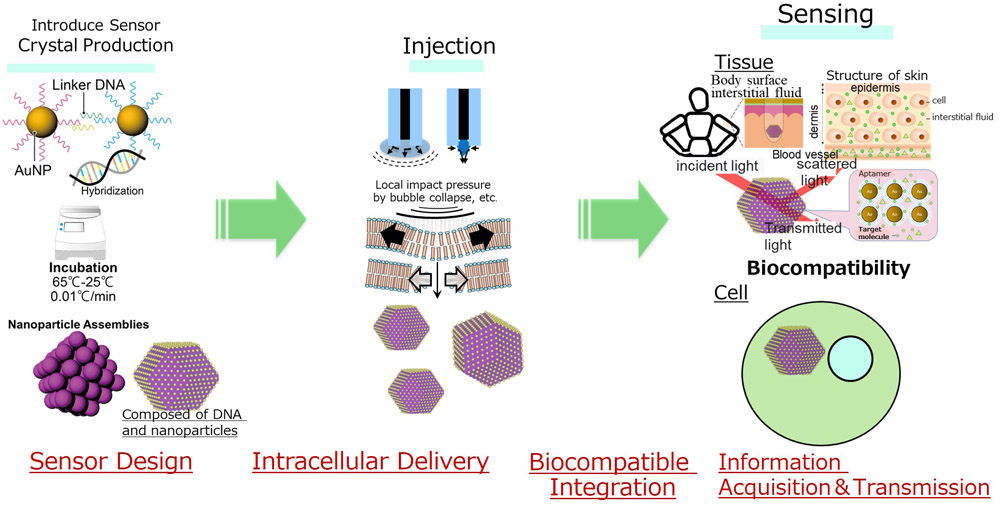
Concept of sensor introduction into cells and tissues
1. Introduction
The direct sensing of intracellular states is a prerequisite for the accurate transmission of biological information. Recent advances in nanotechnology and synthetic biology have enabled the development of artificial intracellular structures that function as biosensors, reporters, or information-processing modules. This review focuses on the introduction of artificial structures into cells and tissues for information acquisition and signal transmission.
2. Intracellular Delivery Technologies for Artificial Structures
This section provides an overview of intracellular delivery technologies for introducing relatively large artificial structures ranging in size from several hundred nanometers to several micrometers into cultured cells or cellular tissues.
2.1. Microinjection Method
Features: Because the structure is directly injected into individual cells, even relatively large structures of the order of several micrometers can be introduced.
Advantages: It enables highly precise and localized delivery to targeted cells.
Disadvantages: It has low throughput and requires considerable technical skill and expertise.
2.2. Electroporation Method
Features: Electroporation enables the delivery of nanoscale particles into cells by applying an electric field to create transient pores in the cell membrane.
Advantages: It allows the simultaneous processing of a large number of cells.
Disadvantages: The delivery efficiency decreases for structures larger than several hundred nanometers. In addition, there is a risk of cell damage caused by the electric fields.
2.3. Endocytosis-Mediated Delivery
Features: This approach utilizes the natural cellular uptake mechanisms. The efficiency of intracellular delivery can be enhanced through the ligand modification of the particle surfaces of artificial structures.
Advantages: It relies on physiological cellular processes, making it inherently biocompatible.
Disadvantages: The size of the artificial structures that can be internalized is typically limited to approximately 500 nm or less. In addition, uptake efficiency depends on the cell type.
2.4. Membrane Disruption Using Microfluidic Devices
Features: Cells are forced through narrow microfluidic channels, causing a temporary disruption of the cell membrane and enabling the introduction of artificial structures into the cell.
Advantages: It offers high throughput and is compatible with the delivery of nano- to submicrometer-sized structures.
Disadvantages: It can impose significant mechanical stress on the cells, potentially affecting their viability and function.
2.5. Cell-Penetrating Peptides (CPPs) and Liposome-Based Delivery
Features: Artificial structures can be encapsulated within lipid membranes or functionalized using specific peptide sequences to enhance their ability to cross cell membranes.
Advantages: Effective intracellular delivery of molecular complexes and other cargo-containing constructs.
Disadvantages: Delivery becomes challenging as the size of the artificial structure increases.
Based on the above discussion, microinjection may currently be the only reliable method for intracellular delivery when artificial structures are several micrometers in size. Electroporation, membrane disruption using microfluidic devices, and endocytosis-mediated uptake are potential options for structures measuring several hundred nanometers.
However, it should be noted that the choice of delivery method depends heavily on the cell type and specific experimental objectives, such as whether cell viability or throughput is the primary consideration. Therefore, the most appropriate delivery strategy must be selected on a case-by-case basis.
3. Recent Trends in Delivered Material in Cells
3.1. Microbeads
Most reports involving microbeads have focused on their use as drug delivery systems or diagnostic applications of magnetic microbeads, whereas there have been very few examples in which microbeads themselves exhibit or perform specific biological functions. However, some studies under the microbead category were related to polystyrene microparticles.
The size (diameter) of polystyrene microparticles typically ranges from approximately 100 nm to 3 μm. The reported mechanisms of intracellular uptake include endocytosis (both clathrin-dependent and clathrin-independent pathways), macropinocytosis-like pathways, and other non-canonical internalization routes.
Roth et al. 1 reported that A549, CaCo-2, THP-1, and other cell types internalize fluorescently labeled polystyrene beads ranging in size from approximately 100 nm to several micrometers depending on their concentration. They further demonstrated that the uptake of these particles can be quantitatively analyzed and visualized using confocal and Raman microscopy.
Hirose et al. 2 demonstrated that 3-μm-diameter beads can be internalized by HeLa cells via a noncanonical endocytic pathway, providing evidence of an uptake mechanism that is distinct from the conventional clathrin-dependent and caveolin-dependent endocytic pathways.
3.2. Nano-/Microcapsules and Nanowires
Similar to microbeads, nano-/microcapsules have been reported primarily as drug delivery vehicles and for diagnostic applications. However, there are relatively few examples in which they perform specific biological functions. Among various nano-and microcapsule-related structures, silicon nanowires have attracted considerable attention.
Silicon nanowires are high-aspect-ratio structures composed of silicon, typically with diameters ranging from several tens to hundreds of nanometers and lengths of several micrometers. In other words, they are extremely slender rod-shaped structures with nanoscale diameters and microscale lengths. Silicon nanowires have been investigated for various biomedical applications, including biosensing (real-time monitoring of local intracellular environments such as pH and ion concentrations), drug delivery (transport of drugs or biomolecules immobilized on their surfaces into cells), and tissue engineering and electrical stimulation (integration into three-dimensional scaffolds for recording and stimulating the electrical activity of neurons and cardiomyocytes).
Silicon nanowires offer several advantages, including ease of surface functionalization (e.g., amine modification and oxide layer formation), high mechanical strength, electrical conductivity, and chemical stability.
The known mechanisms for the cellular uptake of silicon nanowires include membrane wrapping, in which the cell membrane envelops the nanowire and internalizes it through an endocytosis-like process; phagocytosis, which has been reported particularly in macrophages and stem cells; and medium-incubation methods, whereby nanowires dispersed in the culture medium are taken up simply through co-cultivation with cells.
Representative examples of silicon nanowire internalization are described below.
McNear et al. 3 used optical microscopy to confirm that amine-functionalized silicon nanowires (SiNW-NH2) are internalized by CHO cells through a membrane-wrapping-like mechanism, in which the cell membrane progressively envelops the nanowires prior to their uptake.
Zimmerman et al. 4 demonstrated, using live-cell tracking, that silicon nanowires are internalized in multiple cell lines through an active burst-like transport mechanism, in which cargo and organelles do not move continuously at a constant velocity but instead undergo repeated cycles of rapid acceleration and pausing. These findings suggest that silicon nanowires have significant potential as platforms for effective intracellular sensing and drug delivery systems (DDS).
Durán et al. 5 developed a 3 μm \(\times\) 3 μm silicon nanowire chip with two distinct architectures integrating intracellularly deliverable silicon chips and silicon nanowires. These structures were internalized by HeLa cells and compared with non-nanostructured silicon chips.
Both types of chips were successfully taken up by cells without affecting cell viability; however, differences in cellular behavior were observed. In particular, during the early stages of intracellular uptake, the silicon nanowires exhibited superior internalization characteristics compared to bulk silicon. Furthermore, the chips were found to reside within membrane-bound vesicles, whereas some nanowires penetrated the cytosol.
These findings suggest the potential of silicon nanowire chips as a platform for the future development of intracellular sensors and DDS.
3.3. Artificial Organelles
Compartmentalization of intracellular organelles is one of the defining features of living systems. Cells regulate their biological processes more effectively by creating physically separated and specialized microenvironments. Therefore, researchers have explored various technologies aimed at introducing artificial organelles into cells to endow them with functions, such as novel catalytic activities and intracellular monitoring capabilities, which are not found in nature.
Oerlemans et al. 6 conceptually classified artificial organelles into two major categories based on their mode of construction and method of intracellular implementation. The first category consisted of structures that cannot be biosynthesized within cells and therefore must be synthesized externally and subsequently introduced into the cell. Examples include transition metal-containing nanoparticles that exhibit catalytic activity inside cells and nanoreactors that function transiently after intracellular delivery.
The second category of organelles required the introduction of genes encoding the constituent proteins of artificial organelles into cells in the form of viral DNA or plasmid DNA, which was followed by the intracellular production of the artificial organelles through genetic engineering. Here, the cell itself serves as the manufacturing platform for the artificial organelles.
As described below, both strategies have distinct advantages and limitations, making it difficult to conclude that either approach is universally superior to the other.
3.3.1. Structures Synthesized Extracellularly and Subsequently Introduced into Cells
Organometallic Complexes
Organometallic catalysts exhibit a high degree of chemical diversity and can catalyze reactions that do not occur naturally in living systems. Consequently, numerous studies have explored the intracellular introduction of such catalytic systems for various applications, including the labeling of cellular components and the localized activation of prodrugs. For example, transition metals, such as Fe, Cu, and Zn, play essential roles as cofactors in enzymatic reactions, and efforts have been made to introduce these metals into cells to regulate cellular functions.
Semi-Synthetic Artificial Structures
As organometallic complexes are generally hydrophobic, they can traverse cell membranes relatively easily. In contrast, the intracellular uptake of proteins, particularly enzymes, is limited. Enzymes are susceptible to proteolytic degradation and modifications intended to improve their stability or cellular uptake may adversely affect their catalytic activities. Therefore, carrier systems capable of encapsulating proteins are indispensable for delivering protein molecules such as enzymes into cells while preserving their native functionality.
Very few studies have focused on artificial non-protein structures and the technologies required for their introduction into cells remains limited.
3.3.2. Artificial Structures That Can Be Produced Intracellularly Through Genetic Engineering
A greater degree of design flexibility can be achieved with nanoreactors based on proteinaceous architectures than with conventional artificial structures. However, the implementation of this approach requires simultaneous intracellular production of the capsule structure and its contents (e.g., catalytic components) as well as the precise assembly of catalytically functional structures. Ideally, such structures should either confer novel catalytic activities that are absent in the host cell, or restore and enhance existing cellular functions.
Although these genetically encoded structures have the advantage of being inherited by subsequent generations through gene expression, they are extremely challenging to biosynthesize because all the components of the artificial structure must be produced within the cell 6. In other words, successful implementation requires the development of sophisticated systems capable of intracellular self-assembly, autonomous cargo loading, and the expression of catalytic activity; this makes the technical hurdles substantially higher than those associated with extracellularly synthesized artificial structures.
Several studies have reported artificial structures of the order of a few micrometers that can be introduced into cells and thereafter remain functionally active. However, artificial organelles larger than several tens of micrometers that function within cells have not been identified. On the other hand, structures with sizes in the range of approximately 3–10 μm have been developed primarily for extracellular analytical applications, and they do not yet appear to have reached the stage of functioning as intracellular artificial organelles.
Yao et al. 7 constructed an artificial peroxisome (AP) based on a nanohybrid of DNA and ceria (cerium oxide). This DNA–ceria nanohybrid was synthesized from branched DNA containing an i-motif structure that responds to the acidic lysosomal environment, thereby inducing a structural transformation from the nanoscale to bulk scale. Cellular uptake is enhanced when the construct remains in its initial nanoscale form, whereas the bulk-scale AP formed after structural transformation promotes intracellular retention.
The AP exhibits enzyme-mimetic catalytic activity and functions as a reactive oxygen species (ROS) scavenger by decomposing H\(_2\)O\(_2\) into O\(_2\) and H\(_2\)O, thereby eliminating intracellular ROS. Consequently, in living cells, AP efficiently regulates intracellular ROS levels and protects against disruptions in redox homeostasis by preventing glutathione (GSH) depletion.
Furthermore, under AP-mediated protection, cytoskeletal integrity, mitochondrial membrane potential, intracellular calcium levels, and ATPase activity were maintained even under oxidative stress. The energy supply required for cell migration was preserved. The authors concluded that AP suppressed cellular apoptosis and reduced cell death via ROS-scavenging activity.
As an example of intracellular delivery of micrometer-sized artificial structures, Saito et al. 8 demonstrated the intracellular delivery of micrometer-sized structures approximately 1 μm in diameter. In their approach, 1-μm fluorescent beads and DNA origami structures were encapsulated within giant unilamellar vesicles (GUVs), which were subsequently fused with cultured cells, such as HeLa cells, via electrofusion, thereby enabling the intracellular delivery of the encapsulated materials. Furthermore, this study concluded that the introduced structures could be inherited by daughter cells during cell division.
Although electrofusion-based intracellular delivery is unlikely to be readily applicable in clinical setting, it is an effective strategy for generating designer cells and other engineered cellular systems.
3.4. DNA-Containing Structures
Similar to DNA origami, various artificial structures utilize DNA as part or all of their structural framework and can be introduced into cells. Some examples of artificial DNA-containing structures are described below.
It should be noted that the terminology used for DNA-based artificial structures varies among researchers. Consequently, some categories presented here may represent overlapping or closely related technologies. Nevertheless, to comply with the nomenclature adopted by the original authors, the structures were classified according to the names used in the respective publications.
3.4.1. DNA Origami
DNA origami is a class of artificial structures in which long DNA strands are folded and stapled with short DNA oligonucleotides to form precisely defined two-dimensional and three-dimensional architectures. The reported applications include molecular sensing, drug delivery carriers, and targeted therapies, particularly for cancer cells. DNA origami is a nanoscale structure created by folding DNA in an origami-like manner; when introduced into cells, it can be used for the molecular delivery and regulation of cellular functions.
Generally, DNA origami is viewed as a potential alternative to conventional liposomes and lipid nanoparticles. However, recent developments have expanded their functional capabilities beyond those of traditional delivery systems, as described below.
Several approaches have been employed for the intracellular delivery of DNA origami 9. Because DNA is negatively charged, one common strategy is to use complex DNA origami with cationic polymers or lipids using cationic transfection reagents to facilitate its passage across the cell membrane. Another approach involves cell-membrane targeting, in which cell-specific ligands or DNA modifications are used to bind membrane receptors and promote their uptake through endocytosis. In addition, photo responsive control methods have been developed wherein light-responsive elements are incorporated into the DNA origami structure, enabling structural transformations and spatiotemporal control after cellular uptake 9.
The reported functions of intracellularly delivered DNA origami include DDS, in which drugs or RNA molecules are encapsulated within capsule-shaped DNA origami structures and subsequently released inside the cells. Other applications include the regulation of cellular functions by incorporating enzymes or ligands into the structure to manipulate intracellular signaling pathways and metabolic processes. DNA origami has also been used as a mechanical sensor by designing spring-like structures to measure the mechanical behavior of cell-adhesion molecules, such as integrins. Furthermore, DNA origami can play a protective role by forming a nanoscale shell on the cell surface that shields the cells from external stresses.
The size of the DNA origami structures typically ranges from several tens to hundreds of nanometers, which is comparable to that of many viral particles.
3.4.2. DNA Nanocages
DNA nanocages are box-shaped or spherical structures constructed from DNA that are capable of encapsulating cargo such as drugs or enzymes within their interior. A key feature of these structures is their ability to undergo controlled opening and closing in response to specific stimuli such as changes in pH or the presence of particular enzymes. This responsiveness enables the targeted release of encapsulated therapeutics at desired intracellular sites.
Kansara et al. 10 reported that a DNA tetrahedron modified with liposterolized DOTMA (TdN–DOTMA) was efficiently internalized by both cultured cells and zebrafish embryos, and exhibited enhanced stability following uptake. The authors demonstrated that the TdN–DOTMA nanostructure possessed favorable cellular uptake characteristics and maintained its structural integrity in biological environments, highlighting its potential as a platform for intracellular delivery applications.
3.4.3. DNA Hydrogels
DNA hydrogels are gel-like structures formed by crosslinking DNA strands and have been explored for applications, such as sustained intracellular drug delivery. Their key characteristics include biodegradability and responsiveness to external stimuli such as temperature, pH, and enzymes.
Lee et al. 11 provided an overview of stimuli-responsive DNA hydrogels. The authors described how the individual components of these hydrogels can be tailored according to the intended application, with DNA serving as a biocompatible structural framework. Furthermore, the incorporation of inorganic nanomaterials or synthetic polymers enhances the stability and functionality of hydrogel systems.
In addition, stimuli-responsive linkers selected to respond to specific triggers such as changes in pH, temperature, and metal ion concentration can induce structural transformations within the hydrogel. Such systems enable the controlled release of therapeutic agents, target analytes, and signal-generating molecules, making them attractive platforms for biomedical and analytical applications 11.
3.4.4. DNA–Protein Hybrid Structures
The DNA–protein hybrid structures combine programmable DNA scaffolds with functional proteins; they have applications in intracellular sensing, catalysis, and artificial organelles 12.
3.4.5. DNA–Metal Nanoparticle Complexes
By arranging metal nanoparticles (e.g., Au and Ag) using DNA as template, it is possible to construct DNA–metal nanoparticle complexes that can be applied in various biomedical and technological fields, including biosensing, imaging based on fluorescent and optical properties, and photothermal therapy 13.
3.4.6. DNA-Lipid Nanostructures
This type of structure consists of DNA encapsulated within a lipid membrane that forms a liposome-like architecture. Its applications include enhancing cell membrane permeability and serving as a delivery vehicle for siRNAs, drugs, and other therapeutic agents.
Peng et al. 14 designed a DNA nanopore (DNP) by modifying a conventional DNA nanopore using hydrophobic cholesterol groups. They also developed a model system in which a polymer layer was inserted between a lipid bilayer membrane and a supporting substrate (silicon dioxide, SiO\(_2\)), thereby altering the membrane support environment. Using this system, they were able to monitor in real time the process by which DNP was adsorbed and inserted into the lipid membrane.
The results showed that DNA nanopores functionalized with three cholesterol moieties exhibited a higher level of membrane adsorption than DNA nanopores containing only a single cholesterol group, leading to an overall increase in membrane rigidity. Furthermore, when the SiO\(_2\) support was coated with polyethylene glycol (PEG) polymer, the rate of insertion of the DNA nanopores into the lipid membrane increased substantially. These findings demonstrate that both the degree of cholesterol functionalization and the properties of the membrane-support environment strongly influence the membrane interaction and insertion behavior of DNA nanopores.
3.4.7. DNA–Polymer Complexes
DNA–polymer complexes are structures in which DNA is combined with polymers to enhance their stability. Potential applications include improving the long-term intracellular stability and serving as platforms for gene therapy.
For example, Ismail et al. 15 reported that DNA complexes with polymers such as polyethyleneimine (PEI) and poly-L-lysine are widely used as tools for intracellular gene delivery. In addition, Roy et al. 16 successfully employed polymer-containing delivery systems for the intracellular delivery of large nucleic acids, including plasmid DNA (pDNA), messenger RNA (mRNA), and Cas9, highlighting their potential as safe and highly efficient gene transfer carriers.
It should be noted that, for intracellular delivery, these structures are often combined with cationic polymers, lipid nanoparticles, endocytosis-promoting peptides, or related delivery-enhancing agents to facilitate cellular uptake.
3.5. Nano-/Microrobots and Microswimmers
The technological foundation for the intracellular delivery of nanorobots is steadily being established, and interdisciplinary applications are emerging, including localized manipulation using magnetic field guidance, in-vivo navigation under MRI control, and membrane engineering based on DNA origami structures. As reviewed by Tang et al. 17, advances in propulsion mechanisms and surface functionalization have enabled nanorobots to rapidly penetrate cell membranes and achieve efficient intracellular uptake.
However, it should be noted that intracellular localization is not necessarily a requirement for all nanorobot applications, and care must therefore be taken when interpreting the reported results. Specifically, several publications have not provided definitive evidence that nanorobots are present within cells. For example, in studies on nanorobots designed to deliver drugs to cancer cells, the release of therapeutic agents at the cell surface rather than after intracellular entry cannot be ruled out.
Future challenges in the development of nanorobots include improving intracellular uptake efficiency, establishing long-term safety, and achieving organ- or tissue-specific targeting. Addressing these issues is an essential step toward the clinical translation of nanorobot-based technologies.
Mukherjee et al. 18 reported magnetic nanobots capable of probing intracellular mechanical properties.
The authors reported that the nanobots exhibited orientational changes and variations in hydrodynamic pitch within the cellular environment. Based on these behaviors, they concluded that the nanobots were capable of directly sensing intracellular mechanical properties, including non-Newtonian characteristics and structural anisotropy of the cytoplasmic environment.
Behera et al. 19 demonstrated that nanorobots incorporating superparamagnetic iron oxide nanoparticles (SPIONs) into a polymer matrix and possessing a helical structure (approximately 2.8–3.0 μm in length and approximately 400 nm in diameter) can be magnetically manipulated within highly dense biological matrices. The propulsion of these nanorobots was achieved through rotation driven by an external magnetic field. Specifically, the rotating magnetic field generated by the three-axis Helmholtz coil induces a corkscrew-like motion that propels the nanorobot forward.
The study showed that magnetic responsiveness and biocompatibility could be achieved simultaneously, enabling nanorobots to navigate through dense extracellular matrices and other extracellular biological environments. Furthermore, the authors demonstrated the feasibility of localized magnetic hyperthermia therapy against certain types of cancer cells in vitro.
However, the study did not provide data demonstrating the intracellular internalization of these nanorobots. Therefore, it is highly likely that the nanorobots exerted their influence without entering the cells. Nevertheless, if the intended therapeutic or diagnostic objectives can be achieved by simply delivering appropriate stimuli to cells without requiring intracellular entry, the development and practical application of nanorobot technologies in this field may progress rapidly in the future.
3.6. Microchips / Silicon Chips
Several techniques have been developed to embed microchips directly into living cells without compromising cell viability. Functional validation studies have already demonstrated the use of intracellular microchips such as barcodes, biochemical sensors, mechanical sensors, and electrical devices within living cells. Technologically, these studies suggest that nanoscale microchips can be integrated into cells and serve as platforms for sensing and controlling intracellular functions.
Therefore, further advances in biomicrorobotics and intracellular nanosensors are anticipated. Recent representative studies are summarized below.
Arjona et al. 20 demonstrated that the silicon chip device SL-500, once internalized by HeLa cells, induces abnormalities in cell cycle progression and promotes cell death. Specifically, 90.38% of cells containing SL-500 exhibited characteristics of a mechanically altered cell cycle (MACC). Furthermore, 31.35\(\pm\)9.98% of SL-500-containing cells underwent cell death, whereas the incidence of cell death was less than 1% in cells that did not contain the device.
These findings suggest that the observed cell death was caused by device internalization and was a direct consequence of the increased occurrence of MACC events. Because the shape and rigidity of the device were identified as the major determinants of the resulting mechanical disturbances, the authors further demonstrated that the intracellular confinement effects could be mitigated by repositioning the device to locations within the cell that were less likely to cause damage.
Arjona et al. 21 developed an intracellular nickel-based chip that integrated both encoding and magnetic manipulation capabilities into a single device, enabling the capture and enrichment of cells through magnetic control. Using this technology, cells can be selectively trapped and concentrated in a noncontact manner.
The authors demonstrated that the device could be internalized by HeLa cells without adversely affecting cell viability. Furthermore, the incorporated barcode functionality enabled the identification of specific cell subpopulations, whereas the magnetic properties of the chip allowed barcode-containing cells to be magnetically trapped and enriched.
This study is expected to pave the way for the development of intracellular chips that combine magnetic cell manipulation with multifunctional capabilities, thereby expanding the potential applications of intracellular microdevices in cell engineering and biomedical research.
Torras et al. 22 developed a suspension planar array (SPA)-based device equipped with a multiplexed assay system for intracellular sensing. As a proof of concept, they successfully used the SPA chip to detect intracellular pH changes in living HeLa cells.
The developed SPA chip is a powerful tool for obtaining direct information about the cellular microenvironment. The real-time monitoring of intracellular parameters provides a valuable platform for investigating cellular physiology and dynamics. Consequently, this technology has the potential to offer new insights into fundamental cell biology and advance the development of intracellular sensing methodologies.
3.7. Artificial Cytoskeletal Structures (Hydrogel Microparticles)
Hydrogel microparticles and microgels have been studied primarily as carriers for the intracellular delivery of drugs and biomolecules. However, there have been relatively few reports of these materials performing specific biological functions within cells. The examples discussed below represent applications that differ somewhat from the conventional use of hydrogels and illustrate more unconventional functional roles.
Lin et al. 23 demonstrated that synthetic hydrogel polymer networks could be formed within intracellular compartments using photoactivated crosslinking chemistry, thereby preserving the fluidity of the cell membrane and conferring remarkable stability to the resulting intracellular gelated cells. Evaluation of various adherent and suspended cell types across a wide range of hydrogel crosslinking densities revealed that the gelated cells retained their surface properties, membrane lipid fluidity, lipid order, and protein mobility.
Furthermore, experiments using gelated antigen-presenting cells showed that their interactions with antigen-specific T lymphocytes were preserved, leading to effective cell proliferation both in vitro and in vivo while maintaining cell-surface functionality. These findings suggest that intracellular hydrogelation technology is a versatile cell immobilization strategy that can be broadly applied in biomembrane research and development of biomedical devices.
Han et al. 24 developed exosome-coated nanoparticles consisting of oxygen nanobubbles coated with exosomes and incorporated them into a poly(vinyl alcohol)/gelatin hybrid hydrogel. Wound healing can be impaired owing to insufficient angiogenesis, inflammation, or chronic hypoxia. Although exosomes derived from adipose tissue-derived stem cells show considerable promise for promoting wound repair, their intracellular transport is compromised in hypoxic tissues.
To address this limitation, the authors developed exosome-coated nanoparticles capable of alleviating wound hypoxia while efficiently delivering exosomal cargo under low-oxygen conditions. This multifunctional hydrogel demonstrated several therapeutic benefits in a full-thickness wound model using male rats. The self-healing properties of the hydrogel and gelatin components promoted hemostasis, whereas the crosslinked network facilitated hydrogen peroxide decomposition, thereby reducing wound-associated inflammation.
This multifunctional hydrogel showed the potential to promote wound healing, stimulate angiogenesis, enhance exosome delivery, alleviate hypoxia, and suppress inflammation, highlighting its potential as a therapeutic platform for regenerative medicine and wound repair.
3.8. Biosensing Technologies
The Center for Research and Development Strategy (CRDS) of the Japan Science and Technology Agency (JST) provides the following description of biosensing: according to the JST-CRDS, biosensing encompasses technologies for detecting biomolecules, biological signals, and cellular responses for applications in medicine and healthcare a.
Artificial proteins and engineered fluorescent protein-based constructs are the smallest and most biocompatible biosensing platforms. Zhong et al. 25 designed an artificial protein (an intracellularly deliverable biosensor) in which a sensor protein that underwent a conformational change upon binding to a target analyte was inserted into a fluorescent protein.
This engineered protein enabled quantitative measurement of intracellular metabolites, including ATP, cAMP, citrate, and glucose. By visualizing concentration changes in signaling molecules and metabolites involved in intracellular communication as alterations in fluorescence lifetime, intensity, or color, the authors demonstrated a sensing strategy that was less susceptible to experimental conditions and provided high measurement accuracy. This approach offers a versatile design platform that can be adapted to a wide range of target molecules, thereby facilitating the development of broadly applicable intracellular biosensors.
An example of a silicon chip exhibiting biosensing functionality within cells was reported by Gómez-Martínez et al. 26. In this study, the authors investigated whether silicon-based intracellular chips (ICCs) with dimensions smaller than 3 μm could be internalized by living cells and function as intracellular sensors.
To evaluate their sensing capability, ICCs were directly embedded within cells, and changes in their properties under intracellular conditions were examined. The results showed that ICCs located inside the cells emitted green fluorescence, whereas those remaining outside the cells exhibited no detectable fluorescence. These findings indicated that the intracellular environment can induce measurable changes in the optical properties of ICCs.
This study is noteworthy because it demonstrates the potential of the proposed ICCs as devices for detecting and monitoring intracellular processes, thereby highlighting their potential as intracellular biosensing platforms.
Kimura et al. 27 visualized the intercellular communication using nanoscale sensor particles fabricated on a microfluidic chip. By introducing these sensor particles into neutrophils, they demonstrated the ability to monitor and visualize in real time distinct patterns of communication between neutrophils and other cell types, including macrophages, senescent cells, and mesenchymal stem cells, with high spatial and temporal resolutions.
Their findings revealed characteristic differences in the interactions of neutrophils with each cell populations, highlighting the capability of sensor particles to track the dynamic cellular communication processes in detail. This study will contribute to the development of novel technologies for the design, analysis, and control of communication networks.
4. Summary
Among the currently available platforms, DNA-based nanostructures, artificial organelles, intracellular chips, and nanosensor particles have emerged as promising approaches for intracellular sensing and information transmission. Although a wide variety of artificial intracellular structures have been proposed, platforms capable of consistently achieving both intracellular information acquisition and transmission to external systems remain limited. To advance this field toward practical applications, it is essential to integrate and further develop three key technologies: efficient intracellular delivery, long-term biocompatibility, and robust signal transduction systems capable of accurately transferring intracellular information to external devices and platforms.
Acknowledgments
This research project is funded by JSPS (JP22H00198, 25H00410) and JST CREST (JPMJCR19S6) and JST Moonshot Research and Development Program (Grant Number: JPMJMS2217-3-1). The author would like to express sincere gratitude to Intellectual Property Strategy Network Inc. (IPSN) and Dr. Shintani of IPSN for their substantial contributions to the preparation of this review article.
- [1] A. Roth et al., “Quantification of polystyrene uptake by different cell lines using fluorescence microscopy and label-free visualization of intracellular polystyrene particles by raman microspectroscopic imaging,” Cells, Vol.13, No.5, Article No.454, 2024. https://doi.org/10.3390/cells13050454
- [2] H. Hirose et al., “A noncanonical endocytic pathway is involved in the internalization of 3 μm polystyrene beads into HeLa cells,” Biomater. Sci., Vol.10, No.24, pp. 7093-7102, 2022. https://doi.org/10.1039/d2bm01353c
- [3] K. McNear et al., “Understanding cellular internalization pathways of silicon nanowires,” J. Nanobiotechnol., Vol.15 No.1, Article No.17, 2017. https://doi.org/10.1186/s12951-017-0250-0
- [4] J. F. Zimmerman et al., “Cellular uptake and dynamics of unlabeled freestanding silicon nanowires,” Sci. Adv., Vol.2, No.12, Article No.e1601039, 2016. https://doi.org/10.1126/sciadv.1601039
- [5] S. Durán et al., “Internalization and viability studies of suspended nanowire silicon chips in HeLa cells,” Nanomaterials, Vol.10, No.5, Article No.893, 2020. https://doi.org/10.3390/nano10050893
- [6] R. A. J. F. Oerlemans et al., “Artificial organelles: Towards adding or restoring intracellular activity,” ChemBioChem, Vol.22, No.12, pp. 2051-2078, 2021. https://doi.org/10.1002/cbic.202000850
- [7] C. Yao et al., “Dynamic assembly of DNA-ceria nanocomplex in living cells generates artificial peroxisome,” Nat. Commun., Vol.13, Article No.7739, 2022. https://doi.org/10.1038/s41467-022-35472-2
- [8] A. C. Saito et al., “Introducing micrometer-sized artificial objects into live cells: A method for cell–giant unilamellar vesicle electrofusion,” Plos One, Vol.9, No.9, Article No.e106853, 2014. https://doi.org/10.1371/journal.pone.0106853
- [9] D. Singh et al., “Smart nanofluidic systems powered by DNA origami for targeted intracellular delivery: A newer approach,” Explor. Target. Antitumor Ther., Vol.6, Article No.1002349, 2025. https://doi.org/10.37349/etat.2025.1002349
- [10] K. Kansara et al., “Lipid modification of DNA nanocages enhances cellular uptake, migration, and in vivo uptake,” bioRxiv, 2023. https://doi.org/10.1101/2023.05.06.539685
- [11] M. Lee et al., “Stimuli-responsive DNA hydrogel design strategies for biomedical applications,” Biosensors, Vol.15, No.6, Article No.355, 2025. https://doi.org/10.3390/bios15060355
- [12] H. Al-Zarah et al., “A versatile approach for geometry-based self-assembly of DNA-protein hybrid nanostructures using histone-DNA interactions,” Chem. Commun., Vol.61, No.3, pp. 532-535, 2025. https://doi.org/10.1039/d4cc05253f
- [13] X. Ma et al., “DNA-functionalized gold nanoparticles: Modification, characterization, and biomedical applications,” Front. Chem., Vol.10, Article No.1095488, 2022. https://doi.org/10.3389/fchem.2022.1095488
- [14] Z. Peng et al., “Unraveling the time course of interaction between DNA nanopores and lipid bilayers using QCM-D: Role of cholesterol anchors and bilayer supporting substrates,” Nanoscale, Vol.17, No.18, pp. 11668-11678, 2025. https://doi.org/10.1039/d5nr01299f
- [15] A. Ismail and S.-F. Chou, “Polyethylenimine Carriers for Drug and Gene Delivery,” Polymers, Vol.17, No.15, Article No.2150, 2025. https://doi.org/10.3390/polym17152150
- [16] P. Roy et al., “Quinine-Based Polymers Are Versatile and Effective Vehicles for Intracellular pDNA, mRNA, and Cas9 Protein Delivery,” Biomacromolecules, Vol.25, No.10, pp. 6693-6707, 2024. https://doi.org/10.1021/acs.biomac.4c00925
- [17] D. Tang et al., “Autonomous nanorobots as miniaturized surgeons for intracellular applications,” Nanomaterials, Vol.14, No.7, Article No.595, 2024. https://doi.org/10.3390/nano14070595
- [18] S. Mukherjee et al., “Anisotropy, topography and non-newtonian properties of cellular interiors probed by helical magnetic nanobots,” J. Microbio Robot., Vol.21, No.1, Article No.6, 2025. https://doi.org/10.1007/s12213-024-00176-x
- [19] J. Behera et al., “Stable and highly steerable magnetic nanorobots demonstrate therapeutical functionality in crowded biological media,” ACS Nano, Vol.19, No.18, pp. 17785-17795, 2025. https://doi.org/10.1021/acsnano.5c03144
- [20] M. I. Arjona et al., “Intracellular mechanical drugs induce cell-cycle altering and cell death,” Adv. Mater., Vol.34, No.17, Article No.2109581, 2022. https://doi.org/10.1002/adma.202109581
- [21] M. I. Arjona et al., “Integrating magnetic capabilities to intracellular chips for cell trapping,” Sci. Rep., Vol.11, Article No.18495, 2021. https://doi.org/10.1038/s41598-021-98095-5
- [22] N. Torras et al., “Suspended planar-array chips for molecular multiplexing at the microscale,” Adv. Mater., Vol.28, No.7, pp. 1449-1454, 2016.
- [23] J.-C. Lin et al., “Intracellular hydrogelation preserves fluid and functional cell membrane interfaces for biological interactions,” Nat. Commun., Vol.10, Article No.1057, 2019. https://doi.org/10.1038/s41467-019-09049-5
- [24] X. Han et al., “Exosome-coated oxygen nanobubble-laden hydrogel augments intracellular delivery of exosomes for enhanced wound healing,” Nat. Commun., Vol.15, Article No.3435, 2024. https://doi.org/10.1038/s41467-024-47696-5
- [25] C. Zhong et al., “Development of fluorescence lifetime biosensors for ATP, cAMP, citrate, and glucose using the mTurquoise2-based platform,” Cell Rep. Methods, Vol.4, No.11, Article No.100902, 2024. https://doi.org/10.1016/j.crmeth.2024.100902
- [26] R. Gómez-Martínez et al., “Intracellular silicon chips in living cells,” Small, Vol.6, No.4, pp. 499-502, 2010. https://doi.org/10.1002/smll.200901041
- [27] N. Kimura et al., “Cytotransducers for Visualization of Spatiotemporal Intercellular Communication,” Small, Vol.21, No.35, Article No.e2503749, 2025. https://doi.org/10.1002/smll.202503749
- [a] CRDS Research and Development Overview Report (2024), CRDS-FY2024-FR-04 (in Japanese). https://www.jst.go.jp/crds/pdf/2024/FR/CRDS-FY2024-FR-04/CRDS-FY2024-FR-04_20203.pdf [Accessed January 10, 2026]
This article is published under a Creative Commons Attribution-NoDerivatives 4.0 Internationa License.