Paper:
Development of a Microfluidic Ion Current Measurement System for Single-Microplastic Detection
Yuta Kishimoto*, Sachiko Ide*, Toyohiro Naito*
 , Yuta Nakashima**,***
, Yoshitaka Nakanishi**
, and Noritada Kaji*,†
, Yuta Nakashima**,***
, Yoshitaka Nakanishi**
, and Noritada Kaji*,†
*Department of Applied Chemistry, Graduate School of Engineering, Kyushu University
744 Motooka, Nishi-ku, Fukuoka 819-0395, Japan
†Corresponding author
**Faculty of Advanced Science and Technology, Kumamoto University
2-39-1 Kurokami, Chuo-ku, Kumamoto 860-8555, Japan
***International Research Organization for Advanced Science and Technology, Kumamoto University
2-39-1 Kurokami, Chuo-ku, Kumamoto 860-8555, Japan

Microplastics (MPs) can adsorb heavy metals and metalloids and may cause a potential health hazard. Precise measurements of their size, shape, composition, and concentration at a single-MP level are important to evaluate their potential toxicity and identify their original source. However, current single-MP analytical methods such as micro-Raman spectroscopy and scanning electron microscopy have low throughput. Therefore, in this study, we applied the ion current sensing method, which has been used for single cell analysis, to single-MP analysis and examined whether size measurement and composition analysis of MPs at the single particle level are possible. In single-MP measurements, plastic particles must be mono-dispersed in solution at least within the measurement time. The agglomeration behavior was carefully observed after adding sodium dodecyl sulfate to tris-borate-EDTA buffer at 2–16 mM. Under these conditions, the size of polystyrene beads could be measured using the ion current sensing under the mono-dispersed condition. Next, ion current sensing was performed on four pseudo MPs fabricated from different materials (polyethylene, polyethylene terephthalate, polypropylene, and polyvinyl chloride) that were mechanically grazed and UV-irradiated to imitate real marine MPs. Although significant differences in the ion current signals from different material MPs were not observed, fast (100 MPs within 2 s) and precise measurements in the MPs’ sizes at a single-MP level were successfully achieved.
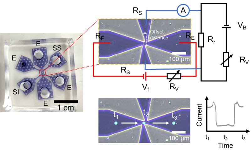
Overview of the microfluidic ion current measurement system
- [1] A. L. Andrady, “Microplastics in the marine environment,” Marine Pollution Bulletin, Vol.62, Issue 8, pp. 1596-1605, 2011. https://doi.org/10.1016/j.marpolbul.2011.05.030
- [2] H. Yang, G. Chen, and J. Wang, “Microplastics in the Marine Environment: Sources, Fates, Impacts and Microbial Degradation,” Toxics, Vol.9, No.2, Article No.41, 2021. https://doi.org/10.3390/toxics9020041
- [3] X. Guo and J. Wang, “The chemical behaviors of microplastics in marine environment: A review,” Marine Pollution Bulletin, Vol.142, pp. 1-14, 2019. https://doi.org/10.1016/j.marpolbul.2019.03.019
- [4] M. Cole, P. Lindeque, C. Halsband, and T. S. Galloway, “Microplastics as contaminants in the marine environment: A review,” Marine Pollution Bulletin, Vol.62, Issue 12, pp. 2588-2597, 2011. https://doi.org/10.1016/j.marpolbul.2011.09.025
- [5] S. M. Praveena, S. N. M. Shaifuddin, and S. Akizuki, “Exploration of microplastics from personal care and cosmetic products and its estimated emissions to marine environment: An evidence from Malaysia,” Marine Pollution Bulletin, Vol.136, pp. 135-140, 2018. https://doi.org/10.1016/j.marpolbul.2018.09.012
- [6] C. M. Rochman, M. A. Browne, B. S. Halpern, B. T. Hentschel, E. Hoh, H. K. Karapanagioti, L. M. Rios-Mendoza, H. Takada, S. Teh, and R. C. Thompson, “Classify plastic waste as hazardous,” Nature, Vol.494, No.7436, pp. 169-171, 2013. https://doi.org/10.1038/494169a
- [7] V. Stock, L. Böhmert, E. Lisicki, R. Block, J. Cara-Carmona, L. K. Pack, R. Selb, D. Lichtenstein, L. Voss, C. J. Henderson, E. Zabinsky, H. Sieg, A. Braeuning, and A. Lampen, “Uptake and effects of orally ingested polystyrene microplastic particles in vitro and in vivo,” Archives of Toxicology, Vol.93, No.7, pp. 1817-1833, 2019. https://doi.org/10.1007/s00204-019-02478-7
- [8] S. E. Nelms, T. S. Galloway, B. J. Godley, D. S. Jarvis, and P. K. Lindeque, “Investigating microplastic trophic transfer in marine top predators,” Environmental Pollution, Vol.238, pp. 999-1007, 2018. https://doi.org/10.1016/j.envpol.2018.02.016
- [9] D. Schymanski, C. Goldbeck, H.-U. Humpf, and P. Fürst, “Analysis of microplastics in water by micro-Raman spectroscopy: Release of plastic particles from different packaging into mineral water,” Water Research, Vol.129, pp. 154-162, 2018. https://doi.org/10.1016/j.watres.2017.11.011
- [10] M. Cole, P. Lindeque, E. Fileman, C. Halsband, R. Goodhead, J. Moger, and T. S. Galloway, “Microplastic Ingestion by Zooplankton,” Environmental Science & Technology, Vol.47, No.12, pp. 6646-6655, 2013. https://doi.org/10.1021/es400663f
- [11] A. Karami, N. Romano, T. Galloway, and H. Hamzah, “Virgin microplastics cause toxicity and modulate the impacts of phenanthrene on biomarker responses in African catfish (Clarias gariepinus),” Environmental Research, Vol.151, pp. 58-70, 2016. https://doi.org/10.1016/j.envres.2016.07.024
- [12] A. M. Elert, R. Becker, E. Duemichen, P. Eisentraut, J. Falkenhagen, H. Sturm, and U. Braun, “Comparison of different methods for MP detection: What can we learn from them, and why asking the right question before measurements matters?,” Environmental Pollution, Vol.231, Part 2, pp. 1256-1264, 2017. https://doi.org/10.1016/j.envpol.2017.08.074
- [13] M. Sano, N. Kaji, Q. Wu, T. Naito, T. Yasui, M. Taniguchi, T. Kawai, and Y. Baba, “Quantitative Evaluation of Dielectric Breakdown of Silicon Micro- and Nanofluidic Devices for Electrophoretic Transport of a Single DNA Molecule,” Micromachines-Basel, Vol.9, Issue 4, Article No.180, 2018. https://doi.org/10.3390/mi9040180
- [14] M. Terada, S. Ide, T. Naito, N. Kimura, M. Matsusaki, and N. Kaji, “Label-free cancer stem-like cell assay conducted at a single cell level using microfluidic mechanotyping devices,” Anal. Chem., Vol.93, No.43, pp. 14409-14416, 2021. https://doi.org/10.1021/acs.analchem.1c02316
- [15] T. Shimada, H. Yasaki, T. Yasui, T. Yanagida, N. Kaji, M. Kanai, K. Nagashima, T. Kawai, and Y. Baba, “PM2.5 Particle Detection in a Microfluidic Device by Using Ionic Current Sensing,” Analytical Sciences, Vol.34, No.12, pp. 1347-1349, 2018. https://doi.org/10.2116/analsci.18C018
- [16] H. Yasaki, T. Yasui, T. Yanagida, N. Kaji, M. Kanai, K. Nagashima, T. Kawai, and Y. Baba, “Substantial Expansion of Detectable Size Range in Ionic Current Sensing through Pores by Using a Microfluidic Bridge Circuit,” J. Am. Chem. Soc., Vol.139, No.40, pp. 14137-14142, 2017. https://doi.org/10.1021/jacs.7b06440
- [17] Y. Nakanishi, H. Yamaguchi, Y. Hirata, Y. Nakashima, and Y. Fujiwara, “Micro-abrasive glass surface for producing microplastics for biological tests,” Wear, Vol.477, Article No.203816, 2021. https://doi.org/10.1016/j.wear.2021.203816
- [18] Y. Nakanishi, Y. Fujiwara, and Y. Nakashima, “Generation of Nano/Microplastics for Immunological Assessments,” Biotribology, Vols.33-34, Article No.100235, 2023. https://doi.org/10.1016/j.biotri.2023.100235
This article is published under a Creative Commons Attribution-NoDerivatives 4.0 Internationa License.