Paper:
Individualization of Musculoskeletal Model for Analyzing Pelvic Floor Muscles Activity Based on Gait Motion Features
Tomohiro Wakaiki*, Takayuki Tanaka*, Koji Shimatani**, Yuichi Kurita***, and Tadayuki Iida**
*Hokkaido University
Kita 14, Nishi 9, Kita-ku, Sapporo, Hokkaido 060-00814, Japan
**Prefectural University of Hiroshima
1-1 Gakuen-machi, Mihara City, Hiroshima 723-0053, Japan
***Hiroshima University
1-4-1 Kagamiyama, Higashihiroshima, Hiroshima 739-8527, Japan

Stress urinary incontinence (SUI) is a typical quality of life disease in women. The strengthening of the pelvic floor muscle (PFM) is considered effective to prevent this. Specifically, PFM activity is affected by individual pelvic shape and posture. Therefore, it is necessary to analyze muscle activity by considering the individual differences. In this study, individual pelvic alignment was estimated from the feature values of natural gait via multiple regression analysis. In addition, individual pelvic feature points were derived from X-ray images and used to deform the standard model to obtain individual pelvic shapes. Results indicate that the residual averages of the estimated feature angles were less than 2° in most cases. Subsequently, measurements of the pelvis were obtained via MRI to evaluate the estimated pelvis shape. The results indicate that individual adaptation leads to muscle attachment positions, which are important in the muscle activity analysis, and closer to the true MRI value when compared to that of the standard pelvic model.
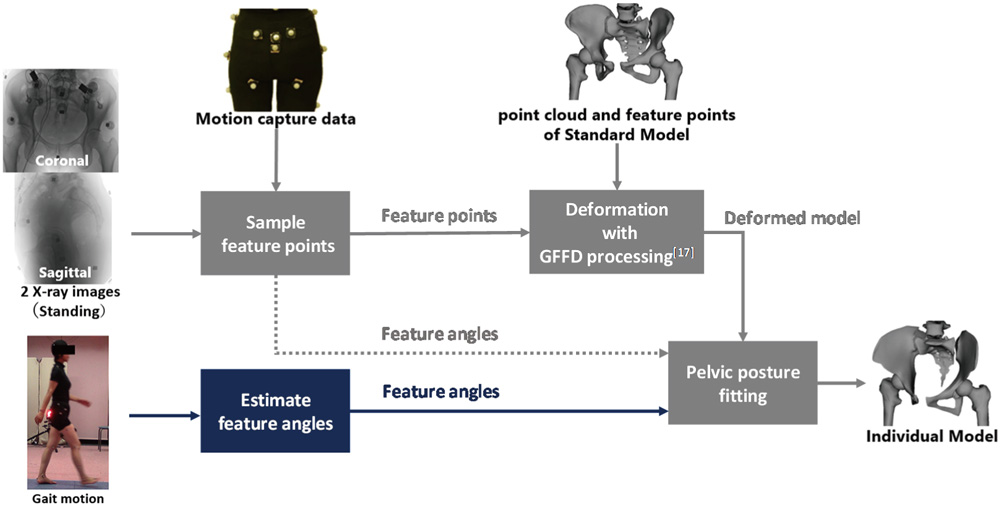
Pelvis individualization method
- [1] M. Okamoto, R. Murayama, and Y. Higuchi, “Development and Evaluation of Pelvic Floor Muscle Exercise Program for Preventing Stress Urinary Incontinence in Postpartum Women,” Research-Aid Report 2012, Vol.27, pp. 23-33, 2012.
- [2] X. Huang et al., “Neurostimulation strategy for stress urinary incontinence,” IEEE Trans. on Neural Systems and Rehabilitation Engineering, Vol.25, No.7, pp. 1068-1078, 2017.
- [3] K. Shimatani et al., “Association between contractions of pelvic floor muscles using X-ray imaging and contractions of transversus abdominis muscles,” ACPT2016, 2016.
- [4] K. Endo et al., “Pelvic Morphologic Angle and Sagittal Spinal Alignment,” The Japanese Clinical Orthopaedic Association, Vol.45, No.5, pp. 443-447, 2010.
- [5] T. Halski et al., “Evaluation of bioelectrical activity of pelvic floor muscles and synergistic muscles depending on orientation of pelvis in menopausal women with symptoms of stress urinary incontinence: a preliminary observational study,” BioMed. Research Int., 2014.
- [6] Y. Imamura et al., “Motion-based-design of elastic material for passive assistive device using musculoskeletal model,” J. Robot. Mechatron., Vol.23, No.6, pp. 978-990, 2011.
- [7] Y. Imamura et al., “Analysis of trunk stabilization effect by passive power-assist device,” J. Robot. Mechatron., Vol.26, No.6, pp. 791-798, 2014.
- [8] S. L. Delp, J. P. Loan, M. G. Hoy, F. E. Zajac, E. L. Topp, and J. M. Rosen, “An interactive graphics-based model of the lower extremity to study orthopaedic surgical procedures,” IEEE Trans. Biomed. Eng., Vol.37, No.8, pp. 757-767, 1990.
- [9] S. S. Blemker et al., “Image-based musculoskeletal modeling: Applications, advances, and future opportunities,” J. of Magnetic Resonance Imaging, Vol.25, No.2, pp. 441-451, 2007.
- [10] J. R. Fielding et al., “MR-based three-dimensional modeling of the normal pelvic floor in women: quantification of muscle mass,” American J. of Roentgenology, Vol.174, No.3, pp. 657-660, 2000.
- [11] A. L. Silva-Filho et al., “Evaluation of pelvic floor muscle cross-sectional area using a 3D computer model based on MRI in women with and without prolapse,” European J. of Obstetrics & Gynecology and Reproductive Biology, Vol.153, No.1, pp. 110-111, 2010.
- [12] K. Mineta et al., “CT-based morphological assessment of the hip joint in Japanese patients: association with radiographic predictors of femoroacetabular impingement,” The Bone and Joint J., Vol.98-B, No.9, pp. 1167-1174, 2016.
- [13] P. Cerveri et al., “2D/3D reconstruction of the distal femur using statistical shape models addressing personalized surgical instruments in knee arthroplasty: A feasibility analysis,” The Int. J. of Medical Robotics and Computer Assisted Surgery, Vol.13, No.4, 2017.
- [14] S. Schumann et al., “Cup implant planning based on 2-D/3-D radiographic pelvis reconstruction – first clinical results,” IEEE Trans. on Biomedical Engineering, Vol.62, No.11, pp. 2665-2673, 2015.
- [15] V. P. Stokes, C. Andersson, and H. Forssberg, “Rotational and translational movement features of the pelvis and thorax during adult human locomotion,” J. of Biomechanics, Vol.22, No.1, pp. 43-50, 1989.
- [16] M. Čadová, and L. M. Gallo, “Is OpenSim suitable for masticatory system analysis?,” Russian J. of Biomechanics, Vol.17, No.3, pp. 53-67, 2013.
- [17] H. Gou et al., “Gait and Posture Analysis Method Based on Genetic Algorithm and Support Vector Machines with Acceleration Data,” J. Robot. Mechatron., Vol.28, No.3, pp. 418-424, 2016.
- [18] N. Baka et al., “2D-3D shape reconstruction of the distal femur from stereo X-ray imaging using statistical shape models,” Medical Image Analysis, Vol.15, No.6, pp. 840-850, 2011.
- [19] L. M. Smoger et al., “Statistical shape modeling predicts patellar bone geometry to enable stereo-radiographic kinematic tracking,” J. of Biomechanics, Vol.58, pp. 187-194, 2017.
- [20] W. Yu, M. Tannast, and G. Zheng, “Non-rigid free-form 2D-3D registration using a B-spline-based statistical deformation model,” Pattern Recognition, Vol.63, pp. 689-699, 2017.
- [21] T. Wakaiki et al., “Individualization of Musculoskeletal Model to Analyze Pelvic Floor Muscles Activity,” Proc. of the 5th Int. Digital Human Modeling Symp., pp. 18-25, 2017.
- [22] K. Hagio et al., “A novel system of four-dimensional motion analysis aftertotal hip arthroplasty,” J. of Orthopaedic Research, Vol.22, No.3, pp. 665-670, 2004.
- [23] K. Kitajima, Y. Akagi, A. Yamauchi, N. Okazawa, and Y. Higuchi, “A Study on Facial Modeling Based on the GFFD Method,” The Japan Society Percision Engineering, Vol.74, No.8, pp. 883-890, 2008.
- [24] N. Yoshida, K. Kanou, and K. Kitajima, “Free-Form Deformations Based on Gaussian Functions Fundamental Theory for Interactive Modeling,” The Japan Society Percision Engineering, Vol.65, No.7, pp. 971-975, 1999.
- [25] J. C. Le Huec, S. Aunoble, L. Philippe, and P. Nicolas, “Pelvic parameters: origin and significance,” European Spine J., Vol.20, No.5, pp. 564-571, 2011.
- [26] H. Koga and J. Murata, “Effects of the Dominant Leg and Leg-crossing Preference on Pelvic Anteversion Angle,” Rigakuryoho Kagaku, Vol.29, No.1, pp. 39-43, 2014.
- [27] H. Kogo et al., “Effects of Dominant Leg and Leg-crossing Preference on Pelvic Anteversion Angle,” European Spine J., Vol.20, No.5, pp. 564-571, 2011.
- [28] S. Takahara, H. Aonuma, and S. Kaneko, “Developing Maximum Likelihood Approach to Three-Dimensional Reconstruction of Musculoskeletal Structure of Invertebrate Animals Based on X-ray Micro CT Data,” The 8th Int. Symp. on Adaptive Motion of Animals and Machines (AMAM2017), pp. 92-93, 2017.
- [29] W. J. Rucklidge, “Locating Objects Using the Hausdorff Distance,” Proc. of IEEE Int. Conf. on Computer Vision, Cambridge, MA, USA, pp.457-464, June 1995.
- [30] D.-G. Sim, O.-K. Kwon, and R.-H. Park, “Object Matching Algorithms Using Robust Hausdorff Distance Measures,” IEEE Trans. on Image Processing, Seoul, Korea, Vol.8, No.3, pp. 425-429, March 1999.
This article is published under a Creative Commons Attribution-NoDerivatives 4.0 Internationa License.