
Research Paper:
MRI Image Segmentation Algorithm for Temporomandibular Joint Discs and Condyles Based on the Improved nnU-Net Framework
Rufu Lin*1
 , Qiang Sun*2,†
, Yan Chen*3
, Zhaohui Zhang*4
, Xiaoyan Zhao*4
, and Qingfeng Wang*1
, Qiang Sun*2,†
, Yan Chen*3
, Zhaohui Zhang*4
, Xiaoyan Zhao*4
, and Qingfeng Wang*1
*1Shunde Innovation School, University of Science and Technology Beijing
No.2 Zhihui Road, Shunde District, Foshan 528399, China
*2China-Japan Friendship Hospital Stomatology Center
No.2 Sakura East Street, Chaoyang District, Beijing 100029, China
†Corresponding author
*3School of Optoelectronic Engineering, Changzhou Institute of Technology
No.666 Liaohe Road, Xinbei District, Changzhou 213032, China
*4School of Automation and Electrical Engineering, University of Science and Technology Beijing
No.30 Xueyuan Road, Haidian District, Beijing 100083, China

Accurate segmentation of the articular disc from MRI is crucial for diagnosing and treating temporomandibular joint disorders (TMD), but it remains challenging because of the small structure size, blurred boundaries, and class imbalance. This study aims to improve articular disc segmentation accuracy for computer-aided TMD diagnosis. We propose an enhanced nnU-Net-based method with three modifications: (1) fewer downsampling layers to preserve spatial detail, (2) a DynFocus module with channel-and-spatial attention at the bottleneck to enhance disc-background discrimination, and (3) a joint loss combining Dice focal and boundary losses to improve sensitivity and boundary delineation. On 50 temporomandibular joint MRI cases, the proposed method improved disc segmentation Dice from 0.69 to 0.71, maintained condyle Dice at 0.89, and achieved an average Dice of 0.80. These results indicate that the proposed enhancements improve disc segmentation performance while preserving condyle segmentation accuracy, supporting their usefulness for MRI-based TMD assessment.
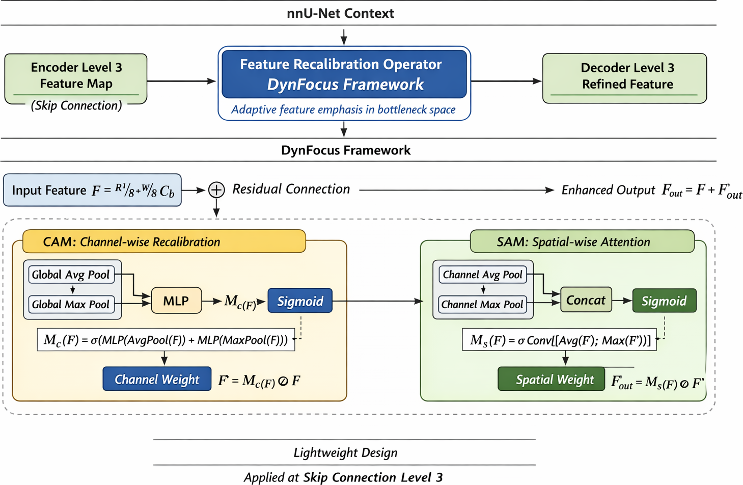
Detailed view of the three proposed modifications to the nnU-Net framework
1. Introduction
Temporomandibular disorders are among the most prevalent conditions affecting the oral and maxillofacial region, with epidemiological studies reporting a mean prevalence of approximately 30%–35% in the general population 1. Structural abnormalities of the temporomandibular joint (TMJ), particularly involving the mandibular condyle and articular disc, play a critical role in the onset and progression of TMD-related symptoms such as pain, joint clicking, and restricted mandibular movement. Accurate morphological assessment of these structures is therefore essential for diagnosis, treatment planning, and outcome evaluation. Recent studies integrating AI tools have shown preliminary promise in improving the diagnostic efficiency 2.
Magnetic resonance imaging (MRI) is widely regarded as the gold standard for TMJ assessment because of its superior soft-tissue contrast, particularly for visualizing the articular disc 3. However, manual delineation of TMJ structures on MRI remains a labor-intensive and highly subjective process, with substantial inter- and intra-observer variability reported even among experienced clinicians. These limitations have motivated an increasing interest in automated segmentation techniques to support objective and reproducible TMJ evaluations. Recent studies have demonstrated the feasibility of automated diagnosis of the specific conditions such as anterior disc displacement using deep learning 4.
In recent years, deep learning-based medical image segmentation methods, particularly convolutional neural networks (CNNs), have demonstrated remarkable performance across a wide range of anatomical structures 5,6. Frameworks such as U-Net and its variants have become de facto standards owing their efficient encoder–decoder architecture and strong representation capability 6,7. Among these methods, nnU-Net has gained prominence as a self-configuring segmentation framework that automatically adapts preprocessing strategies, network architecture, and training protocols to specific datasets, achieving state-of-the-art performance in multiple international challenges 8.
Despite these advances, directly applying existing segmentation frameworks to TMJ MRI remains challenging. The TMJ articular disc is an extremely thin structure, typically measuring only 1–2 mm in thickness, and exhibits blurred boundaries and low contrast relative to the surrounding tissues 9,10. Moreover, severe class imbalance arises because the disc occupies only a small fraction of the image volume, whereas the background and condylar regions dominate. These characteristics often lead to information loss during deep feature downsampling, boundary blurring, and false-positive predictions, even when using advanced frameworks such as nnU-Net are used 8,11.
Several studies have attempted to address TMJ segmentation using deep learning; however, most existing approaches either focus primarily on the condyle or rely on standard network configurations that are not specifically optimized for small and thin anatomical targets 9,12,10. Consequently, the segmentation performance for the articular disc remains suboptimal, limiting the clinical applicability of the current methods.
To address these challenges, this study proposes an improved nnU-Net-based segmentation framework tailored for TMJ MRI. The proposed method introduces three targeted optimizations: (1) a customized downsampling strategy to preserve fine-grained spatial information of the thin articular disc 8,13,14; (2) the design of the DynFocus framework, a specialized bottleneck integration module that leverages dual-path attention mechanisms to selectively amplify informative features of the articular disc while suppressing irrelevant anatomical interference 15,14,16; (3) a joint loss function combining Dice, focal, and boundary losses to simultaneously address class imbalance and boundary ambiguity 13,17,18,19. Comprehensive experiments on a clinical TMJ MRI dataset demonstrated that the proposed approach significantly improved articular disc segmentation accuracy while maintaining robust performance for condylar segmentation.
3. Methods
3.1. Overall Framework
The proposed method is built upon the nnU-Net framework and is specifically designed for simultaneous segmentation of the mandibular condyle and articular disc from TMJ MRI 8. Unlike conventional segmentation tasks that focus on relatively large and homogeneous anatomical structures, TMJ segmentation presents a dual-scale challenge: the condyle occupies a comparatively large region with relatively clear boundaries, whereas the articular disc is a thin, small, and low-contrast structure embedded in complex surrounding tissues.
To address this imbalance, we introduce a task-oriented modification of the nnU-Net framework that preserves the fine spatial details critical for disc segmentation while maintaining sufficient contextual understanding for condylar structures. The overall framework follows an encoder–decoder paradigm with skip connections but incorporates targeted architectural and optimization-level adaptations to mitigate information loss, enhance feature discrimination, and improve boundary delineation.
Figure 1 illustrates the proposed improved nnU-Net framework for simultaneous segmentation of the mandibular condyle and articular disc from TMJ MRI. The architecture reduces the number of downsampling stages from four to three to preserve fine spatial details of the thin articular disc, integrates a CBAM at the bottleneck, and is trained with a joint loss function.
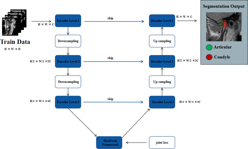
Fig. 1. Network framework. Green: articular disc; red: mandibular condyle.
3.2. Customized nnU-Net Architecture
3.2.1. Reduced Downsampling Strategy
In the standard nnU-Net configuration, four successive downsampling operations are commonly employed, reducing the spatial resolution of the feature maps to one-sixteenth of the original input size. Although this strategy is effective for capturing high-level semantic context in many segmentation tasks, this may be suboptimal for anatomically thin structures such as TMJ articular discs.
The articular disc typically exhibits a thickness of only 1–2 mm on MRI, corresponding to a limited number of voxels. Therefore, excessive downsampling can lead to an irreversible loss of spatial information, causing disc features to vanish in deeper layers and resulting in blurred or incomplete segmentation boundaries. To alleviate this issue, we reduce the number of downsampling stages from four to three, ensuring that the minimum feature map resolution remains at one-eighth of the input size.
This modification preserves the fine-grained spatial details essential for disc localization and boundary definition, while still maintaining sufficient receptive field coverage for condylar segmentation. Importantly, this architectural adjustment introduces only a marginal increase in the computational cost and does not compromise training stability, making it well-suited for small-target segmentation scenarios in clinical MRI data.
3.2.2. DynFocus Framework
To address the inherent challenges of feature ambiguity and the ultrathin morphology (1–2 mm) of the TMJ articular disc, we propose the DynFocus framework. This modular component is strategically integrated at the network bottleneck and serves as a dynamic bridge between the encoder and decoder. Unlike conventional architectures that treat the bottleneck as a simple convolutional transition, DynFocus operates as a semantic refinement zone that selectively amplifies relevant anatomical signals before spatial resolution is restored.
The DynFocus framework achieves high-precision localization through a dual-path attention mechanism 26. By aggregating global spatial information, the framework dynamically re-weights feature channels to prioritize those that are most discriminative for the articular disc, effectively suppressing background noise in low-contrast MRI slices. A spatial attention map is generated to concentrate the model’s computational focus of the model on the precise coordinates of the disc, mitigating the risk of false-positive predictions in the surrounding complex soft tissues.
By deploying DynFocus at the highest receptive field level, the model ensures that the fine-grained spatial details preserved by the reduced downsampling strategy are purified and enhanced. This modular integration is essential for the subsequent joint loss function to achieve sharp boundary delineation and superior segmentation accuracy for small-scale anatomical targets 15,16.
3.3. Joint Loss Function Design
Severe class imbalances and boundary ambiguities present significant challenges in TMJ articular disc segmentation. The disc occupies only a small fraction of the image volume, making standard loss functions prone to bias toward the dominant background and condylar regions. Moreover, partial volume effects and blurred boundaries further complicate accurate delineation.
To address these issues, we design a joint loss function that combines Dice, focal, and boundary losses, each targeting a specific limitation of conventional optimization strategies. The Dice loss directly optimizes region overlap and is effective in mitigating class imbalances at the global level. The focal loss dynamically downweights easily classified samples, encouraging the network to focus on hard-to-segment disc regions. The boundary loss explicitly penalizes discrepancies along object contours, improving boundary sharpness and reducing edge blurring.
The final loss function is defined as a weighted sum of these three components, enabling simultaneous optimization of region accuracy, hard-sample sensitivity, and boundary precision. This joint formulation is particularly well-suited for thin anatomical structures such as TMJ articular discs, in which accurate boundary localization is critical for clinical interpretation.
Figure 2 show the detailed view of the three proposed modifications to the nnU-Net framework.
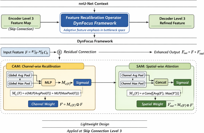
Fig. 2. Detailed view of the three proposed modifications to the nnU-Net framework. (1) Reduced downsampling strategy (3 stages instead of 4) to preserve fine-grained spatial information of the thin articular disc. (2) CBAM integrated at the bottleneck for enhanced feature discrimination. (3) Joint loss function combining Dice, focal, and boundary losses to simultaneously address class imbalance and boundary ambiguity.
4. Experiments
4.1. Dataset and Preprocessing
This study was conducted using a retrospective TMJ MRI dataset collected from patients diagnosed with temporomandibular disorders. The dataset consisted of MRI scans of 50 patients acquired using a 1.5T/3.0T MRI scanner with a dedicated TMJ imaging protocol. All images were obtained in the closed-mouth position that is commonly used for clinical evaluation of TMJ disc morphology.
Manual annotations of the mandibular condyle and articular disc were performed slice-by-slice by experienced oral and maxillofacial radiologists and subsequently reviewed by a senior expert to ensure annotation consistency. These annotations served as the reference standard for supervised learning.
Prior to training, all MRI volumes were resampled to a uniform voxel spacing and intensity-normalized following the default preprocessing pipeline of nnU-Net. Data augmentation strategies, including random rotation, scaling, and elastic deformation, were employed to enhance the model robustness and mitigate overfitting.
4.2. Computational Environment
All experiments were implemented using the PyTorch framework and conducted on a workstation equipped with an NVIDIA GPU. The proposed method was trained following the nnU-Net training strategy, with modifications strictly limited to the architectural components described in Section 3 to ensure a fair comparison.
The dataset was divided into training, validation, and testing sets at the patient level to prevent data leakage. Model training was performed using a fixed number of epochs, and the best-performing model on the validation set was selected for final evaluation. The Adam optimizer was employed with an initial learning rate consistent with the nnU-Net defaults, and learning rate decay was applied during training.
For a baseline comparison, the original nnU-Net framework with the standard configuration was trained under identical experimental conditions, including data preprocessing, augmentation strategies, and training schedules. This setup ensured that the observed performance differences could be attributed to the proposed modifications rather than to implementation or training discrepancies.
The segmentation performance was quantitatively evaluated using the Dice similarity coefficient (Dice) and Hausdorff distance (HD). The Dice measured the volumetric overlap between the predicted segmentation and the ground truth, providing an overall assessment of the region-level accuracy. The HD evaluated the maximum surface distance between two segmentations and was particularly sensitive to boundary errors.
Given the thin morphology of the TMJ articular disc, boundary accuracy is critical for clinical interpretation. Therefore, the HD was included as a metric complementary to the Dice to better capture the segmentation discrepancies along the object contours. All metrics were computed separately for the mandibular condyle and articular disc and averaged to provide an overall performance assessment.
4.3. Comparison with Baseline Methods
The proposed method was compared with the standard nnU-Net framework to evaluate the effectiveness of the introduced modifications. The quantitative results are summarized in Table 1.
| Model | Condyle | Articular disc | Average | IoU | HD | Accuracy | Precision | Recall |
| Our model | 0.89321 | 0.71543 | 0.80036 | 0.66716 | 1.45710 | 0.99936 | 0.86601 | 0.83105 |
| U-Net | 0.81367 | 0.45047 | 0.63539 | 0.46562 | 2.64498 | 0.98890 | 0.77631 | 0.74299 |
| U-NetPlusPlus | 0.87515 | 0.67844 | 0.77164 | 0.62818 | 1.62110 | 0.99728 | 0.83921 | 0.81286 |
| SWin-UNet | 0.88246 | 0.65963 | 0.76371 | 0.61774 | 1.67011 | 0.98926 | 0.84041 | 0.81201 |
| nnU-Net | 0.90457 | 0.69106 | 0.79186 | 0.65543 | 1.55810 | 0.99833 | 0.85937 | 0.83249 |
Both methods achieved high Dice scores for mandibular condyle segmentation, reflecting the relatively large size and clear boundaries of the structure. The proposed method demonstrated a comparable or slightly improved performance, indicating that the architectural modifications did not compromise the segmentation of larger anatomical regions.
In contrast, substantial improvements were observed in articular disc segmentation. The proposed method achieved a higher Dice score and noticeably reduced HD compared with the baseline nnU-Net. These results suggested that the reduced downsampling strategy and attention-enhanced bottleneck effectively preserved the fine structural details and improved boundary delineation for the thin-disc structure.
To further illustrate these improvements, representative segmentation results from the four subjects are shown in Fig. 3. The proposed method demonstrated significantly sharper boundaries and fewer missed thin-disc regions than the baseline nnU-Net and other state-of-the-art models.
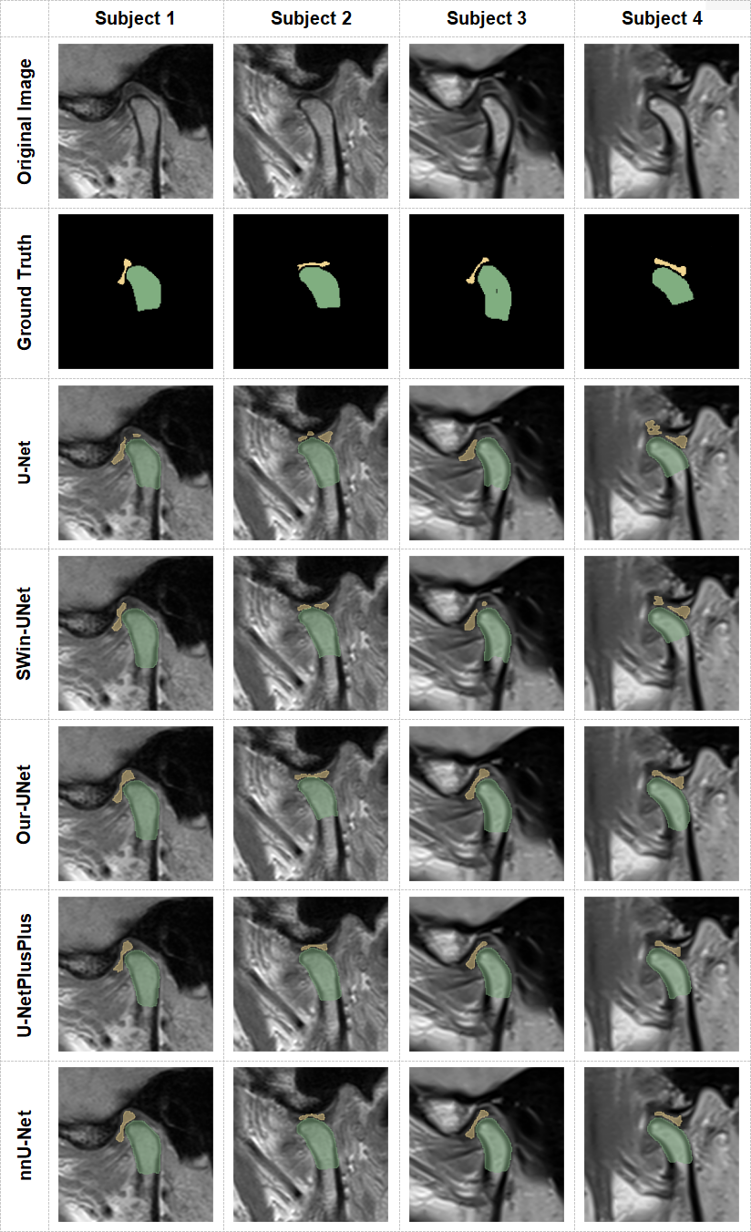
Fig. 3. Performance of different models among four representative subjects.
4.4. Ablation Study
An ablation study was conducted by incrementally introducing the proposed modifications to assess the contribution of each component in the proposed framework 13,14. Specifically, we evaluated (1) the baseline nnU-Net, (2) nnU-Net with reduced downsampling, (3) nnU-Net + DynFocus, and (4) the full model with the joint loss function.
As presented in Table 2, reducing the number of downsampling stages led to noticeable improvements in articular disc Dice and HD, confirming the importance of preserving spatial resolution for thin structures. The ablation results confirmed that the DynFocus framework served as a pivotal component for boundary refinement. By implementing DynFocus at the bottleneck, the model achieved a significantly lower HD (1.45 vs. 1.55), proving its superior capability in delineating the precise contours of the articular disc.
| Model | Condyle | Articular disc | Average | HD |
| Baseline nnU-Net | 0.90457 | 0.69106 | 0.79186 | 1.55810 |
| nnU-Net with reduced downsampling | 0.90180 | 0.70353 | 0.80358 | 1.50504 |
| nnU-Net + DynFocus | 0.89751 | 0.70986 | 0.80368 | 1.47558 |
| Full model with the joint loss function | 0.89321 | 0.71543 | 0.80432 | 1.45710 |
In this study, the Dice coefficient was selected as the evaluation metric. The results obtained from the dataset used in this study are summarized in Table 1. Our model achieved an average Dice coefficient of 0.80, with 0.89 for the condylar region and 0.71 for the articular disc. In comparison, U-Net yielded a lower average Dice coefficient of 0.63, with 0.81 for the condylar region and only 0.45 for the articular disc. These results were significant because they demonstrated that the proposed model detected the presence of the articular disc and successfully located it. The proposed model consistently outperformed U-Net, U-Net++, and SWin-UNet in terms of segmentation accuracy. For both the condyle and articular disc, the Dice coefficient of the proposed model exceeded those of the other models. Although the Dice coefficient of the proposed model for the condylar region was slightly lower than that of nnU-Net, it outperformed for the articular disc, the target area of this study.
5. Conclusions
In this study, we proposed an improved nnU-Net-based framework for automatic segmentation of the TMJ articular disc and mandibular condyle from MRI 8,13. The proposed method effectively addressed the challenges associated with thin morphology, low contrast, and class imbalance by tailoring the network architecture and optimization strategy to the structural characteristics of the TMJ articular disc.
Specifically, reducing the degree of downsampling helped to preserve the fine spatial details critical for disc segmentation 13,14. The proposed DynFocus framework effectively bridged the gap between high-level semantics and fine-grained spatial localization. Its modular design demonstrated that targeted bottleneck optimization was essential for clinical tasks involving ultra-thin anatomical targets 15,16. In addition, the joint loss function combining Dice, focal, and boundary losses improved region overlap and boundary accuracy 17,18. The experimental results demonstrated that the proposed method consistently outperformed the standard nnU-Net, particularly in articular disc segmentation, without compromising the performance on the mandibular condyle 9,12,10,13.
These findings suggest that task-oriented customization of generic segmentation frameworks can substantially improve the performance for small and thin anatomical structures in TMJ MRI. The proposed approach has the potential to support objective and reproducible TMJ assessments and may serve as a practical reference for future studies on TMJ image analysis.
Acknowledgments
This study was supported by the Beijing Natural Science Foundation Joint Fund (Grant No.4242014).
- [1] G. Zieliński, B. Pająk-Zielińska, and M. Ginszt, “A meta-analysis of the global prevalence of temporomandibular disorders,” J. Clin. Med., Vol.13, No.5, Article No.1365, 2024. https://doi.org/10.3390/jcm13051365
- [2] O. Almășan, L. Mureșanu, I. A. Mureșanu et al., “An examination of temporomandibular joint disc displacement through magnetic resonance imaging by integrating artificial intelligence: Preliminary findings,” Medicina, Vol.60, No.9, Article No.1396, 2024. https://doi.org/10.3390/medicina60091396
- [3] M. Qian and M. Wu, “Research status of three-dimensional model reconstruction of temporomandibular joint based on magnetic resonance imaging,” J. Stomatol., Vol.43, No.10, pp. 930-934, 2023 (in Chinese). https://doi.org/10.13591/j.cnki.kqyx.2023.10.013
- [4] Y. Yu et al., “Deep learning-based automated diagnosis of temporomandibular joint anterior disc displacement and its clinical application,” Front. Physiol., Vol.15, Article No.1445258, 2024. https://doi.org/10.3389/fphys.2024.1445258
- [5] L. C. Chen, G. Papandreou, I. Kokkinos et al., “DeepLab: Semantic image segmentation with deep convolutional nets, atrous convolution, and fully connected CRFs,” IEEE Trans. Pattern Anal. Mach. Intell., Vol.40, No.4, pp. 834-848, 2018. https://doi.org/10.1109/TPAMI.2017.2699184
- [6] O. Ronneberger, P. Fischer, and T. Brox, “U-Net: Convolutional networks for biomedical image segmentation,” Proc. Int. Conf. on Medical Image Computing and Computer-Assisted Intervention (MICCAI 2015), pp. 234-241, 2015. https://doi.org/10.1007/978-3-319-24574-4_28
- [7] Ö. Çiçek, A. Abdulkadir, S. S. Lienkamp et al., “3D U-Net: Learning dense volumetric segmentation from sparse annotation,” Proc. Int. Conf. on Medical Image Computing and Computer-Assisted Intervention (MICCAI 2016), pp. 424-432, 2016. https://doi.org/10.1007/978-3-319-46723-8_49
- [8] F. Isensee, P. F. Jaeger, A. A. Kohl et al., “nnU-Net: A self-configuring method for deep learning-based biomedical image segmentation,” Nat. Methods, Vol.18, No.2, pp. 203-211, 2021. https://doi.org/10.1038/s41592-020-01008-z
- [9] S. Ito, Y. Mine, Y. Yoshimi et al., “Automated segmentation of articular disc of the temporomandibular joint on magnetic resonance images using deep learning,” Sci. Rep., Vol.12, No.1, Article No.221, 2022. https://doi.org/10.1038/s41598-021-04354-w
- [10] M. Li, K. Punithakumar, P. W. Major et al., “Temporomandibular joint segmentation in MRI images using deep learning,” J. Dent., Vol.127, Article No.104345, 2022. https://doi.org/10.1016/j.jdent.2022.104345
- [11] A. Celaya, B. Riviere, and D. Fuentes, “A generalized surface loss for reducing the Hausdorff distance in medical imaging segmentation,” arXiv:2302.03868, 2023. https://doi.org/10.48550/arXiv.2302.03868
- [12] S. Vinayahalingam, B. Berends, F. Baan et al., “Deep learning for automated segmentation of the temporomandibular joint,” J. Dent., Vol.132, Article No.104475, 2023. https://doi.org/10.1016/j.jdent.2023.104475
- [13] H. He, C. Sun, Z. Wang, and Y. Dan, “Linear Transformer based U-shaped lightweight segmentation network,” J. Adv. Comput. Intell. Intell. Inform., Vol.29, No.6, pp. 1319-1328, 2025. https://doi.org/10.20965/jaciii.2025.sd0000
- [14] Z. Fu et al., “An enhanced U-network by combining PPM and CBAM for medical image segmentation,” IEEE Access, Vol.12, pp. 107098-107112, 2024. https://doi.org/10.1109/access.2024.3426518
- [15] S. Woo, J. Park, J. Y. Lee et al., “CBAM: Convolutional block attention module,” Proc. European Conf. on Computer Vision (ECCV 2018), pp. 3-19, 2018. https://doi.org/10.1007/978-3-030-01234-2_1
- [16] J. Wu, “Enhancing nnU-Net for improved medical image segmentation: A comparative study with total segmentator,” Proc. 2nd Int. Conf. Data Anal. Mach. Learn. (DAML 2024), pp. 246-251, 2024. https://doi.org/10.5220/0013515100004619
- [17] J. Du, K. Guan, P. Liu et al., “Boundary-sensitive loss function with location constraint for hard region segmentation,” IEEE J. Biomed. Health Inform., Vol.27, No.2, pp. 992-1003, 2022. https://doi.org/10.1109/jbhi.2022.3222390
- [18] M. F. Ahamed et al., “Rethinking U-Net architecture in medical imaging: Advancing the efficient and interpretable UKAN-CBAM framework for colorectal polyp segmentation,” Artif. Intell. Med., Vol.157, Article No.102978, 2026. https://doi.org/10.1016/j.artmed.2026.103352
- [19] P. Shi et al., “Centerline boundary dice loss for vascular segmentation,” Proc. Int. Conf. on Medical Image Computing and Computer-Assisted Intervention (MICCAI 2024), pp. 458-467, 2024. https://doi.org/10.1007/978-3-031-72111-3_5
- [20] J. Schlemper, O. Oktay, M. Schaap et al., “Attention gated networks: Learning to leverage salient regions in medical images,” Med. Image Anal., Vol.53, pp. 197-207, 2019. https://doi.org/10.1016/j.media.2019.01.012
- [21] F. Liu et al., “The automatic segmentation of the temporomandibular joint based on MRI using deep learning method,” Stomatology, 2025 (in Chinese). https://doi.org/10.13591/j.cnki.kqyx.2025.06.009
- [22] H. Kervadec, J. Bouchtiba, C. Desrosiers et al., “Boundary loss for highly unbalanced segmentation,” Medical Image Analysis, Article No.101851, 2019. https://doi.org/10.1016/j.media.2020.101851
- [23] J. Chen, Y. Lu, Q. Yu et al., “TransUNet: Transformers make strong encoders for medical image segmentation,” arXiv:2102.04306, 2021. https://doi.org/10.48550/arXiv.2102.04306
- [24] H. Cao, Y. Wang, J. Chen et al., “Swin-Unet: Unet-like pure transformer for medical image segmentation,” Proc. European Conf. on Computer Vision (ECCV 2022), pp. 205-218, 2022. https://doi.org/10.1007/978-3-031-25066-8_9
- [25] Z. Liu, Y. Lin, Y. Cao et al., “Swin Transformer: Hierarchical vision Transformer using shifted Windows,” Proc. 2021 IEEE/CVF Int. Conf. on Computer Vision (ICCV 2021), pp. 10012-10022, 2021. https://doi.org/10.1109/ICCV48922.2021.00986
- [26] K. Becker et al., “Digitale workflows in der kieferorthopädie,” Quintessenz Zahnmed., Vol.76, No.11, 2025 (in German). https://doi.org/10.1007/978-3-658-45141-7_17
This article is published under a Creative Commons Attribution-NoDerivatives 4.0 Internationa License.