
Research Paper:
MRI Image Segmentation Algorithm for Temporomandibular Joint Discs and Condyles Based on the Improved nnU-Net Framework
Rufu Lin*1
 , Qiang Sun*2,†
, Yan Chen*3
, Zhaohui Zhang*4
, Xiaoyan Zhao*4
, and Qingfeng Wang*1
, Qiang Sun*2,†
, Yan Chen*3
, Zhaohui Zhang*4
, Xiaoyan Zhao*4
, and Qingfeng Wang*1
*1Shunde Innovation School, University of Science and Technology Beijing
No.2 Zhihui Road, Shunde District, Foshan 528399, China
*2China-Japan Friendship Hospital Stomatology Center
No.2 Sakura East Street, Chaoyang District, Beijing 100029, China
†Corresponding author
*3School of Optoelectronic Engineering, Changzhou Institute of Technology
No.666 Liaohe Road, Xinbei District, Changzhou 213032, China
*4School of Automation and Electrical Engineering, University of Science and Technology Beijing
No.30 Xueyuan Road, Haidian District, Beijing 100083, China

Accurate segmentation of the articular disc from MRI is crucial for diagnosing and treating temporomandibular joint disorders (TMD), but it remains challenging because of the small structure size, blurred boundaries, and class imbalance. This study aims to improve articular disc segmentation accuracy for computer-aided TMD diagnosis. We propose an enhanced nnU-Net-based method with three modifications: (1) fewer downsampling layers to preserve spatial detail, (2) a DynFocus module with channel-and-spatial attention at the bottleneck to enhance disc-background discrimination, and (3) a joint loss combining Dice focal and boundary losses to improve sensitivity and boundary delineation. On 50 temporomandibular joint MRI cases, the proposed method improved disc segmentation Dice from 0.69 to 0.71, maintained condyle Dice at 0.89, and achieved an average Dice of 0.80. These results indicate that the proposed enhancements improve disc segmentation performance while preserving condyle segmentation accuracy, supporting their usefulness for MRI-based TMD assessment.
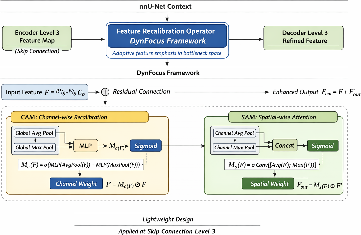
Detailed view of the three proposed modifications to the nnU-Net framework
- [1] G. Zieliński, B. Pająk-Zielińska, and M. Ginszt, “A meta-analysis of the global prevalence of temporomandibular disorders,” J. Clin. Med., Vol.13, No.5, Article No.1365, 2024. https://doi.org/10.3390/jcm13051365
- [2] O. Almășan, L. Mureșanu, I. A. Mureșanu et al., “An examination of temporomandibular joint disc displacement through magnetic resonance imaging by integrating artificial intelligence: Preliminary findings,” Medicina, Vol.60, No.9, Article No.1396, 2024. https://doi.org/10.3390/medicina60091396
- [3] M. Qian and M. Wu, “Research status of three-dimensional model reconstruction of temporomandibular joint based on magnetic resonance imaging,” J. Stomatol., Vol.43, No.10, pp. 930-934, 2023 (in Chinese). https://doi.org/10.13591/j.cnki.kqyx.2023.10.013
- [4] Y. Yu et al., “Deep learning-based automated diagnosis of temporomandibular joint anterior disc displacement and its clinical application,” Front. Physiol., Vol.15, Article No.1445258, 2024. https://doi.org/10.3389/fphys.2024.1445258
- [5] L. C. Chen, G. Papandreou, I. Kokkinos et al., “DeepLab: Semantic image segmentation with deep convolutional nets, atrous convolution, and fully connected CRFs,” IEEE Trans. Pattern Anal. Mach. Intell., Vol.40, No.4, pp. 834-848, 2018. https://doi.org/10.1109/TPAMI.2017.2699184
- [6] O. Ronneberger, P. Fischer, and T. Brox, “U-Net: Convolutional networks for biomedical image segmentation,” Proc. Int. Conf. on Medical Image Computing and Computer-Assisted Intervention (MICCAI 2015), pp. 234-241, 2015. https://doi.org/10.1007/978-3-319-24574-4_28
- [7] Ö. Çiçek, A. Abdulkadir, S. S. Lienkamp et al., “3D U-Net: Learning dense volumetric segmentation from sparse annotation,” Proc. Int. Conf. on Medical Image Computing and Computer-Assisted Intervention (MICCAI 2016), pp. 424-432, 2016. https://doi.org/10.1007/978-3-319-46723-8_49
- [8] F. Isensee, P. F. Jaeger, A. A. Kohl et al., “nnU-Net: A self-configuring method for deep learning-based biomedical image segmentation,” Nat. Methods, Vol.18, No.2, pp. 203-211, 2021. https://doi.org/10.1038/s41592-020-01008-z
- [9] S. Ito, Y. Mine, Y. Yoshimi et al., “Automated segmentation of articular disc of the temporomandibular joint on magnetic resonance images using deep learning,” Sci. Rep., Vol.12, No.1, Article No.221, 2022. https://doi.org/10.1038/s41598-021-04354-w
- [10] M. Li, K. Punithakumar, P. W. Major et al., “Temporomandibular joint segmentation in MRI images using deep learning,” J. Dent., Vol.127, Article No.104345, 2022. https://doi.org/10.1016/j.jdent.2022.104345
- [11] A. Celaya, B. Riviere, and D. Fuentes, “A generalized surface loss for reducing the Hausdorff distance in medical imaging segmentation,” arXiv:2302.03868, 2023. https://doi.org/10.48550/arXiv.2302.03868
- [12] S. Vinayahalingam, B. Berends, F. Baan et al., “Deep learning for automated segmentation of the temporomandibular joint,” J. Dent., Vol.132, Article No.104475, 2023. https://doi.org/10.1016/j.jdent.2023.104475
- [13] H. He, C. Sun, Z. Wang, and Y. Dan, “Linear Transformer based U-shaped lightweight segmentation network,” J. Adv. Comput. Intell. Intell. Inform., Vol.29, No.6, pp. 1319-1328, 2025. https://doi.org/10.20965/jaciii.2025.sd0000
- [14] Z. Fu et al., “An enhanced U-network by combining PPM and CBAM for medical image segmentation,” IEEE Access, Vol.12, pp. 107098-107112, 2024. https://doi.org/10.1109/access.2024.3426518
- [15] S. Woo, J. Park, J. Y. Lee et al., “CBAM: Convolutional block attention module,” Proc. European Conf. on Computer Vision (ECCV 2018), pp. 3-19, 2018. https://doi.org/10.1007/978-3-030-01234-2_1
- [16] J. Wu, “Enhancing nnU-Net for improved medical image segmentation: A comparative study with total segmentator,” Proc. 2nd Int. Conf. Data Anal. Mach. Learn. (DAML 2024), pp. 246-251, 2024. https://doi.org/10.5220/0013515100004619
- [17] J. Du, K. Guan, P. Liu et al., “Boundary-sensitive loss function with location constraint for hard region segmentation,” IEEE J. Biomed. Health Inform., Vol.27, No.2, pp. 992-1003, 2022. https://doi.org/10.1109/jbhi.2022.3222390
- [18] M. F. Ahamed et al., “Rethinking U-Net architecture in medical imaging: Advancing the efficient and interpretable UKAN-CBAM framework for colorectal polyp segmentation,” Artif. Intell. Med., Vol.157, Article No.102978, 2026. https://doi.org/10.1016/j.artmed.2026.103352
- [19] P. Shi et al., “Centerline boundary dice loss for vascular segmentation,” Proc. Int. Conf. on Medical Image Computing and Computer-Assisted Intervention (MICCAI 2024), pp. 458-467, 2024. https://doi.org/10.1007/978-3-031-72111-3_5
- [20] J. Schlemper, O. Oktay, M. Schaap et al., “Attention gated networks: Learning to leverage salient regions in medical images,” Med. Image Anal., Vol.53, pp. 197-207, 2019. https://doi.org/10.1016/j.media.2019.01.012
- [21] F. Liu et al., “The automatic segmentation of the temporomandibular joint based on MRI using deep learning method,” Stomatology, 2025 (in Chinese). https://doi.org/10.13591/j.cnki.kqyx.2025.06.009
- [22] H. Kervadec, J. Bouchtiba, C. Desrosiers et al., “Boundary loss for highly unbalanced segmentation,” Medical Image Analysis, Article No.101851, 2019. https://doi.org/10.1016/j.media.2020.101851
- [23] J. Chen, Y. Lu, Q. Yu et al., “TransUNet: Transformers make strong encoders for medical image segmentation,” arXiv:2102.04306, 2021. https://doi.org/10.48550/arXiv.2102.04306
- [24] H. Cao, Y. Wang, J. Chen et al., “Swin-Unet: Unet-like pure transformer for medical image segmentation,” Proc. European Conf. on Computer Vision (ECCV 2022), pp. 205-218, 2022. https://doi.org/10.1007/978-3-031-25066-8_9
- [25] Z. Liu, Y. Lin, Y. Cao et al., “Swin Transformer: Hierarchical vision Transformer using shifted Windows,” Proc. 2021 IEEE/CVF Int. Conf. on Computer Vision (ICCV 2021), pp. 10012-10022, 2021. https://doi.org/10.1109/ICCV48922.2021.00986
- [26] K. Becker et al., “Digitale workflows in der kieferorthopädie,” Quintessenz Zahnmed., Vol.76, No.11, 2025 (in German). https://doi.org/10.1007/978-3-658-45141-7_17
This article is published under a Creative Commons Attribution-NoDerivatives 4.0 Internationa License.