Letter:
Exploring the Bio-Functional Breaking Point of Living Tissue Subjected to External Physical Pressure
Shotaro Tanaka and Fumio Nakamura
Department of Biochemistry, School of Medicine, Tokyo Women’s Medical University
8-1 Kawada-cho, Shinjuku-ku, Tokyo 162-8666, Japan

Long before reaching its mechanical breaking point, a bio-system begins responding to stress at its own “bio-functional breaking point,” a phase of life activity dysfunction. However, little is known about the correlation between tissue flexibility and the conditions under which cellular response, damage, and death occur. We are now developing a new confocal microscopy-based observation method to analyze cell aggregates (spheroids) that are under physical pressure. The method concomitantly assesses cellular responses, stress levels, and cellular structure changes. Using this method, we found that the artificial suppression of the gene expression of fibronectin, a major component of the extracellular matrix, provides different mechanical characteristics to hepatoma-derived cell line spheroids than does the control wild type. This study may aid in the prediction of the characteristics of a tissue of interest by simply analyzing the tissue gene expression pattern, providing valuable information for the development and operation of wearable devices. It may also help in the preparation of custom devices that suit specific individuals.
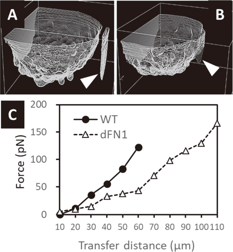
Deformation analysis of cancer cell line spheroid
- [1] W. Mueller-Klieser, “Multicellular spheroids,” J. Cancer Res. Clin. Oncol., Vol.113, Issue 2, pp. 101-122, 1987.
- [2] S. Gunti et al., “Organoid and spheroid tumor models: Techniques and applications,” Cancers (Basel), Vol.13, Issue 4, 874, 2021.
- [3] G. Venugopalan et al., “Multicellular architecture of malignant breast epithelia influences mechanics,” PLoS ONE, Vol.9, Issue 8, e101955, 2014.
- [4] Y. Abidine et al., “Viscoelastic Properties in Cancer: From Cells to Spheroids,” Cells, Vol.10, Issue 7, 1704, 2021.
- [5] Y. Ida et al., “ROCK inhibitors enhance the production of large lipid-enriched 3D organoids of 3T3-L1 cells,” Sci. Rep., Vol.11, 5479, 2021.
- [6] S. Sakuma et al., “Cellular force measurement using a nanometric-probe-integrated microfluidic chip with a displacement reduction mechanism,” J. Robot. Mechatron., Vol.25, No.2, pp. 277-284, 2013.
This article is published under a Creative Commons Attribution-NoDerivatives 4.0 Internationa License.