Paper:
A New Ultrasonographic Image Displaying System to Support Vein Detection
Shuhei Noyori*, Gojiro Nakagami*, Hiroshi Noguchi**, Koichi Yabunaka*, Taketoshi Mori**, and Hiromi Sanada*
*Departments of Gerontological Nursing/Wound Care Management, Graduate School of Medicine, The University of Tokyo
7-3-1 Hongo, Bunkyo-ku, Tokyo 113-0033, Japan
**Department of Life Support Technology (Molten), Graduate School of Medicine, The University of Tokyo
7-3-1 Hongo, Bunkyo-ku, Tokyo 113-0033, Japan

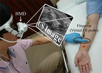
Virtual ultrasonic probe system
- [1] W. Zingg and D. Pittet, “Peripheral venous catheters: an under-evaluated problem,” Int. J. Antimicrob. Agents, Vol.34, No.4, pp. S38-42, 2009.
- [2] C. M. Rickard, J. Webster, M. C. Wallis, N. Marsh, M. R. McGrail, V. French, L. Foster, P. Gallagher, J. R. Gowardman, L. Zhang, A. McClymont, and M. Whitby, “Routine versus clinically indicated replacement of peripheral intravenous catheters: a randomised controlled equivalence trial,” Lancet, Vol.380, No.9847, pp. 1066-1074, 2012.
- [3] T. Takahashi, R. Murayama, M. Oe, G. Nakagami, H. Tanabe, K. Yabunaka, R. Arai, C. Komiyama, M. Uchida, and H. Sanada, “Thrombus with subcutaneous edema detected by ultrasonography related to peripheral intravenous catheter failure,” Congress brochure of The 4th World Congress on Vascular Access, p. 20, 2016.
- [4] L. C. Hadaway and D. A. Millam, “On the road to successful I.V. starts,” Nursing (Lond)., Vol.35, No.1, pp. S1-14, 2005.
- [5] P. Szmuk, J. Steiner, R. B. Pop, A. Farrow-Gillespie, E. J. Mascha, and D. I. Sessler, “The VeinViewer vascular imaging system worsens first-attempt cannulation rate for experienced nurses in infants and children with anticipated difficult intravenous access,” Anesth. Analg., Vol.116, No.5, pp. 1087-1092, 2013.
- [6] M. J. Kim, J. M. Park, N. Rhee, S. M. Je, S. H. Hong, Y. M. Lee, S. P. Chung, and S. H. Kim, “Efficacy of VeinViewer in pediatric peripheral intravenous access: a randomized controlled trial.,” Eur. J. Pediatr., Vol.171, No.7, pp. 1121-1125, 2012.
- [7] R. N. Kaddoum, D. L. Anghelescu, M. E. Parish, B. B. Wright, L. Trujillo, J. Wu, Y. Wu, and L. L. Burgoyne, “A randomized controlled trial comparing the AccuVein AV300 device to standard insertion technique for intravenous cannulation of anesthetized children,” Paediatr. Anaesth., Vol.22, No.9, pp. 884-889, 2012.
- [8] M. D. Witting, S. M. Schenkel, B. J. Lawner, and B. D. Euerle, “Effects of Vein Width and Depth on Ultrasound-Guided Peripheral Intravenous Success Rates,” J. Emerg. Med., Vol.39, No.1, pp. 70-75, 2010.
- [9] J. M. Fields, A. J. Dean, R. W. Todman, A. K. Au, K. L. Anderson, B. S. Ku, J. M. Pines, and N. L. Panebianco, “The effect of vessel depth, diameter, and location on ultrasound-guided peripheral intravenous catheter longevity,” Am. J. Emerg. Med., Vol.30, No.7, pp. 1134-1140, 2012.
- [10] H. Tanabe, T. Takahashi, R. Murayama, K. Yabunaka, M. Oe, Y. Matsui, R. Arai, M. Uchida, C. Komiyama, and H. Sanada, “Using Ultrasonography for Vessel Diameter Assessment to Prevent Infiltration,” J. Infus. Nurs., Vol.39, No.2, pp. 105-111, 2016.
- [11] American Institute of Ultrasound in Medicine, “Use of Ultrasound to Guide Vascular Access Procedures,” AIUM Pract. Guidel., 2012.
- [12] Y. Kobayashi, R. Hamano, H. Watanabe, T. Koike, J. Hong, K. Toyoda, M. Uemura, S. Ieiri, M. Tomikawa, T. Ohdaira, M. Hashizume, and M. G. Fujie, “Preliminary in vivo evaluation of a needle insertion manipulator for central venous catheterization,” ROBOMECH J., Vol.1, No.18, 2014.
- [13] N. Kaneko, M. Sato, T. Takeshima, Y. Sehara, and E. Watanabe, “Ultrasound-guided central venous catheterization using an optical see-through head-mounted display: A pilot study,” J. Clin. Ultrasound, Vol.44, No.8, pp. 487-491, 2016.
- [14] N. Hanafusa, T. Torato, and T. Katano, “Future Blood Vessel Puncture Procedure With Use of Head Mount Display,” Ther. Apher. Dial., Vol.20, No.1, pp. 88-89, 2016.
- [15] D. Lee and S. Lee, “Vision-Based Finger Action Recognition by Angle Detection and Contour Analysis,” ETRI J., Vol.33, No.3, pp. 415-422, 2011.
- [16] G. Welch and G. Bishop, “An Introduction to the Kalman Filter,” Course Notes 8 ACM SIGGRAPH 2001, 2001.
- [17] Y. Aoki, K. Kaneko, T. Sakai, and K. Masuda, “A study of scanning the ultrasound probe on body surface and construction of visual servo system based on echogram,” J. Robot. Mechatronics, Vol.22, No.3, pp. 273-279, 2010.
- [18] K. Ito, S. Sugano, R. Takeuchi, K. Nakamura, and H. Iwata, “Usability and performance of a wearable tele-echography robot for focused assessment of trauma using sonography,” Med. Eng. Phys., Vol.35, No.2, pp. 165-171, 2013.
- [19] Y. Minami, H. Chung, M. Kudo, S. Kitai, S. Takahashi, T. Inoue, K. Ueshima, and H. Shiozaki, “Radiofrequency ablation of hepatocellular carcinoma: value of virtual CT sonography with magnetic navigation,” AJR. Am. J. Roentgenol., Vol.190, No.6, pp. W335-W341, 2008.
This article is published under a Creative Commons Attribution-NoDerivatives 4.0 Internationa License.