
Paper:
Deep Learning-Based Morphological Analysis for Identifying Culture Status and Contractile Motion in Skeletal Muscle Tissues
Yudai Kizure, Daiki Miyashita, and Yuya Morimoto

Faculty of Science and Engineering, Waseda University
3-4-1 Okubo, Shinjuku-ku, Tokyo 169-8555, Japan

This study investigates a deep learning-based framework that uses static microscopic images to identify culture status and contractile motion in cultured skeletal muscle tissues. Noninvasive functional evaluation is important for advancements in tissue engineering and biohybrid robotics. However, acquiring large-scale labeled datasets is often difficult owing to the time-consuming nature of functional measurements. To explore the feasibility of image-based evaluation under the data constraints, we implemented a structured image processing and validation pipeline. First, tissue regions were isolated using U2-Net, achieving a Dice coefficient of 0.984 to reduce background interference. Second, an ensemble learning strategy was examined via a culture-day identification task using 528 images. A soft-voting ensemble of convolutional neural networks achieved a Macro F1-score of 0.817, indicating that morphological features associated with culture progression could be effectively captured. Additionally, self-supervised pre-training via SimSiam was tested to extract domain-specific representations from unlabeled images. For contractile motion identification, the ImageNet-pre-trained EfficientNetV2-S paired with a random forest classifier attained an accuracy and F1-score of 75.0% and 0.800, respectively. These results suggest that functional properties of cultured tissues can be partially inferred from static morphological characteristics. While the limited size of the dataset and low identification confidence pose challenges, this study provides a baseline methodology for noninvasive, image-based cellular sensing in engineered biological systems.
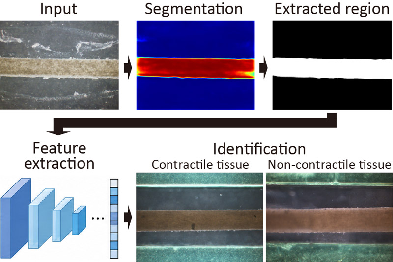
Image-based muscle tissue analysis
- [1] Z. Lin, T. Jiang, and J. Shang, “The emerging technology of biohybrid micro-robots: A review,” Bio-Des. Manuf., Vol.5, pp. 107-132, 2022. https://doi.org/10.1007/s42242-021-00135-6
- [2] R. Raman, “Biofabrication of living actuators,” Annu. Rev. Biomed. Eng., Vol.26, pp. 223-245, 2024. https://doi.org/10.1146/annurev-bioeng-110122-013805
- [3] J. S. H. Seah, S. Singh, L. P. Tan, and D. Choudhury, “Scaffolds for the manufacture of cultured meat,” Crit. Rev. Biotechnol., Vol.42, No.2, pp. 311-323, 2022. https://doi.org/10.1080/07388551.2021.1931803
- [4] D. Wilson, O. Tsigkou, L. Bosworth, and C. Jones, “Leather-like materials by cellular agriculture,” Text. Prog., Vol.55, No.1, 2023. https://doi.org/10.1080/00405167.2023.2188835
- [5] Y. Hirata, Y. Morimoto, E. Nam, and S. Takeuchi, “Portable biohybrid odorant sensors using cell-laden collagen micropillars,” Lab. on a Chip., Vol.19, No.11, pp. 1971-1976, 2019. https://doi.org/10.1039/c9lc00131j
- [6] M. Kawai, M. H. Nie, H. Oda, Y. Morimoto, and S. Takeuchi, “Living skin on a robot,” Matter-Us, Vol.5, No.7, pp. 2190-2208, 2022. https://doi.org/10.1016/j.matt.2022.05.019
- [7] M. Furuhashi, Y. Morimoto, A. Shima, F. Nakamura, H. Ishikawa, and S. Takeuchi, “Formation of contractile 3D bovine muscle tissue for construction of millimetre-thick cultured steak,” Npj. Sci. Food., Vol.5, Article No.6, 2021. https://doi.org/10.1038/s41538-021-00090-7
- [8] F. Louis, M. Furuhashi, H. Yoshinuma, S. Takeuchi, and M. Matsusaki, “Mimicking Wagyu beef fat in cultured meat: Progress in edible bovine adipose tissue production with controllable fatty acid composition,” Mater. Today Bio., Vol.21, Article No.100720, 2023. https://doi.org/10.1016/j.mtbio.2023.100720
- [9] X. Z. Ren, Y. Morimoto, and S. Takeuchi, “Biohybrid hand actuated by multiple human muscle tissues,” Sci. Robot., Vol.10, No.99, Article No.eadr5512, 2025. https://doi.org/10.1126/scirobotics.adr5512
- [10] Y. Morimoto, H. Yamada, B. Jo, M. H. Nie, and S. Takeuchi, “Multipole electrodes for driving biohybrid robots via selective muscle contraction,” Sensor Actuat B-Chem., Vol.448 (Part 2), Article No.138924, 2026. https://doi.org/10.1016/j.snb.2025.138924
- [11] X. K. Gao, K. Okasaki, H. Ohashi, T. Sakurai, and Y. Morimoto, “Jointless tongue-like bioactuator for multidirectional motion through directional electrical stimulation,” Adv. Intell. Syst.-Ger., Vol.8, No.4, Article No.e202501213, 2026. https://doi.org/10.1002/aisy.202501213
- [12] W. N. Bian, M. Juhas, T. W. Pfeiler, and N. Bursac, “Local tissue geometry determines contractile force generation of engineered muscle networks,” Tissue Engineering Part A, Vol.18, Nos.9-10, pp. 957-967, 2012. https://doi.org/10.1089/ten.tea.2011.0313
- [13] M. Juhas, G. C. Engelmayr, A. N. Fontanella, G. M. Palmer, and N. Bursac, “Biomimetic engineered muscle with capacity for vascular integration and functional maturation in vivo,” Proc. Natl. Acad. Sci. U.S.A., Vol.111, No.15, pp. 5508-5513, 2014. https://doi.org/10.1073/pnas.1402723111
- [14] H. Niioka, S. Asatani, A. Yoshimura, H. Ohigashi, S. Tagawa, and J. Miyake, “Classification of C2C12 cells at differentiation by convolutional neural network of deep learning using phase contrast images,” Hum. Cell, Vol.31, pp. 87-93, 2018. https://doi.org/10.1007/s13577-017-0191-9
- [15] A. Shakarchy, G. Zarfati, A. Hazak, R. Mealem, K. Huk, T. Ziv, O. Avinoam, and A. Zaritsky, “Machine learning inference of continuous single-cell state transitions during myoblast differentiation and fusion,” Mol. Syst. Biol., Vol.20, Article No.6, 2024. https://doi.org/10.1038/s44320-024-00010-3
- [16] M. Ali, V. Benfante, G. Basirinia, P. Alongi, A. Sperandeo, A. Quattrocchi, A. G. Giannone, D. Cabibi, A. Yezzi, D. D. Raimondo, A. Tuttolomondo, and A. Comelli, “Applications of artificial intelligence, deep learning, and machine learning to support the analysis of microscopic images of cells and tissues,” J. Imaging, Vol.11, No.2, Article No.59, 2025. https://doi.org/10.3390/jimaging11020059
- [17] Y. L. Wang and Y. C. Lin, “Traction force microscopy by deep learning,” Biophys. J., Vol.120, No.15, pp. 3079-3090, 2021. https://doi.org/10.1016/j.bpj.2021.06.011
- [18] H. H. Li, D. Matsunaga, T. S. Matsui, H. Aosaki, G. Kinoshita, K. Inoue, A. Doostmohammadi, and S. Deguchi, “Wrinkle force microscopy: A machine learning based approach to predict cell mechanics from images,” Commun. Biol., Vol.5, Article No.361, 2022. https://doi.org/10.1038/s42003-022-03288-x
- [19] Z. W. Lin, J. C. Garbern, R. Liu, Q. Li, E. M. Juncosa, H. L. T. Elwell, M. Sokol, J. Aoyama, U. S. Deumer, E. M. Hsiao, H. Sheng, R. T. Lee, and J. Liu, “Tissue-embedded stretchable nanoelectronics reveal endothelial cell-mediated electrical maturation of human 3D cardiac microtissues,” Sci. Adv., Vol.9, No.10, Article No.eade8513, 2023. https://doi.org/10.1126/sciadv.ade8513
- [20] W. Lee, C. M. Boghdady, V. Lelarge, R. L. Leask, L. McCaffrey, and C. Moraes, “Ultrasoft edge-labelled hydrogel sensors reveal internal tissue stress patterns in invasive engineered tumors,” Biomaterials, Vol.296, Article No.122073, 2023. https://doi.org/10.1016/j.biomaterials.2023.122073
- [21] J. Lee, H. Kim, H. R. Lim, Y. S. Kim, T. T. T. Hoang, J. Choi, G. J. Jeong, H. Kim, R. Herbert, I. Soltis, K. R. Kim, S. H. Lee, Y. Kwon, Y. Lee, Y. C. Jang, and W. H. Yeo, “Large-scale smart bioreactor with fully integrated wireless multivariate sensors and electronics for long-term in situ monitoring of stem cell culture,” Sci. Adv., Vol.10, No.7, Article No.eadk6714, 2024. https://doi.org/10.1126/sciadv.adk6714
- [22] Y. Morimoto, M. Kato-Negishi, H. Onoe, and S. Takeuchi, “Three-dimensional neuron-muscle constructs with neuromuscular junctions,” Biomaterials, Vol.34, No.37, pp. 9413-9419, 2013. https://doi.org/10.1016/j.biomaterials.2013.08.062
- [23] X. B. Qin, Z. C. Zhang, C. Y. Huang, M. Dehghan, O. R. Zaiane, and M. Jagersand, “U-Net: Going deeper with nested U-structure for salient object detection,” Pattern Recogn., Vol.106, Article No.107404, 2020. https://doi.org/10.1016/j.patcog.2020.107404
- [24] Z. Liu, H. Mao, C.-Y. Wu, C. Feichtenhofer, T. Darrell, and S. Xie, “A ConvNet for the 2020s,” Proc. of the IEEE/CVF Conf. on Computer Vision and Pattern Recognition (CVPR), pp. 11966-11976, 2022. https://doi.org/10.1109/CVPR52688.2022.01167
- [25] M. Tan and Q. V. Le, “EfficientNetV2: Smaller models and faster training,” Proc. of the 38th Int. Conf. on Machine Learning (ICML), Vol.139, pp. 10096-10106, 2021. https://doi.org/10.48550/arXiv.2104.00298
- [26] G. Huang, Z. Liu, L. V. D. Maaten, and K. Q. Weinberger, “Densely connected convolutional networks,” Proc. of the IEEE Conf. on Computer Vision and Pattern Recognition (CVPR), pp. 2261-2269, 2017. https://doi.org/10.1109/CVPR.2017.243
- [27] R. Díaz and A. Marathe, “Soft labels for ordinal regression,” Proc. of the IEEE/CVF Conf. on Computer Vision and Pattern Recognition (CVPR), pp. 4733-4742, 2019. https://doi.org/10.1109/CVPR.2019.00487
- [28] T.-Y. Lin, P. Goyal, R. Girshick, K. He, and P. Dollár, “Focal loss for dense object detection,” Proc. of the IEEE Int. Conf. on Computer Vision (ICCV), pp. 2999-3007, 2017. https://doi.org/10.1109/ICCV.2017.324
- [29] E. D. Cubuk, B. Zoph, J. Shlens, and Q. V. Le, “RandAugment: Practical automated data augmentation with a reduced search space,” Proc. of the IEEE/CVF Conf. on Computer Vision and Pattern Recognition Workshops (CVPRW), pp. 3008-3017, 2020. https://doi.org/10.1109/CVPRW50498.2020.00359
- [30] Z. Zhong, L. Zheng, G. Kang, S. Li, and Y. Yang, “Random erasing data augmentation,” Proc. of the AAAI Conf. on Artificial Intelligence, Vol.34, No.7, pp. 13001-13008, 2020. https://doi.org/10.1609/aaai.v34i07.7000
- [31] H. Zhang, M. Cisse, Y. N. Dauphin, and D. Lopez-Paz, “mixup: Beyond empirical risk minimization,” Proc. of the Int. Conf. on Learning Representations (ICLR), 2018.
- [32] S. Yun, D. Han, S. J. Oh, S. Chun, J. Choe, and Y. Yoo, “CutMix: Regularization strategy to train strong classifiers with localizable features,” Proc. of the IEEE/CVF Int. Conf. on Computer Vision (ICCV), pp. 6022-6031, 2019. https://doi.org/10.1109/ICCV.2019.00612
- [33] N. Moshkov, B. Mathe, A. Kertesz-Farkas, R. Hollandi, and P. Horvath, “Test-time augmentation for deep learning-based cell segmentation on microscopy images,” Sci. Rep-Uk, Vol.10, Article No.5068, 2020. https://doi.org/10.1038/s41598-020-61808-3
- [34] I. Loshchilov and F. Hutter, “Decoupled weight decay regularization,” Proc. of the Int. Conf. on Learning Representations (ICLR), 2019.
- [35] I. Loshchilov and F. Hutter, “SGDR: Stochastic Gradient Descent with Warm Restarts,” Proc. of the Int. Conf. on Learning Representations (ICLR), 2017.
- [36] X. Chen and K. He, “Exploring simple siamese representation learning,” Proc. of the IEEE/CVF Conf. on Computer Vision and Pattern Recognition (CVPR), pp. 15745-15753, 2021. https://doi.org/10.1109/CVPR46437.2021.01549
- [37] M. Lin, Q. Chen, and S. Yan, “Network in network,” Proc. of the Int. Conf. on Learning Representations (ICLR), 2014.
This article is published under a Creative Commons Attribution-NoDerivatives 4.0 Internationa License.