
Review:
Ultrasonic Sensors and Actuators for Cell/Tissue Culture in Biomedical Applications
Shun Koda
 and Yuta Kurashina†
Department of Mechanical Systems Engineering, Graduate School of Engineering, Tokyo University of Agriculture and Technology
2-24-16 Nakacho, Koganei, Tokyo 184-8588, Japan
†Corresponding author

“Sensor introduction into cells and tissues for information transmission” is crucial for understanding cellular and tissue states, as it allows real-time, noninvasive monitoring of dynamic physiological changes. In this review article, we highlight ultrasound technologies for cells and tissues as noninvasive, highly penetrative mechanical modalities for both sensor introduction and information retrieval. In both ultrasonic sensors and actuators, a persistent trade-off remains between achieving higher resolution through increased frequencies and the resultant significant signal attenuation. Ultimately, innovations in optical technologies play a significant role in advancing ultrasonic systems, and vice versa, fostering cross-disciplinary breakthroughs in biomedical engineering.
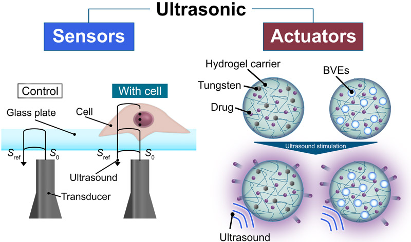
Single-cell ultrasonic microscopy and ultrasound-mediated drug release
- [1] P. G. Auran and O. Silven, “Underwater sonar range sensing and 3D image formation,” Control Eng. Pract., Vol.4, No.3, pp. 393-400, 1996. https://doi.org/10.1016/0967-0661(96)00017-2
- [2] N. K. Mutlib, S. B. Baharom, A. El-Shafie, and M. Z. Nuawi, “Ultrasonic health monitoring in structural engineering: Buildings and bridges,” Struct. Control Health Monit., Vol.23, No.3, pp. 409-422, 2016. https://doi.org/10.1002/stc.1800
- [3] D. Rouseff et al., “Underwater acoustic communication by passive-phase conjugation: Theory and experimental results,” IEEE J. Ocean. Eng., Vol.26, No.4, pp. 821-831, 2001. https://doi.org/10.1109/48.972122
- [4] B. Verhaagen and D. Fernández Rivas, “Measuring cavitation and its cleaning effect,” Ultrason. Sonochem., Vol.29, pp. 619-628, 2016. https://doi.org/10.1016/j.ultsonch.2015.03.009
- [5] J. Carpenter and V. K. Saharan, “Ultrasonic assisted formation and stability of mustard oil in water nanoemulsion: Effect of process parameters and their optimization,” Ultrason. Sonochem., Vol.35, Part A, pp. 422-430, 2017. https://doi.org/10.1016/j.ultsonch.2016.10.021
- [6] S. Mostafavi, D. F. Hesser, and B. Markert, “Effect of process parameters on the interface temperature in ultrasonic aluminum wire bonding,” J. Manuf. Process., Vol.36, pp. 104-114, 2018. https://doi.org/10.1016/j.jmapro.2018.09.020
- [7] A. Brotchie, F. Grieser, and M. Ashokkumar, “Effect of power and frequency on bubble-size distributions in acoustic cavitation,” Phys. Rev. Lett., Vol.102, No.8, Article No.084302, 2009. https://doi.org/10.1103/PhysRevLett.102.084302
- [8] D. Kong, K. Nishio, and M. K. Kurosawa, “Surface acoustic wave propulsion system with acoustic radiation force,” Sens. Actuators A: Phys., Vol.309, Article No.111943, 2020. https://doi.org/10.1016/j.sna.2020.111943
- [9] N. Zhang et al., “Microliter ultrafast centrifuge platform for size-based particle and cell separation and extraction using novel omnidirectional spiral surface acoustic waves,” Lab Chip, Vol.21, No.5, pp. 904-915, 2021. https://doi.org/10.1039/d0lc01012j
- [10] L. Mancia et al., “Modeling tissue-selective cavitation damage,” Phys. Med. Biol., Vol.64, No.22, Article No.225001, 2019. https://doi.org/10.1088/1361-6560/ab5010
- [11] R. A. Marklein, J. Lam, M. Guvendiren, K. E. Sung, and S. R. Bauer, “Functionally-relevant morphological profiling: A tool to assess cellular heterogeneity,” Trends Biotechnol., Vol.36, No.1, pp. 105-118, 2018. https://doi.org/10.1016/j.tibtech.2017.10.007
- [12] J. Guck et al., “Optical deformability as an inherent cell marker for testing malignant transformation and metastatic competence,” Biophys. J., Vol.88, No.5, pp. 3689-3698, 2005. https://doi.org/10.1529/biophysj.104.045476
- [13] E. van Vliet et al., “Electrophysiological recording of re-aggregating brain cell cultures on multi-electrode arrays to detect acute neurotoxic effects,” NeuroToxicology, Vol.28, No.6, pp. 1136-1146, 2007. https://doi.org/10.1016/j.neuro.2007.06.004
- [14] X. Bao et al., “Capturing the interactome of newly transcribed RNA,” Nat. Methods, Vol.15, No.3, pp. 213-220, 2018. https://doi.org/10.1038/nmeth.4595
- [15] E. Bagus Prastika et al., “Time and frequency domain deconvolution for cross-sectional cultured cell observation using an acoustic impedance microscope,” Ultrasonics, Vol.119, Article No.106601, 2022. https://doi.org/10.1016/j.ultras.2021.106601
- [16] E. M. Strohm, G. J. Czarnota, and M. C. Kolios, “Quantitative measurements of apoptotic cell properties using acoustic microscopy,” IEEE Trans. Ultrason. Ferroelectr. Freq. Control, Vol.57, No.10, pp. 2293-2304, 2010. https://doi.org/10.1109/TUFFC.2010.1690
- [17] N. Hozumi, S. Yoshida, and K. Kobayashi, “Three-dimensional acoustic impedance mapping of cultured biological cells,” Ultrasonics, Vol.99, Article No.105966, 2019. https://doi.org/10.1016/j.ultras.2019.105966
- [18] C. L. M. Lee et al., “Noninvasive imaging of rat-derived microglia and its reactivity to inflammatory molecules via acoustic impedance microscopy,” J. Med. Ultrason., Vol.51, No.1, pp. 29-37, 2024. https://doi.org/10.1007/s10396-023-01379-8
- [19] K. Takanashi et al., “Quantitative evaluation method for differentiation of C2C12 myoblasts by ultrasonic microscopy,” Jpn. J. Appl. Phys., Vol.56, No.7S1, Article No.07JF11, 2017. https://doi.org/10.7567/JJAP.56.07JF11
- [20] N. Fujiwara et al., “Deep-learning generation of high-resolution images of live cells in culture using tri-frequency acoustic images,” Phys. Rev. X, Vol.15, No.2, Article No.021015, 2025. https://doi.org/10.1103/PhysRevX.15.021015
- [21] R. T. Tjeung, M. S. Hughes, L. Y. Yeo, and J. R. Friend, “Surface acoustic wave micromotor with arbitrary axis rotational capability,” Appl. Phys. Lett., Vol.99, No.21, Article No.214101, 2011. https://doi.org/10.1063/1.3662931
- [22] Z. Li, Y. Jones, J. Hossenlopp, R. Cernosek, and F. Josse, “Analysis of liquid-phase chemical detection using guided shear horizontal-surface acoustic wave sensors,” Anal. Chem., Vol.77, No.14, pp. 4595-4603, 2005. https://doi.org/10.1021/ac0504621
- [23] F. Lunardelli, M. Agostini, E. Corradi, and M. Cecchini, “Dual-mode shear horizontal / Rayleigh-like surface-acoustic-wave configuration on lithium niobate 64° Y-cut,” Sens. Actuators A: Phys., Vol.378, Article No.115780, 2024. https://doi.org/10.1016/j.sna.2024.115780
- [24] K. Chang et al., “Label-free and high-sensitive detection of human breast cancer cells by aptamer-based leaky surface acoustic wave biosensor array,” Biosens. Bioelectron., Vol.60, pp. 318-324, 2014. https://doi.org/10.1016/j.bios.2014.04.027
- [25] A. Link and T. Franke, “Acoustic erythrocytometer for mechanically probing cell viscoelasticity,” Lab Chip, Vol.20, No.11, pp. 1991-1998, 2020. https://doi.org/10.1039/c9lc00999j
- [26] T. Dehoux et al., “All-optical broadband ultrasonography of single cells,” Sci. Rep., Vol.5, Article No.8650, 2015. https://doi.org/10.1038/srep08650
- [27] L. Liu, L. Plawinski, M.-C. Durrieu, and B. Audoin, “Label-free multi-parametric imaging of single cells: Dual picosecond optoacoustic microscopy,” J. Biophotonics, Vol.12, No.8, Article No.e201900045, 2019. https://doi.org/10.1002/jbio.201900045
- [28] F. Pérez-Cota et al., “Picosecond ultrasonics for elasticity-based imaging and characterization of biological cells,” J. Appl. Phys., Vol.128, No.16, Article No.160902, 2020. https://doi.org/10.1063/5.0023744
- [29] T. Dehoux et al., “Probing single-cell mechanics with picosecond ultrasonics,” Ultrasonics, Vol.56, pp. 160-171, 2015. https://doi.org/10.1016/j.ultras.2014.07.010
- [30] V. Gnyawali, E. M. Strohm, J.-Z. Wang, S. S. H. Tsai, and M. C. Kolios, “Simultaneous acoustic and photoacoustic microfluidic flow cytometry for label-free analysis,” Sci. Rep., Vol.9, Article No.1585, 2019. https://doi.org/10.1038/s41598-018-37771-5
- [31] C. Walker et al., “Liquid-like condensates that bind actin promote assembly and bundling of actin filaments,” Dev. Cell, Vol.60, No.11, pp. 1550-1567.e4, 2025. https://doi.org/10.1016/j.devcel.2025.01.012
- [32] X. Wang et al., “Focal adhesion and actin orientation regulated by cellular geometry determine stem cell differentiation via mechanotransduction,” Acta Biomater., Vol.182, pp. 81-92, 2024. https://doi.org/10.1016/j.actbio.2024.05.017
- [33] J. Alsved, M. Rezayati Charan, P. Ohlsson, A. Urbansky, and P. Augustsson, “Label-free separation of peripheral blood mononuclear cells from whole blood by gradient acoustic focusing,” Sci. Rep., Vol.14, Article No.8748, 2024. https://doi.org/10.1038/s41598-024-59156-7
- [34] P. Li et al., “Acoustic separation of circulating tumor cells,” Proc. Natl. Acad. Sci. U.S.A., Vol.112, No.16, pp. 4970-4975, 2015. https://doi.org/10.1073/pnas.1504484112
- [35] A. A. Nawaz et al., “Acoustofluidic fluorescence activated cell sorter,” Anal. Chem., Vol.87, No.24, pp. 12051-12058, 2015. https://doi.org/10.1021/acs.analchem.5b02398
- [36] M. Wu et al., “Circulating tumor cell phenotyping via high-throughput acoustic separation,” Small, Vol.14, No.32, Article No.1801131, 2018. https://doi.org/10.1002/smll.201801131
- [37] T. Franke, S. Braunmüller, L. Schmid, A. Wixforth, and D. A. Weitz, “Surface acoustic wave actuated cell sorting (SAWACS),” Lab Chip, Vol.10, No.6, pp. 789-794, 2010. https://doi.org/10.1039/b915522h
- [38] M. Miansari et al., “Inducing mild traumatic brain injury in C. elegans via cavitation-free surface acoustic wave-driven ultrasonic irradiation,” Sci. Rep., Vol.9, Article No.12775, 2019. https://doi.org/10.1038/s41598-019-47295-1
- [39] Y. Kurashina, T. Yamashita, S. Kurabayashi, K. Takemura, and K. Ando, “Growth control of leaf lettuce with exposure to underwater ultrasound and dissolved oxygen supersaturation,” Ultrason. Sonochem., Vol.51, pp. 292-297, 2019. https://doi.org/10.1016/j.ultsonch.2018.10.005
- [40] Y. Kurashina, K. Takemura, and J. Friend, “Cell agglomeration in the wells of a 24-well plate using acoustic streaming,” Lab Chip, Vol.17, No.5, pp. 876-886, 2017. https://doi.org/10.1039/c6lc01310d
- [41] M. Nakao et al., “Formation of large scaffold-free 3-D aggregates in a cell culture dish by ultrasound standing wave trapping,” Ultrasound Med. Biol., Vol.45, No.5, pp. 1306-1315, 2019. https://doi.org/10.1016/j.ultrasmedbio.2019.01.013
- [42] S. Koda, K. Wakayama, S. Takahashi, and Y. Kurashina, “Rayleigh-type surface acoustic wave-driven shooting for directional artificial pollination of strawberry flowers,” Sens. Actuators A: Phys., Vol.405, Article No.117774, 2026. https://doi.org/10.1016/j.sna.2026.117774
- [43] K. Melde et al., “Ultrasound-assisted tissue engineering,” Nat. Rev. Bioeng., Vol.2, No.6, pp. 486-500, 2024. https://doi.org/10.1038/s44222-024-00166-5
- [44] C. Imashiro et al., “Cell patterning method on a clinically ubiquitous culture dish using acoustic pressure generated from resonance vibration of a disk-shaped ultrasonic transducer,” IEEE Trans. Biomed. Eng., Vol.66, No.1, pp. 111-118, 2019. https://doi.org/10.1109/TBME.2018.2835834
- [45] C. Imashiro, Y. Kurashina, and K. Takemura, “Cell patterning method using resonance vibration of a metallic cell cultivation substrate,” Adv. Biomed. Eng., Vol.5, pp. 142-148, 2016. https://doi.org/10.14326/abe.5.142
- [46] Z. Ma et al., “Acoustic holographic cell patterning in a biocompatible hydrogel,” Adv. Mater., Vol.32, No.4, Article No.1904181, 2020. https://doi.org/10.1002/adma.201904181
- [47] S. Li et al., “Standing surface acoustic wave based cell coculture,” Anal. Chem., Vol.86, No.19, pp. 9853-9859, 2014. https://doi.org/10.1021/ac502453z
- [48] K. Martinez Villegas, R. Rasouli, and M. Tabrizian, “Enhancing metabolic activity and differentiation potential in adipose mesenchymal stem cells via high-resolution surface-acoustic-wave contactless patterning,” Microsyst. Nanoeng., Vol.8, Article No.79, 2022. https://doi.org/10.1038/s41378-022-00415-w
- [49] D. J. Collins et al., “Two-dimensional single-cell patterning with one cell per well driven by surface acoustic waves,” Nat. Commun., Vol.6, Article No.8686, 2015. https://doi.org/10.1038/ncomms9686
- [50] J. Huang et al., “ZnO/glass-based SAW tweezer for dexterous particle patterning and patterned cell culturing,” Microfluid. Nanofluid., Vol.27, No.5, Article No.34, 2023. https://doi.org/10.1007/s10404-023-02646-3
- [51] Y. Kurashina et al., “Enzyme-free release of adhered cells from standard culture dishes using intermittent ultrasonic traveling waves,” Commun. Biol., Vol.2, Article No.393, 2019. https://doi.org/10.1038/s42003-019-0638-5
- [52] A. Salari, S. Appak-Baskoy, I. R. Coe, S. S. H. Tsai, and M. C. Kolios, “An ultrafast enzyme-free acoustic technique for detaching adhered cells in microchannels,” RSC Adv., Vol.11, No.52, pp. 32824-32829, 2021. https://doi.org/10.1039/d1ra04875a
- [53] H. Tauchi et al., “Effective and intact cell detachment from a clinically ubiquitous culture flask by combining ultrasonic wave exposure and diluted trypsin,” Biotechnol. Bioprocess Eng., Vol.24, No.3, pp. 536-543, 2019. https://doi.org/10.1007/s12257-018-0491-2
- [54] A. Bussonnière et al., “Cell detachment and label-free cell sorting using modulated surface acoustic waves (SAWs) in droplet-based microfluidics,” Lab Chip, Vol.14, No.18, pp. 3556-3563, 2014. https://doi.org/10.1039/c4lc00625a
- [55] K. Fujishiro, S. Okada, F. Rossi, T. Kuchimaru, and Y. Kurashina, “Intracellular reactive oxygen species generation induced by high-frequency ultrasound in thickness vibration mode,” Adv. Sci., Vol.13, No.28, Article No.e19582, 2026. https://doi.org/10.1002/advs.202519582
- [56] X. Yang, X. Sun, D. Zhou, Y. Li, and H. Liu, “Towards wearable A-mode ultrasound sensing for real-time finger motion recognition,” IEEE Trans. Neural Syst. Rehabil. Eng., Vol.26, No.6, pp. 1199-1208, 2018. https://doi.org/10.1109/TNSRE.2018.2829913
- [57] E. Sassaroli, A. Scorza, C. Crake, S. A. Sciuto, and M.-A. Park, “Breast ultrasound technology and performance evaluation of ultrasound equipment: B-mode,” IEEE Trans. Ultrason. Ferroelectr. Freq. Control, Vol.64, No.1, pp. 192-205, 2017. https://doi.org/10.1109/TUFFC.2016.2619622
- [58] J. Li, K. Zhu, and L. Pan, “Wrist and finger motion recognition via M-mode ultrasound signal: A feasibility study,” Biomed. Signal Process. Control, Vol.71, Part A, Article No.103112, 2022. https://doi.org/10.1016/j.bspc.2021.103112
- [59] Y. Jin et al., “Estimation of joint torque in dynamic activities using wearable A-mode ultrasound,” Nat. Commun., Vol.15, Article No.5756, 2024. https://doi.org/10.1038/s41467-024-50038-0
- [60] H. Hu et al., “A wearable cardiac ultrasound imager,” Nature, Vol.613, No.7945, pp. 667-675, 2023. https://doi.org/10.1038/s41586-022-05498-z
- [61] M. Lin et al., “A fully integrated wearable ultrasound system to monitor deep tissues in moving subjects,” Nat. Biotechnol., Vol.42, No.3, pp. 448-457, 2024. https://doi.org/10.1038/s41587-023-01800-0
- [62] X. Gao et al., “A wearable echomyography system based on a single transducer,” Nat. Electron., Vol.7, No.11, pp. 1035-1046, 2024. https://doi.org/10.1038/s41928-024-01271-4
- [63] S. Zhou et al., “Clinical validation of a wearable ultrasound sensor of blood pressure,” Nat. Biomed. Eng., Vol.9, No.6, pp. 865-881, 2025. https://doi.org/10.1038/s41551-024-01279-3
- [64] J. Park et al., “Opto-ultrasound biosensor for wearable and mobile devices: Realization with a transparent ultrasound transducer,” Biomed. Opt. Express, Vol.13, No.9, pp. 4684-4692, 2022. https://doi.org/10.1364/BOE.468969
- [65] M. Angerer et al., “A polymer-based CMUT probe for imaging the spinal cord in rats,” IEEE Trans. Ultrason. Ferroelectr. Freq. Control, Vol.72, No.11, pp. 1437-1447, 2025. https://doi.org/10.1109/TUFFC.2025.3605440
- [66] I. M. Suarez-Castellanos et al., “Dynamic ultrasound focusing and centimeter-scale ex vivo tissue ablations with a CMUT probe developed for endocavitary HIFU therapies,” IEEE Trans. Ultrason. Ferroelectr. Freq. Control, Vol.70, No.11, pp. 1470-1481, 2023. https://doi.org/10.1109/TUFFC.2023.3301977
- [67] M. Wang and J. Chen, “Volumetric flow measurement using an implantable CMUT array,” IEEE Trans. Biomed. Circuits Syst., Vol.5, No.3, pp. 214-222, 2011. https://doi.org/10.1109/TBCAS.2010.2095848
- [68] M. Qu et al., “Imaging of simulated muscle based on single chip of AlN piezoelectric micromachined ultrasonic transducer,” J. Micromech. Microeng., Vol.32, No.7, Article No.075003, 2022. https://doi.org/10.1088/1361-6439/ac70a6
- [69] D. E. Dausch et al., “In vivo real-time 3-D intracardiac echo using PMUT arrays,” IEEE Trans. Ultrason. Ferroelectr. Freq. Control, Vol.61, No.10, pp. 1754-1764, 2014. https://doi.org/10.1109/TUFFC.2014.006452
- [70] X. Jiang, V. Perrot, F. Varray, S. Bart, and P. G. Hartwell, “Piezoelectric micromachined ultrasonic transducer for arterial wall dynamics monitoring,” IEEE Trans. Ultrason. Ferroelectr. Freq. Control, Vol.69, No.1, pp. 291-298, 2022. https://doi.org/10.1109/TUFFC.2021.3120283
- [71] C. Errico et al., “Ultrafast ultrasound localization microscopy for deep super-resolution vascular imaging,” Nature, Vol.527, No.7579, pp. 499-502, 2015. https://doi.org/10.1038/nature16066
- [72] T. Opacic et al., “Motion model ultrasound localization microscopy for preclinical and clinical multiparametric tumor characterization,” Nat. Commun., Vol.9, Article No.1527, 2018. https://doi.org/10.1038/s41467-018-03973-8
- [73] S.-H. Lee, J. Y. Shin, A. Lee, and C. Bustamante, “Counting single photoactivatable fluorescent molecules by photoactivated localization microscopy (PALM),” Proc. Natl. Acad. Sci. U.S.A., Vol.109, No.43, pp. 17436-17441, 2012. https://doi.org/10.1073/pnas.1215175109
- [74] M. J. Rust, M. Bates, and X. Zhuang, “Sub-diffraction-limit imaging by stochastic optical reconstruction microscopy (STORM),” Nat. Methods, Vol.3, No.10, pp. 793-796, 2006. https://doi.org/10.1038/nmeth929
- [75] C. Demené et al., “Transcranial ultrafast ultrasound localization microscopy of brain vasculature in patients,” Nat. Biomed. Eng., Vol.5, No.3, pp. 219-228, 2021. https://doi.org/10.1038/s41551-021-00697-x
- [76] K. Kataoka, A. Harada, and Y. Nagasaki, “Block copolymer micelles for drug delivery: Design, characterization and biological significance,” Adv. Drug Deliv. Rev., Vol.47, No.1, pp. 113-131, 2001. https://doi.org/10.1016/S0169-409X(00)00124-1
- [77] R. R. López et al., “The effect of different organic solvents in liposome properties produced in a periodic disturbance mixer: Transcutol®, a potential organic solvent replacement,” Colloids Surf. B: Biointerfaces, Vol.198, Article No.111447, 2021. https://doi.org/10.1016/j.colsurfb.2020.111447
- [78] Y. Kurashina et al., “Adeno-associated virus-encapsulated alginate microspheres loaded in collagen gel carriers for localized gene transfer,” Adv. Healthc. Mater., Vol.13, No.12, Article No.2303546, 2024. https://doi.org/10.1002/adhm.202303546
- [79] T. Kubota, Y. Kurashina, J. Zhao, K. Ando, and H. Onoe, “Ultrasound-triggered on-demand drug delivery using hydrogel microbeads with release enhancer,” Mater. Des., Vol.203, Article No.109580, 2021. https://doi.org/10.1016/j.matdes.2021.109580
- [80] R. Yamakawa, H. Onoe, and Y. Kurashina, “Hydrogel carrier with bubble vibration enhancer for ultrasound-triggered drug release,” Ultrason. Sonochem., Vol.112, Article No.107173, 2025. https://doi.org/10.1016/j.ultsonch.2024.107173
- [81] T. Jamneala et al., “Ultra-miniature coupled resonator filter with single-layer acoustic coupler,” IEEE Trans. Ultrason. Ferroelectr. Freq. Control, Vol.56, No.11, pp. 2553-2558, 2009. https://doi.org/10.1109/TUFFC.2009.1342
- [82] S. Takatsuka, T. Kubota, Y. Kurashina, and H. Onoe, “Near-infrared-triggered on-demand controlled release of adeno-associated virus from alginate hydrogel microbeads with heat transducer for gene therapy,” Small, Vol.19, No.7, Article No.2204139, 2023. https://doi.org/10.1002/smll.202204139
- [83] A. Hioki, S. Takatsuka, Y. Kurashina, and H. Onoe, “Alginate hydrogel microbeads with different mesh structures enable controlled release of adeno-associated virus for gene therapy,” Proc. 2023 22nd Int. Conf. Solid-State Sens., Actuators Microsyst. (Transducers), pp. 482-485, 2023.
- [84] Y. Bando et al., “Synthesis of hydrogel microparticles from ultra-high-viscosity pre-gel solution by combining centrifugal force and ultrasonic vibration,” Small, Vol.21, No.34, Article No.2412715, 2025. https://doi.org/10.1002/smll.202412715
- [85] O. Al-Bataineh, R. Abdallat, T. Momani, S. Alzghoul, and O. Smadi, “In-vivo evaluation of ultrasound transducers with sweep-frequency excitation for noninvasive transdermal insulin delivery,” J. Drug Deliv. Sci. Technol., Vol.112, Article No.107258, 2025. https://doi.org/10.1016/j.jddst.2025.107258
- [86] K. Matsubara et al., “Transdermal administration of macromolecular drugs by multi-frequency sequential ultrasonic irradiation,” Int. J. Pharm., Vol.675, Article No.125560, 2025. https://doi.org/10.1016/j.ijpharm.2025.125560
- [87] J. Zhang et al., “Flexible surface acoustic wave technology for enhancing transdermal drug delivery,” Drug Deliv. Transl. Res., Vol.15, No.4, pp. 1363-1375, 2025. https://doi.org/10.1007/s13346-024-01682-y
- [88] A. Boucaud, M. A. Garrigue, L. Machet, L. Vaillant, and F. Patat, “Effect of sonication parameters on transdermal delivery of insulin to hairless rats,” J. Control. Release, Vol.81, Nos.1-2, pp. 113-119, 2002. https://doi.org/10.1016/S0168-3659(02)00054-8
- [89] C. M. Schoellhammer et al., “Applicability and safety of dual-frequency ultrasonic treatment for the transdermal delivery of drugs,” J. Control. Release, Vol.202, pp. 93-100, 2015. https://doi.org/10.1016/j.jconrel.2015.02.002
- [90] X. Xie et al., “Transdermal delivery of bFGF with sonophoresis facilitated by chitosan nanocarriers,” J. Drug Deliv. Sci. Technol., Vol.75, Article No.103675, 2022. https://doi.org/10.1016/j.jddst.2022.103675
- [91] C. Yu et al., “A conformable ultrasound patch for cavitation-enhanced transdermal cosmeceutical delivery,” Adv. Mater., Vol.35, No.23, Article No.2300066, 2023. https://doi.org/10.1002/adma.202300066
- [92] T. Asakura, K. Yoshioka, A. Ogawa, and Y. Kurashina, “Effect of particle size distribution on the transmission efficiency of atomized water to the tracheal tube,” Sens. Actuators A: Phys., Vol.370, Article No.115270, 2024. https://doi.org/10.1016/j.sna.2024.115270
- [93] K. Yoshioka, Y. Kurashina, A. Ogawa, and T. Asakura, “Effect of the area of a lithium niobate transducer on the efficiency of ultrasonic atomization driven by resonance vibration,” Ultrason. Sonochem., Vol.86, Article No.106019, 2022. https://doi.org/10.1016/j.ultsonch.2022.106019
- [94] K. Wakayama, S. Kurihara, and Y. Kurashina, “Narrow-width surface acoustic wave device-driven olfactory epithelium-targeted intranasal atomization,” Int. J. Pharm., Vol.692, Article No.126630, 2026. https://doi.org/10.1016/j.ijpharm.2026.126630
- [95] H. Chen and E. E. Konofagou, “The size of blood–brain barrier opening induced by focused ultrasound is dictated by the acoustic pressure,” J. Cereb. Blood Flow Metab., Vol.34, No.7, pp. 1197-1204, 2014. https://doi.org/10.1038/jcbfm.2014.71
- [96] C.-H. Fan et al., “Drug-loaded bubbles with matched focused ultrasound excitation for concurrent blood–brain barrier opening and brain-tumor drug delivery,” Acta Biomater., Vol.15, pp. 89-101, 2015. https://doi.org/10.1016/j.actbio.2014.12.026
- [97] R. Ji et al., “Cavitation-modulated inflammatory response following focused ultrasound blood-brain barrier opening,” J. Control. Release, Vol.337, pp. 458-471, 2021. https://doi.org/10.1016/j.jconrel.2021.07.042
- [98] D. Omata et al., “Effects of encapsulated gas on stability of lipid-based microbubbles and ultrasound-triggered drug delivery,” J. Control. Release, Vols.311-312, pp. 65-73, 2019. https://doi.org/10.1016/j.jconrel.2019.08.023
- [99] S. Kim et al., “Localized ultrasonic stimulation using a piezoelectric micromachined ultrasound transducer array for selective neural differentiation of magnetic cell-based robots,” Microsyst. Nanoeng., Vol.11, Article No.52, 2025. https://doi.org/10.1038/s41378-025-00900-y
- [100] T. Luo and M. Wu, “Biologically inspired micro-robotic swimmers remotely controlled by ultrasound waves,” Lab Chip, Vol.21, No.21, pp. 4095-4103, 2021. https://doi.org/10.1039/d1lc00575h
- [101] N. Mahkam et al., “Acoustic streaming-induced multimodal locomotion of bubble-based microrobots,” Adv. Sci., Vol.10, No.35, Article No.2304233, 2023. https://doi.org/10.1002/advs.202304233
This article is published under a Creative Commons Attribution-NoDerivatives 4.0 Internationa License.