
Review:
Ultrasonic Sensors and Actuators for Cell/Tissue Culture in Biomedical Applications
Shun Koda
 and Yuta Kurashina†
Department of Mechanical Systems Engineering, Graduate School of Engineering, Tokyo University of Agriculture and Technology
2-24-16 Nakacho, Koganei, Tokyo 184-8588, Japan
†Corresponding author

“Sensor introduction into cells and tissues for information transmission” is crucial for understanding cellular and tissue states, as it allows real-time, noninvasive monitoring of dynamic physiological changes. In this review article, we highlight ultrasound technologies for cells and tissues as noninvasive, highly penetrative mechanical modalities for both sensor introduction and information retrieval. In both ultrasonic sensors and actuators, a persistent trade-off remains between achieving higher resolution through increased frequencies and the resultant significant signal attenuation. Ultimately, innovations in optical technologies play a significant role in advancing ultrasonic systems, and vice versa, fostering cross-disciplinary breakthroughs in biomedical engineering.
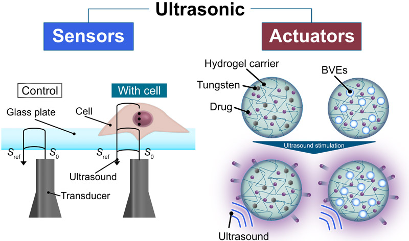
Single-cell ultrasonic microscopy and ultrasound-mediated drug release
1. Introduction
“Sensor introduction into cells and tissues for information transmission” is crucial for understanding cellular and tissue states, as it enables real-time, noninvasive monitoring of dynamic physiological changes. In the introduction of both sensors, ultrasound serves as a highly effective mechanical force due to its noninvasive nature and deep tissue permeability. Furthermore, when transmitting information from cells and tissues, ultrasound is expected to enable noninvasive signal transmission and acquisition by leveraging its piezoelectric and inverse piezoelectric effects. Hence, in this review article, we highlight ultrasound technologies that contribute to “sensor introduction into cells and tissues for information transmission.” Specifically, we introduce various technologies surrounding ultrasound sensors and actuators for cells and tissues. Owing to its high penetration depth through diverse matter and its ability to exert noncontact mechanical forces, ultrasound has long been used in various industrial applications, such as sensors and actuators. For sensors, some applications of ultrasound include the detection of underwater objects 1, structural health monitoring of aging infrastructure 2, and underwater communication 3. For actuators, some applications of ultrasound include ultrasonic cleaning 4, emulsification 5, and wire bonding processes 6. Recently, advances in microelectromechanical systems (MEMS) and the growing demand for medical solutions have spurred the development of ultrasonic sensors and actuators for cells and tissues. Ultrasonic sensors and actuators for cells and tissues utilize a diverse range of phenomena. Ultrasonic sensors primarily rely on the piezoelectric and inverse piezoelectric effects for the reception and transmission of ultrasound, respectively. By contrast, ultrasonic actuators interact with biological targets through mechanical effects, including cavitation 7, acoustic radiation force 8, and acoustic streaming 9. Among these phenomena, cavitation has raised particular concern in previous studies due to the risk of unintended tissue damage 10. Given the risk and critical role of biocompatibility with cells and tissues, ultrasonic sensors and actuators require optimization of the device design, including the transducer material, transducer shape, frequency, and acoustic pressure.
2. Ultrasonic Sensors for Evaluating Cell Properties
2.1. Background of Ultrasonic Sensors for Evaluating Cell Properties
The function and state of cells are defined by multiple parameters, including morphology 11, mechanical properties 12, electrical response 13, and molecular expression 14. Technologies that enable high-precision and dynamic measurements of these cellular parameters constitute a core foundation not only for elucidating biological phenomena in basic biology but also for supporting next-generation disease diagnostics, drug discovery, and regenerative medicine. Although optical cell evaluation is widely adopted, it is expected that ultrasound cell observation will provide real-time assessment due to its noninvasive and safe characteristics. Therefore, in recent years, ultrasonic-based approaches have been actively explored for noninvasive, noncontact, and deep measurement of cellular parameters. Various ultrasonic sensors, including acoustic microscopy, surface acoustic wave (SAW) biosensors, and optoacoustic systems, have been adapted to measure cellular parameters. These sensors transmit and receive ultrasonic waves via inverse piezoelectric and piezoelectric effects, respectively. When ultrasound penetrates materials with different impedances, changes in the amplitude attenuation and phase delay occur. Ultrasonic sensors can be used to detect these parameters to perform sensing.
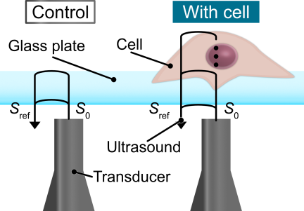
Fig. 1. Overview of acoustic microscopy. The physical properties are evaluated by calculating the impedance from the acoustic waves reflected and transmitted at each layer. The glass surface, where no cells are attached, is evaluated as a control for comparison with the cell adhesion areas. This figure was redrawn based on 15.
2.2. Acoustic Microscopy
Owing to the limited penetration capability of optical microscopy, acoustic microscopy has been extensively explored due to its superior penetrability and noninvasiveness. The basic principle of acoustic microscopy is to receive reflected waves after ultrasound irradiation from a transducer to the bottom surface of a cell-culturing substrate. The acoustic impedance is then calculated from the amplitude and phase of the reflected wave for each layer (Fig. 1) 15. The impedance measured from a substrate without cells, consisting only of the substrate and culture medium, is used as a reference for control. The variables detected by acoustic microscopy include impedance, phase shifting, and amplitude attenuation. Based on these variables, the stiffness 16 and morphology 17 of the single cells are evaluated. The experimental setup consists of a piezo stage, pulser–receiver, transducer, and cell culture dish arranged from bottom to top 18. Because the size of a single cell is \({\sim}20\) μm, high-resolution observation is required. Therefore, the position of the transducer equipped with a sapphire lens for ultrasonic focusing is precisely controlled using a piezoelectric stage to obtain high-resolution scanning. The cell types studied include glial cells 17, C2C12 cells 19, mesenchymal stem cells 20, human-induced pluripotent stem cells 20, and MCF-7 cells 16. The lateral resolution in the \(xy\)-direction is of the order of several micrometers due to the diffraction limit of ultrasound 16. By contrast, in one case, the axial resolution along the beam direction of the ultrasound was improved to \({\sim}0.2\) μm 15. As a result, a cross-sectional impedance map of glial cells was obtained, enabling visualization of the gap between the cell and the substrate to which the cell adheres 15. In addition, for lateral resolution in the \(xy\)-direction, although achieving a resolution comparable to the wavelength of light remains difficult, methods based on deep-learning-based image processing have been explored to enhance the resolution 20. These studies indicate that acoustic microscopy is a highly useful method for evaluating single cells not only in the \(xy\)-direction but also in the \(z\)-direction. Therefore, it is expected that acoustic microscopy will be a key technique for real-time evaluation of cell morphology and mechanical properties. One of the fundamental physical limitations of acoustic microscopy is noise and signal degradation. In complex imaging targets featuring numerous small scattering structures, including the intracellular environment or entire apoptotic cells, the resolution degradation caused by backscattering poses a significant challenge 16. Consequently, achieving high-resolution imaging of internal cellular structures requires advancements in noise reduction techniques, including deep learning.
2.3. Surface Acoustic Wave
SAW is an elastic wave that travels across the surface of a solid. SAW is excited onto a piezoelectric substrate using an interdigital transducer (IDT). There are several modes of SAW. Rayleigh SAW (R-SAW) exhibits a vertical particle displacement 21, whereas shear-horizontal SAW (SH-SAW) exhibits particle displacement parallel to the piezoelectric substrate surface 22. R-SAW is suitable for use in dry environments and has been widely used in gas sensing. By contrast, for biosensing applications where the device typically operates in aqueous and wet conditions, the use of R-SAW leads to significant attenuation due to the leakage of acoustic waves into the surrounding fluid 23. Because of these characteristics, SH-SAW has become the predominant choice for biosensing applications. In a previous study, label-free and highly sensitive detection of breast cancer cells has been developed using Leaky SAW, a type of SH-SAW, as a biosensor 24. In this case, the sensing device had two IDTs positioned face-to-face as the transmitting and receiving ports, with the region between them defined as the sensing area. Gold was sputter-deposited onto the sensing area. The gold surface was then functionalized with aptamers that specifically bound mucin 1 (MUC1), a protein highly expressed on the surface of MCF-7 breast cancer cells. When MCF-7 cells were captured by the aptamers, the phase of the leaky SAW shifted due to mass loading in the sensing region. This phase shift enabled the detection of MCF-7 cells with extremely high sensitivity and a detection limit as low as 32 cells/mL.
In recent years, the leaky waves of R-SAW in fluid, which were previously considered to be problematic, have also been used for biosensors. For biosensors using R-SAW, the mechanical properties of the cells are predominantly evaluated, rather than the mass loading associated with phase shifts. One previous application is the measurement of the viscoelasticity of red blood cells using standing waves generated by traveling R-SAW 25. Within a polydimethylsiloxane (PDMS) microchannel, red blood cells were trapped at the pressure nodes of standing waves generated by R-SAW and were deformed by the acoustic radiation force. The mechanical properties of the red blood cells, elasticity and viscosity, were evaluated using the Taylor deformation parameter (\(D\)), which represents the magnitude of cell deformation, and relaxation times (\(\tau_{1}\) and \(\tau_{2}\)), which describe the rate of deformation. Consequently, the viscoelastic properties of individual cells were successfully evaluated, highlighting the strong potential of this approach for the diagnosis of hematological disorders. Although the surface-concentrated energy of the SH-SAW makes 3D sensing at the tissue scale challenging, it is highly effective for 2D sensing at the cellular scale. Therefore, SAW devices have been widely used for chip-scale sensing platforms for cell evaluation. Based on previous studies on SAW-based cell sensing, SH-SAW is suitable for measuring cells via changes in acoustic propagation characteristics, whereas R-SAW is better suited for applying mechanical effects, including cell deformation.
2.4. Optoacoustic Systems
Laser-induced optoacoustic microscopy has been developed to enhance axial resolution. Picosecond ultrasonic waves (PUs) excited by lasers are pulsed waves containing broadband frequency components ranging from GHz 26 to THz 27. By utilizing PUs with their characteristic extremely short wavelengths, it is possible to achieve a higher axial resolution in cellular observations compared with conventional acoustic microscopy. The pump-probe technique has been proposed as a method for detecting laser-induced PUs, which involves irradiating cells with a probe beam to detect minute changes in the reflectivity caused by acoustic waves 28. A proper device design enables noninvasive cellular observation using PUs. The acoustic waves generated by PUs do not possess sufficient energy to cause molecular-level damage to cells 28. In one case, it has been suggested that the evaluation was entirely noninvasive, given that the acoustic stress of PUs was \(\sim\)200 Pa, whereas osteoblasts exhibited an adhesion stress in the range of several kPa 29. To prevent thermal damage to cells, the temperature rise can be suppressed using substrates such as sapphire or diamond 28. These results demonstrate that PUs are a promising noninvasive and label-free technique for super-resolution visualization of cellular elasticity, with potential applications in diagnostics and regenerative medicine.
Another application involves combining it with a flow cytometry system. In a previous study, a label-free acoustic flow cytometer was developed that simultaneously detected ultrahigh-frequency ultrasound (375 MHz) backscatter and optoacoustic waves generated by nanosecond laser pulses in single cells flowing through a microchannel 30. Using this system, the authors distinguished and counted differently colored polystyrene beads, as well as red and white blood cells in human blood samples. Red blood cells generated both ultrasound and strong optoacoustic signals, whereas white blood cells produced only ultrasound signals, enabling accurate identification without labeling or cell lysis. The results showed excellent agreement with those obtained using conventional fluorescence-activated cell sorting with a label-free sensing platform for samples such as red and white blood cells without compromising their integrity. These findings demonstrate that multiple physical, morphological, and optical absorption properties can be obtained simultaneously and nondestructively from single cells, highlighting the potential of this technique for biomedical and diagnostic applications. Optoacoustic systems achieve the high-resolution characteristics of optical microscopy by leveraging the fact that the wavelength of light is significantly shorter than that of sound. However, a major technical challenge is the significant attenuation caused by light scattering, which is a limitation shared with conventional optical microscopy. Consequently, optoacoustic systems are more advantageous for cellular-scale imaging than macroscopic observations at the tissue level.
2.5. Future Prospects of Ultrasonic Sensors for Evaluating Cell Properties
Ultrasonic sensors have attracted attention for three-dimensional evaluation of single cells in a living state and are highly suitable for evaluating the physical properties of single cells, such as stiffness. However, this resolution is insufficient for the evaluation of single cells. At the single-cell scale, molecular-level resolution is often required to visualize actin filaments 31,32. In addition, noise processing is critical for ultrasound sensing. As the frequency increases, the received signal weakens, resulting in a lower signal-to-noise ratio. Therefore, further improvements in the resolution and noise processing are necessary to advance ultrasound-based single-cell evaluations towards practical applications. Increasing the ultrasonic frequency is an effective approach to enhance the resolution of ultrasonic sensors. However, there is an inherent trade-off: as the frequency increases, the stronger attenuation of ultrasonic waves inevitably limits the spatial resolution. As demonstrated in many studies, deep-learning-based approaches have been actively investigated for noise reduction and resolution enhancement. Further resolution enhancement is expected through improvements in the reliability of the images generated by deep learning. Moreover, synergistic integration with optics holds great promise for establishing new characterization frameworks. Effectively incorporating and integrating the advantages of optical observation is the key to overcoming the current limitations.
3. Ultrasonic Actuators for Cell Manipulation
3.1. Backgrounds of Ultrasonic Actuators for Cell Manipulation
In life sciences research, the spatial and functional control of living cells serves as a fundamental technology spanning from basic research to clinical applications. In particular, the isolation of target cells from heterogeneous populations, cell patterning for tissue engineering, and external remote manipulation of intracellular signaling play an important role in advancing drug discovery, regenerative medicine, and neuroscience. In this context, ultrasonic actuators have been actively explored due to their biocompatibility. Ultrasonic actuators enable label-free manipulation while maintaining excellent biocompatibility through noncontact and noninvasive characteristics. Moreover, ultrasound enables exceptionally high penetration into deep tissues compared with stimulus sources such as electricity or light. Thus, ultrasonic actuators have the potential to extend beyond in vitro manipulation of microfluidic channels to in vivo cell manipulation, including therapeutic applications.
3.2. Cell Sorting
Cell-sorting techniques have seen significant advancements for the detection of biomarkers at extremely low concentrations in samples. Cell sorting is a cell manipulation technique based on ultrasonic actuators. In several previous studies, specific cells have been separated and extracted from cell populations injected into PDMS microchannels using continuous ultrasound. Although bulk acoustic waves generated by lead zirconate titanate (PZT) have also been used for sorting devices 33, SAWs have been used because of their compatibility with lab-on-chip devices 34,35,36,37. Acoustic streaming and acoustic radiation pressure have been primarily employed for the manipulation of cells or biological organisms using ultrasonic actuators 38,39,40. The basic mechanism behind cell manipulation, including cell sorting, involves the alignment of cells at the pressure nodes of standing ultrasound using acoustic radiation forces 33,36,41. R-SAW can generate an acoustic radiation pressure that is sufficiently high to eject droplets containing microparticles 42. Sorting methods based on acoustic streaming require a fluorescence detection system because acoustic streaming is selectively applied after verifying the fluorescence of the target cells in the microchannel 37. The acoustic radiation force that drives cell sorting can be expressed as
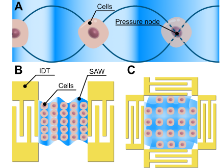
Fig. 2. Cell patterning variations using R-SAW. (A) Principle of cell patterning using R-SAW. Cells are trapped at pressure nodes. (B, C) The IDT designs for each patterning: (B) linear patterning and (C) grid-like patterning.
3.3. Cell Patterning
In tissue engineering aimed at developing various organs in vitro, it is essential to achieve precise spatiotemporal control over the positioning of diverse cell types 43. To achieve this, ultrasound-based cell patterning has been actively investigated using an acoustic radiation force based on a principle similar to that of cell sorting (Fig. 2(A)). Ultrasonic actuators have been widely applied in cell-patterning techniques because they enable noninvasive and contactless manipulation of cells. Taking advantage of the contactless manipulation offered by acoustics, a circular cell pattern was achieved by applying a 7.7-kHz resonant vibration to a ubiquitous dish using a disk-shaped ultrasonic transducer 44. Similarly, a cross-circular nodal pattern of cells was obtained using resonant vibrations at 26.9 kHz 45. Although these methods directly apply transducer resonance, another study has achieved more complex patterning by incorporating acoustic holograms 46. In addition, Rayleigh-type SAW devices capable of generating high-frequency ultrasound on a two-dimensional array have been widely used. The configuration of the IDTs formed on the piezoelectric substrate differs depending on the desired patterning geometry. For patterning aimed at aligning cells in a line, two IDTs are fabricated facing each other (Fig. 2(B)) 47,48. In a previous study, a cell co-culture system for two types of line-patterned cells was developed by controlling the relative phase of the SAWs generated from one of the IDTs 47. By contrast, for a uniform arrangement of cells on a two-dimensional array, two pairs of opposing IDTs were fabricated on the substrate such that the propagation directions of the SAWs were orthogonal to each other. In a previous study, single cells were patterned into an isolated grid structure using high-frequency SAWs of over 100 MHz (Fig. 2(C)) 49. In this case, human peripheral blood mononuclear cells patterned in a grid maintained viability comparable to that of the control for more than 6 h under a SAW input power of 0.22 W. Although 128° Y-cut lithium niobate (LN) is the gold standard piezoelectric substrate for R-SAW devices for biomedical applications, 128° Y-cut LN has anisotropy, which restricts its orientation for IDT fabrication. Therefore, ZnO is a suitable piezoelectric material for forming IDTs with various orientations to achieve more complex cell patterning 50.
3.4. Cell Detachment
Cell detachment is an essential step in the cell passaging process and is therefore important in various studies involving adherent cells. In standard adherent cell cultures, the cells are grown on a dish surface. Upon reaching confluency, the cells are detached and harvested to regulate their density for subsequent steps. Enzymes such as trypsin are widely used to detach cells. However, the invasiveness of trypsinization is a major concern because it degrades proteins on the cell surface 51. Therefore, in recent years, noninvasive cell detachment using ultrasound has been actively studied. PZT 52 and Langevin transducers incorporating PZT 53 have been widely used to excite ultrasound in the kHz range in ultrasound-based cell detachment devices. The shear stress generated by ultrasound-induced shear flow is one of the major mechanisms of ultrasound-induced cell detachment. The shear flow arises not from acoustic streaming but from sloshing induced by frequency-swept 51 or amplitude-modulated ultrasound 54. Another major mechanism of ultrasound-induced cell detachment is acoustic pressure 51. These ultrasound-induced effects do not act independently; rather, they contribute collectively to cell detachment. Ultrasound-induced cell detachment exhibits greater noninvasiveness than conventional methods. In one study, cell proliferation 48 h after detachment increased to 130% of that observed after trypsin treatment 51. In addition, cell detachment in a microchannel using pulsed ultrasound generated by a PZT transducer showed high cell viability of 86%–91%, indicating low invasiveness 52. Thus, ultrasound-based cell detachment contributes to noninvasive operations and has great potential for practical applications.
3.5. Future Prospects of Ultrasonic Actuators for Cell Manipulation
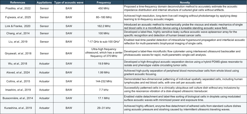
Owing to its noninvasive and label-free characteristics, ultrasound has been applied for cell sorting, cell patterning, and cell detachment. Ultrasonic sensors and actuators for cells have been appropriately designed depending on their purpose (Table 1). Previous studies have reported long-term evaluations of cell viability following exposure to kHz-range ultrasound and have demonstrated the effectiveness of pulsed kHz-range ultrasound. In the future, high-frequency ultrasound may provide an alternative for achieving more localized and selective cellular manipulation. Although the effects of low-frequency ultrasound on cells have been investigated, the effects of high-frequency ultrasound remain insufficiently evaluated. A recent study has indicated that high-frequency ultrasound (6.5 MHz) induced the generation of intracellular reactive oxygen species, potentially leading to apoptosis 55. Elucidating the effects of high-frequency ultrasound on cells will advance the development of localized and selective cellular manipulation techniques that exploit its advantages.
4. Ultrasonic Sensors for Evaluating Tissue Properties
4.1. Background of Ultrasonic Sensors for Evaluating Tissue Properties
Owing to its noninvasive nature and high tissue penetration, ultrasound has been widely applied as a biosensing modality for both single cells and tissues. Conventionally, large and rigid probes are manually operated by technicians. However, this conventional approach has limitations in terms of real-time measurement and microscale evaluation of tissues. In recent years, real-time ultrasound imaging and high-resolution contrast techniques for microscale evaluation of tissues have been actively developed, including wearable sensors, capacitive/piezoelectric micromachined ultrasonic transducers (MUTs), and ultrasound localization microscopy (ULM). The principle of ultrasonic sensing is based on the characterization of material properties through the reflection and transmission ratios resulting from differences in the acoustic impedance of the target. Ultrasonic sensors used for evaluating tissues have several sensing modes: amplitude mode (A-mode), brightness mode (B-mode), and motion mode (M-mode). The A-mode acquires one-dimensional echo signals along the depth 56. The B-mode generates two-dimensional cross-sectional images 57. The M-mode continuously tracks tissue motion over time 58.
4.2. Flexible/Compact Wearable Sensors
The dynamics and morphological changes in deep tissues sensitively reflect physiological processes, such as cardiovascular health and mechanical loading of muscles 59,60. Traditionally, these parameters have been assessed using bulky ultrasound imaging systems that require skilled operators 61. By contrast, recent efforts have focused on the development of flexible/compact wearable sensors for ultrasonic sensing (FCWSs). FCWSs are compact and attachable to patients, enabling continuous ultrasonic sensing of tissues. The design specifications of wearable sensors vary significantly, depending on the sensing mode. The A-mode typically uses a single transducer 59, whereas the B-mode employs an array of multiple transducers 60. The operating frequencies are determined based on the trade-off between the required penetration depth and spatial resolution. In previous studies, frequencies of 2–3 MHz were typically employed for deep visceral organs, such as the heart 60,61, whereas frequencies of 4–5 MHz were used for central arteries or deep musculature 60,62. Concurrently, higher frequencies ranging from 6 to 7 MHz were reserved for high-resolution tracking of superficial peripheral arteries 61,63. The basic structure of FCWSs comprises a backing layer that absorbs backward-propagating acoustic waves, an electrode layer, a transducer layer, an acoustic impedance-matching layer, and a protective layer for device encapsulation. In array-type FCWS devices, a patterned multilayer gallium–indium liquid metal electrode is used to supply electrical signals to each transducer while maintaining the overall softness of the device 60. Furthermore, a previous study reported a transparent FCWS by selecting light-transmitting materials (LN disk and indium tin oxide) for all primary layers (piezoelectric and electrode layers, respectively) 64. The development of diverse FCWS models and their compact designs have enabled the widespread clinical use of ultrasonic sensors for tissues.
4.3. Capacitive/Piezoelectric Micromachined Ultrasonic Transducers
Conventional bulk piezoelectric transducers have several fundamental limitations in medical ultrasound applications, including diagnostic imaging and therapeutic modalities. These limitations include constraints in miniaturization and high-density array integration, acoustic impedance mismatch, and the inherent trade-off between spatial resolution and penetration depth 65. MUTs have attracted considerable attention to address these limitations. Two major types of MUTs have emerged as the dominant ones, including capacitive MUTs (CMUTs) 65,66,67 and piezoelectric MUTs (PMUTs) 68,69,70. The CMUT operates based on a principle similar to that of a parallel-plate capacitor. A large DC voltage (typically \(\sim\)50–200 V) must be continuously applied to maintain the electrostatic attraction that pulls the membrane, which is composed of silicon-based materials or polymers 65. Ultrasound is generated by applying an AC voltage to the CMUT while maintaining the DC bias voltage. By contrast, the PMUT utilizes the flexural vibration of a piezoelectric thin film formed on a membrane to generate ultrasound. Because the PMUT does not require a large DC bias voltage and can be driven at low voltages (typically below 20 V) 68, it exhibits low power consumption and improved safety. Therefore, PMUTs are suitable for wearable and portable devices 68,69,70. As a portable device using PMUT, an integrated device with a steerable catheter has been reported for the acquisition of 3-D intracardiac echo 69. In this integrated device for 3-D intracardiac echo, full \(60° \times 60°\) volume sectors were obtained with a penetration depth of 8–10 cm at 26 to 31 frames per second 69. These results indicate that PMUTs provide high sensing performance despite their compact size.
4.4. Ultrasound Localization Microscopy
The spatial resolution of conventional ultrasound imaging is limited by the wavelengths of the acoustic waves used. At the wavelengths typically employed in clinical applications, the resolution is generally of the order of 200 μm to 1 mm 71, resulting in a long-standing trade-off between spatial resolution and penetration depth. Consequently, it has been difficult for conventional ultrasound techniques to individually resolve capillary networks with diameters on the order of a few to several tens of micrometers (\(\sim5\) μm–80 μm) and to evaluate blood flow 72. ULM is an emerging technique that overcomes these physical limitations by combining contrast agents (microbubbles) with ultrafast imaging and advanced localization algorithms. The principle of super-resolution in ULM is inspired by optical super-resolution techniques, such as photoactivated localization microscopy 73 and stochastic optical reconstruction microscopy 74. In ULM, microbubbles, which are contrast agents intravenously injected into the vasculature, are used as the point sources. By maintaining a sufficiently low microbubble concentration to ensure spatial separation, the precise positions of the individual microbubbles can be accurately localized. Furthermore, by tracking the trajectories of microbubbles and accumulating their positional information, ULM enables not only the reconstruction of capillary-level vascular maps but also the quantification of functional parameters, including blood flow velocity 71 and direction within vessels 72. Using ULM, blood flow dynamics have been evaluated transcranially at a spatial resolution of up to 25 μm, enabling the detection of vortical flow within small deep-seated aneurysms 75. In conventional ULM, a low microbubble concentration is required to accurately localize their positions, which often results in prolonged imaging times. To solve this problem, a recent study has proposed a deep-learning-based approach called Localization with Context Awareness-ultrasound localization microscopy to improve the localization performance even at high microbubble concentrations.
4.5. Future Prospects of Ultrasonic Sensors for Evaluating Tissue Properties
Real-time ultrasound imaging and structural visualization techniques for tissues have been actively developed over the years. Integration with MEMS fabrication has enabled the miniaturization of ultrasound devices, making wearable applications feasible. In addition, super-resolution analysis of capillaries has been achieved through continuous tracking of microbubble trajectories. In the future, it is expected that super-resolution imaging without microbubbles will be realized. Super-resolution visualization of capillaries is still invasive for biological tissues due to the need for intravenous injection of microbubbles. If this super-resolution analysis by ULM can be made wearable, it will enable simpler and noninvasive detection of tumors and aneurysms.
5. Ultrasonic Actuators for Tissue Treatment
5.1. Background of Ultrasonic Actuators for Tissue Treatment
Over the past decades, the application of ultrasound technology to biological tissues has established its position as a diagnostic modality. By contrast, recent advances in actuator technologies have rapidly increased its importance as a means of active therapeutic intervention. Because of its strong potential for clinical applications, ultrasound-based actuators for biological tissues have been extensively studied, including in vivo evaluation. By leveraging the high tissue penetrability of ultrasound, its applications have been explored for therapeutic purposes, including drug delivery to deep tissues and induction of stable cavitation.
5.2. Drug Delivery System
Because of the significant side effects of anticancer drugs and related agents, systemic administration is highly invasive for patients. Therefore, drug delivery systems (DDSs) have been extensively explored for selective and efficient drug administration. DDSs are designed to achieve localized administration by encapsulating drugs with significant side effects within carriers and releasing them specifically at the affected site. Biocompatible materials, including micelles 76, liposomes 77, and hydrogels 78, have been used as drug carriers. Among these carriers, DDSs that utilize hydrogels as drug carriers enable the sustained release of therapeutic agents over time. Drug release from the carriers is triggered by external stimuli. Ultrasound has been widely used as a release trigger because it can deliver mechanical stimulation to deep tissues in a noninvasive manner. To enhance the responsiveness of drug carriers to ultrasound, nanoparticles have been incorporated into carriers, including tungsten 79 and bubble vibration enhancers (BVEs) 80. Tungsten particles with high acoustic impedance 81 and biodegradability have been suggested as suitable release enhancers (Fig. 3(A)). Although the biosafety of tungsten has never been fully elucidated to date, BVEs, which are used as contrast agents, have been proposed as highly biocompatible release enhancers (Fig. 3(B)) 80. In another study, the drugs were coated with gold to prevent leakage in the absence of ultrasound irradiation 81. The sonothermal effect induced the heating of gold upon ultrasound exposure, which led to the dissociation of gold clusters acting as crosslinking points and the subsequent release of the model drug. Because these hydrogel carriers must be miniaturized, various techniques for generating microscale hydrogel particles have been developed 79,82,83. By combining centrifugal force and ultrasonic vibration, hydrogel microparticles were successfully generated from an ultrahigh-viscosity pregel solution 84.
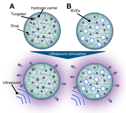
Fig. 3. Ultrasound-triggered drug-releasing hydrogel drug carriers containing different enhancers: (A) tungsten and (B) bubble vibration enhancers (BVEs). This figure was redrawn based on 79 and 80.
5.3. Transdermal/Transnasal Non-Invasive Drug Administration
Recently, with the increasing demand for macromolecular drugs such as mRNA and viral vector vaccines, various administration methods for these drugs have been developed. Ultrasound-driven transdermal drug delivery is a less invasive alternative to conventional injections. Macromolecular drugs face the challenge of limited absorption into the body because of barriers such as the stratum corneum of the skin. Drug permeation through the stratum corneum is enhanced by ultrasound-induced effects generated at the skin surface, including cavitation 85, acoustic streaming 86, and thermal effects 87. This drug administration method is known as sonophoresis 88. Various ultrasound irradiation strategies have been employed in sonophoresis, including frequency sweeping 85 and simultaneous 89 and sequential 86 irradiation at kHz- and MHz-band frequencies. In sonophoresis using sequential irradiation with kHz- and MHz-band frequencies, drug permeation is enhanced by acoustic streaming, which is prominent in the MHz range 86. Biomacromolecular therapeutics administered via sonophoresis are not limited to drugs, such as insulin. In one study, the basic fibroblast growth factor-loaded chitosan nanoparticles have also been administered 90. Since the skin surface has the property of adsorbing positively charged particles, this study enhanced skin permeability by loading the basic fibroblast growth factor onto carboxymethyl chitosan nanoparticles to give them a negative charge 90. Wearable ultrasound-mediated transdermal drug delivery devices fabricated by integrating miniaturized PZT transducers into PDMS patches have also been reported 91.
Another noninvasive drug administration method using ultrasound is intranasal delivery via drug atomization. When high-frequency ultrasound at an appropriate power is applied to a droplet, the droplet becomes atomized 92,93. In particular, droplet atomization using SAW devices exhibits directivity along the Rayleigh angle. The directional atomization of drugs using a narrow-width SAW device has enabled targeted delivery to the olfactory epithelium in a transparent silicone nasal cavity model 94.
5.4. Blood–Brain Barrier Opening
The blood–brain barrier (BBB) is a specialized protective system that tightly regulates the transport of substances between the bloodstream and brain tissue through tight junctions in the vascular endothelial cells. This system protects the brain from toxins and maintains an appropriate neural environment. However, the BBB is also a major hindrance to the delivery of most therapeutic agents for neurological diseases, particularly those with molecular weights exceeding 400 Da. Therefore, the transient opening of the BBB using focused ultrasound in combination with microbubbles has been proposed as a promising actuator technology with the potential to fundamentally transform the treatment of central nervous system disorders. The BBB is primarily opened by inducing inertial and stable cavitation of microbubbles in the vasculature using ultrasound 95. Inertial cavitation is employed to deliver extremely large molecules (e.g., \({>}500\) kDa) to the brain 95. However, they can also generate microjets and shock waves due to bubble collapse. Because of microjets and shock waves, BBB opening induced by inertial cavitation is associated with significant risks, including microhemorrhage, cell membrane damage, and acute and chronic brain tissue injury 96. Therefore, most studies have focused on improving the efficiency of drug delivery via BBB opening mediated by stable cavitation 95,96,97. When a gas with high solubility in water is encapsulated within microbubbles, the bubbles rapidly dissolve in the blood. Therefore, the gases with low solubilities in water are recommended to form microbubbles 98. As described, ultrasonic actuators have attracted considerable attention for their permeability and noninvasive characteristics that enable highly precise control of the BBB opening.
5.5. Future Prospects of Ultrasonic Actuators for Tissue Treatment
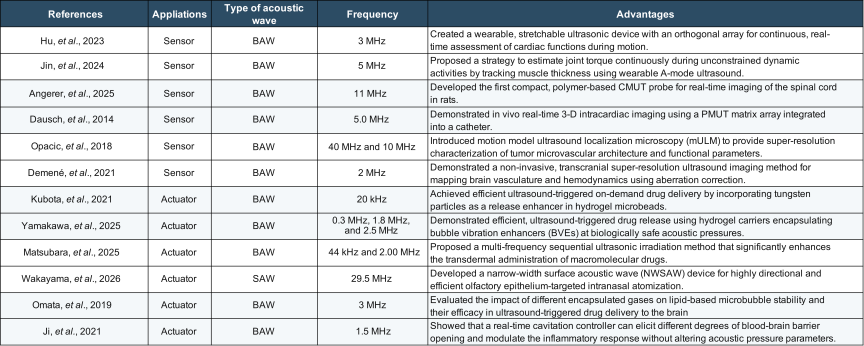
The use of ultrasound actuators for tissue treatment has been extensively investigated. In DDSs, ultrasound is expected to serve as a noninvasive external stimulus that can reach deep tissues. Furthermore, transdermal and intranasal drug delivery methods have been actively developed based on the mechanical effects of ultrasound, including cavitation, thermal effects, and acoustic streaming. In the context of BBB opening, drug delivery to the brain via expansion of intercellular tight junctions induced by stable cavitation has been proposed. The characteristics of the ultrasonic sensors and actuators for tissues, which represent a larger scale than cells, have been appropriately designed, depending on their purpose (Table 2). These ultrasound-driven therapeutic approaches may further enhance treatment efficacy when combined with micro- and nanorobots (MNrobots) 99,100,101. In current DDSs, drug-loaded capsules are mostly limited to simple responses, such as the release of their cargo only in response to specific stimuli. Integrating these drug-loaded capsules with MNrobots to incorporate various functions will enable more efficient and localized drug delivery. Recently, MNrobots have emerged as an actively explored field for medical applications. These MNrobots are typically driven by externally applied magnetic or acoustic fields. MNrobots with bubble membranes actuated by ultrasound have also been reported 100,101. Although MNrobots still require further miniaturization to operate in highly confined environments such as blood vessels, they can enable transformative active therapeutic strategies if they achieve functions for drug delivery and the expansion of intercellular tight junctions.
6. Conclusion
Based on the reviewed literature, we discovered that ultrasonic sensors and actuators have been adapted for cells and tissues using a variety of methodologies to meet the ever-evolving medical needs. Owing to its superior penetration depth, ultrasound can reach deep-seated tissues while ensuring noninvasive sensing of cells and tissues with minimal invasion. Ultrasonic sensors have been primarily used to obtain impedance mapping and characterize the mechanical properties of cells and tissues, including viscoelasticity. To enhance the resolution of ultrasonic sensors, some approaches have been integrated with and inspired by optical sensing techniques, including optoacoustic imaging and ULM. Ultrasonic actuators have also been widely adopted for cell and tissue manipulation due to their noncontact mechanical functionality and noninvasiveness. Various phenomena, such as cavitation, acoustic radiation force, and acoustic streaming, have been applied to ultrasonic actuators. The amplitude and frequency of ultrasound must be carefully optimized to maintain noninvasiveness during the manipulation of cells and tissues. In both ultrasonic sensors and actuators, there is a trade-off between achieving higher resolution through increased frequencies and the resultant significant attenuation. One effective approach to overcome this trade-off is to draw inspiration from optical sensors and actuators, as demonstrated by ULM and optoacoustic imaging. Innovations in optical technologies hold significant potential for advancing ultrasonic technologies, and vice versa. The field of ultrasound sensors and actuators for cells and tissues has experienced remarkable growth. Looking ahead, it is anticipated that numerous innovative ultrasonic technologies will emerge to further advance the acquisition of cell/tissue information from intracellular/intratissue sensors.
- [1] P. G. Auran and O. Silven, “Underwater sonar range sensing and 3D image formation,” Control Eng. Pract., Vol.4, No.3, pp. 393-400, 1996. https://doi.org/10.1016/0967-0661(96)00017-2
- [2] N. K. Mutlib, S. B. Baharom, A. El-Shafie, and M. Z. Nuawi, “Ultrasonic health monitoring in structural engineering: Buildings and bridges,” Struct. Control Health Monit., Vol.23, No.3, pp. 409-422, 2016. https://doi.org/10.1002/stc.1800
- [3] D. Rouseff et al., “Underwater acoustic communication by passive-phase conjugation: Theory and experimental results,” IEEE J. Ocean. Eng., Vol.26, No.4, pp. 821-831, 2001. https://doi.org/10.1109/48.972122
- [4] B. Verhaagen and D. Fernández Rivas, “Measuring cavitation and its cleaning effect,” Ultrason. Sonochem., Vol.29, pp. 619-628, 2016. https://doi.org/10.1016/j.ultsonch.2015.03.009
- [5] J. Carpenter and V. K. Saharan, “Ultrasonic assisted formation and stability of mustard oil in water nanoemulsion: Effect of process parameters and their optimization,” Ultrason. Sonochem., Vol.35, Part A, pp. 422-430, 2017. https://doi.org/10.1016/j.ultsonch.2016.10.021
- [6] S. Mostafavi, D. F. Hesser, and B. Markert, “Effect of process parameters on the interface temperature in ultrasonic aluminum wire bonding,” J. Manuf. Process., Vol.36, pp. 104-114, 2018. https://doi.org/10.1016/j.jmapro.2018.09.020
- [7] A. Brotchie, F. Grieser, and M. Ashokkumar, “Effect of power and frequency on bubble-size distributions in acoustic cavitation,” Phys. Rev. Lett., Vol.102, No.8, Article No.084302, 2009. https://doi.org/10.1103/PhysRevLett.102.084302
- [8] D. Kong, K. Nishio, and M. K. Kurosawa, “Surface acoustic wave propulsion system with acoustic radiation force,” Sens. Actuators A: Phys., Vol.309, Article No.111943, 2020. https://doi.org/10.1016/j.sna.2020.111943
- [9] N. Zhang et al., “Microliter ultrafast centrifuge platform for size-based particle and cell separation and extraction using novel omnidirectional spiral surface acoustic waves,” Lab Chip, Vol.21, No.5, pp. 904-915, 2021. https://doi.org/10.1039/d0lc01012j
- [10] L. Mancia et al., “Modeling tissue-selective cavitation damage,” Phys. Med. Biol., Vol.64, No.22, Article No.225001, 2019. https://doi.org/10.1088/1361-6560/ab5010
- [11] R. A. Marklein, J. Lam, M. Guvendiren, K. E. Sung, and S. R. Bauer, “Functionally-relevant morphological profiling: A tool to assess cellular heterogeneity,” Trends Biotechnol., Vol.36, No.1, pp. 105-118, 2018. https://doi.org/10.1016/j.tibtech.2017.10.007
- [12] J. Guck et al., “Optical deformability as an inherent cell marker for testing malignant transformation and metastatic competence,” Biophys. J., Vol.88, No.5, pp. 3689-3698, 2005. https://doi.org/10.1529/biophysj.104.045476
- [13] E. van Vliet et al., “Electrophysiological recording of re-aggregating brain cell cultures on multi-electrode arrays to detect acute neurotoxic effects,” NeuroToxicology, Vol.28, No.6, pp. 1136-1146, 2007. https://doi.org/10.1016/j.neuro.2007.06.004
- [14] X. Bao et al., “Capturing the interactome of newly transcribed RNA,” Nat. Methods, Vol.15, No.3, pp. 213-220, 2018. https://doi.org/10.1038/nmeth.4595
- [15] E. Bagus Prastika et al., “Time and frequency domain deconvolution for cross-sectional cultured cell observation using an acoustic impedance microscope,” Ultrasonics, Vol.119, Article No.106601, 2022. https://doi.org/10.1016/j.ultras.2021.106601
- [16] E. M. Strohm, G. J. Czarnota, and M. C. Kolios, “Quantitative measurements of apoptotic cell properties using acoustic microscopy,” IEEE Trans. Ultrason. Ferroelectr. Freq. Control, Vol.57, No.10, pp. 2293-2304, 2010. https://doi.org/10.1109/TUFFC.2010.1690
- [17] N. Hozumi, S. Yoshida, and K. Kobayashi, “Three-dimensional acoustic impedance mapping of cultured biological cells,” Ultrasonics, Vol.99, Article No.105966, 2019. https://doi.org/10.1016/j.ultras.2019.105966
- [18] C. L. M. Lee et al., “Noninvasive imaging of rat-derived microglia and its reactivity to inflammatory molecules via acoustic impedance microscopy,” J. Med. Ultrason., Vol.51, No.1, pp. 29-37, 2024. https://doi.org/10.1007/s10396-023-01379-8
- [19] K. Takanashi et al., “Quantitative evaluation method for differentiation of C2C12 myoblasts by ultrasonic microscopy,” Jpn. J. Appl. Phys., Vol.56, No.7S1, Article No.07JF11, 2017. https://doi.org/10.7567/JJAP.56.07JF11
- [20] N. Fujiwara et al., “Deep-learning generation of high-resolution images of live cells in culture using tri-frequency acoustic images,” Phys. Rev. X, Vol.15, No.2, Article No.021015, 2025. https://doi.org/10.1103/PhysRevX.15.021015
- [21] R. T. Tjeung, M. S. Hughes, L. Y. Yeo, and J. R. Friend, “Surface acoustic wave micromotor with arbitrary axis rotational capability,” Appl. Phys. Lett., Vol.99, No.21, Article No.214101, 2011. https://doi.org/10.1063/1.3662931
- [22] Z. Li, Y. Jones, J. Hossenlopp, R. Cernosek, and F. Josse, “Analysis of liquid-phase chemical detection using guided shear horizontal-surface acoustic wave sensors,” Anal. Chem., Vol.77, No.14, pp. 4595-4603, 2005. https://doi.org/10.1021/ac0504621
- [23] F. Lunardelli, M. Agostini, E. Corradi, and M. Cecchini, “Dual-mode shear horizontal / Rayleigh-like surface-acoustic-wave configuration on lithium niobate 64° Y-cut,” Sens. Actuators A: Phys., Vol.378, Article No.115780, 2024. https://doi.org/10.1016/j.sna.2024.115780
- [24] K. Chang et al., “Label-free and high-sensitive detection of human breast cancer cells by aptamer-based leaky surface acoustic wave biosensor array,” Biosens. Bioelectron., Vol.60, pp. 318-324, 2014. https://doi.org/10.1016/j.bios.2014.04.027
- [25] A. Link and T. Franke, “Acoustic erythrocytometer for mechanically probing cell viscoelasticity,” Lab Chip, Vol.20, No.11, pp. 1991-1998, 2020. https://doi.org/10.1039/c9lc00999j
- [26] T. Dehoux et al., “All-optical broadband ultrasonography of single cells,” Sci. Rep., Vol.5, Article No.8650, 2015. https://doi.org/10.1038/srep08650
- [27] L. Liu, L. Plawinski, M.-C. Durrieu, and B. Audoin, “Label-free multi-parametric imaging of single cells: Dual picosecond optoacoustic microscopy,” J. Biophotonics, Vol.12, No.8, Article No.e201900045, 2019. https://doi.org/10.1002/jbio.201900045
- [28] F. Pérez-Cota et al., “Picosecond ultrasonics for elasticity-based imaging and characterization of biological cells,” J. Appl. Phys., Vol.128, No.16, Article No.160902, 2020. https://doi.org/10.1063/5.0023744
- [29] T. Dehoux et al., “Probing single-cell mechanics with picosecond ultrasonics,” Ultrasonics, Vol.56, pp. 160-171, 2015. https://doi.org/10.1016/j.ultras.2014.07.010
- [30] V. Gnyawali, E. M. Strohm, J.-Z. Wang, S. S. H. Tsai, and M. C. Kolios, “Simultaneous acoustic and photoacoustic microfluidic flow cytometry for label-free analysis,” Sci. Rep., Vol.9, Article No.1585, 2019. https://doi.org/10.1038/s41598-018-37771-5
- [31] C. Walker et al., “Liquid-like condensates that bind actin promote assembly and bundling of actin filaments,” Dev. Cell, Vol.60, No.11, pp. 1550-1567.e4, 2025. https://doi.org/10.1016/j.devcel.2025.01.012
- [32] X. Wang et al., “Focal adhesion and actin orientation regulated by cellular geometry determine stem cell differentiation via mechanotransduction,” Acta Biomater., Vol.182, pp. 81-92, 2024. https://doi.org/10.1016/j.actbio.2024.05.017
- [33] J. Alsved, M. Rezayati Charan, P. Ohlsson, A. Urbansky, and P. Augustsson, “Label-free separation of peripheral blood mononuclear cells from whole blood by gradient acoustic focusing,” Sci. Rep., Vol.14, Article No.8748, 2024. https://doi.org/10.1038/s41598-024-59156-7
- [34] P. Li et al., “Acoustic separation of circulating tumor cells,” Proc. Natl. Acad. Sci. U.S.A., Vol.112, No.16, pp. 4970-4975, 2015. https://doi.org/10.1073/pnas.1504484112
- [35] A. A. Nawaz et al., “Acoustofluidic fluorescence activated cell sorter,” Anal. Chem., Vol.87, No.24, pp. 12051-12058, 2015. https://doi.org/10.1021/acs.analchem.5b02398
- [36] M. Wu et al., “Circulating tumor cell phenotyping via high-throughput acoustic separation,” Small, Vol.14, No.32, Article No.1801131, 2018. https://doi.org/10.1002/smll.201801131
- [37] T. Franke, S. Braunmüller, L. Schmid, A. Wixforth, and D. A. Weitz, “Surface acoustic wave actuated cell sorting (SAWACS),” Lab Chip, Vol.10, No.6, pp. 789-794, 2010. https://doi.org/10.1039/b915522h
- [38] M. Miansari et al., “Inducing mild traumatic brain injury in C. elegans via cavitation-free surface acoustic wave-driven ultrasonic irradiation,” Sci. Rep., Vol.9, Article No.12775, 2019. https://doi.org/10.1038/s41598-019-47295-1
- [39] Y. Kurashina, T. Yamashita, S. Kurabayashi, K. Takemura, and K. Ando, “Growth control of leaf lettuce with exposure to underwater ultrasound and dissolved oxygen supersaturation,” Ultrason. Sonochem., Vol.51, pp. 292-297, 2019. https://doi.org/10.1016/j.ultsonch.2018.10.005
- [40] Y. Kurashina, K. Takemura, and J. Friend, “Cell agglomeration in the wells of a 24-well plate using acoustic streaming,” Lab Chip, Vol.17, No.5, pp. 876-886, 2017. https://doi.org/10.1039/c6lc01310d
- [41] M. Nakao et al., “Formation of large scaffold-free 3-D aggregates in a cell culture dish by ultrasound standing wave trapping,” Ultrasound Med. Biol., Vol.45, No.5, pp. 1306-1315, 2019. https://doi.org/10.1016/j.ultrasmedbio.2019.01.013
- [42] S. Koda, K. Wakayama, S. Takahashi, and Y. Kurashina, “Rayleigh-type surface acoustic wave-driven shooting for directional artificial pollination of strawberry flowers,” Sens. Actuators A: Phys., Vol.405, Article No.117774, 2026. https://doi.org/10.1016/j.sna.2026.117774
- [43] K. Melde et al., “Ultrasound-assisted tissue engineering,” Nat. Rev. Bioeng., Vol.2, No.6, pp. 486-500, 2024. https://doi.org/10.1038/s44222-024-00166-5
- [44] C. Imashiro et al., “Cell patterning method on a clinically ubiquitous culture dish using acoustic pressure generated from resonance vibration of a disk-shaped ultrasonic transducer,” IEEE Trans. Biomed. Eng., Vol.66, No.1, pp. 111-118, 2019. https://doi.org/10.1109/TBME.2018.2835834
- [45] C. Imashiro, Y. Kurashina, and K. Takemura, “Cell patterning method using resonance vibration of a metallic cell cultivation substrate,” Adv. Biomed. Eng., Vol.5, pp. 142-148, 2016. https://doi.org/10.14326/abe.5.142
- [46] Z. Ma et al., “Acoustic holographic cell patterning in a biocompatible hydrogel,” Adv. Mater., Vol.32, No.4, Article No.1904181, 2020. https://doi.org/10.1002/adma.201904181
- [47] S. Li et al., “Standing surface acoustic wave based cell coculture,” Anal. Chem., Vol.86, No.19, pp. 9853-9859, 2014. https://doi.org/10.1021/ac502453z
- [48] K. Martinez Villegas, R. Rasouli, and M. Tabrizian, “Enhancing metabolic activity and differentiation potential in adipose mesenchymal stem cells via high-resolution surface-acoustic-wave contactless patterning,” Microsyst. Nanoeng., Vol.8, Article No.79, 2022. https://doi.org/10.1038/s41378-022-00415-w
- [49] D. J. Collins et al., “Two-dimensional single-cell patterning with one cell per well driven by surface acoustic waves,” Nat. Commun., Vol.6, Article No.8686, 2015. https://doi.org/10.1038/ncomms9686
- [50] J. Huang et al., “ZnO/glass-based SAW tweezer for dexterous particle patterning and patterned cell culturing,” Microfluid. Nanofluid., Vol.27, No.5, Article No.34, 2023. https://doi.org/10.1007/s10404-023-02646-3
- [51] Y. Kurashina et al., “Enzyme-free release of adhered cells from standard culture dishes using intermittent ultrasonic traveling waves,” Commun. Biol., Vol.2, Article No.393, 2019. https://doi.org/10.1038/s42003-019-0638-5
- [52] A. Salari, S. Appak-Baskoy, I. R. Coe, S. S. H. Tsai, and M. C. Kolios, “An ultrafast enzyme-free acoustic technique for detaching adhered cells in microchannels,” RSC Adv., Vol.11, No.52, pp. 32824-32829, 2021. https://doi.org/10.1039/d1ra04875a
- [53] H. Tauchi et al., “Effective and intact cell detachment from a clinically ubiquitous culture flask by combining ultrasonic wave exposure and diluted trypsin,” Biotechnol. Bioprocess Eng., Vol.24, No.3, pp. 536-543, 2019. https://doi.org/10.1007/s12257-018-0491-2
- [54] A. Bussonnière et al., “Cell detachment and label-free cell sorting using modulated surface acoustic waves (SAWs) in droplet-based microfluidics,” Lab Chip, Vol.14, No.18, pp. 3556-3563, 2014. https://doi.org/10.1039/c4lc00625a
- [55] K. Fujishiro, S. Okada, F. Rossi, T. Kuchimaru, and Y. Kurashina, “Intracellular reactive oxygen species generation induced by high-frequency ultrasound in thickness vibration mode,” Adv. Sci., Vol.13, No.28, Article No.e19582, 2026. https://doi.org/10.1002/advs.202519582
- [56] X. Yang, X. Sun, D. Zhou, Y. Li, and H. Liu, “Towards wearable A-mode ultrasound sensing for real-time finger motion recognition,” IEEE Trans. Neural Syst. Rehabil. Eng., Vol.26, No.6, pp. 1199-1208, 2018. https://doi.org/10.1109/TNSRE.2018.2829913
- [57] E. Sassaroli, A. Scorza, C. Crake, S. A. Sciuto, and M.-A. Park, “Breast ultrasound technology and performance evaluation of ultrasound equipment: B-mode,” IEEE Trans. Ultrason. Ferroelectr. Freq. Control, Vol.64, No.1, pp. 192-205, 2017. https://doi.org/10.1109/TUFFC.2016.2619622
- [58] J. Li, K. Zhu, and L. Pan, “Wrist and finger motion recognition via M-mode ultrasound signal: A feasibility study,” Biomed. Signal Process. Control, Vol.71, Part A, Article No.103112, 2022. https://doi.org/10.1016/j.bspc.2021.103112
- [59] Y. Jin et al., “Estimation of joint torque in dynamic activities using wearable A-mode ultrasound,” Nat. Commun., Vol.15, Article No.5756, 2024. https://doi.org/10.1038/s41467-024-50038-0
- [60] H. Hu et al., “A wearable cardiac ultrasound imager,” Nature, Vol.613, No.7945, pp. 667-675, 2023. https://doi.org/10.1038/s41586-022-05498-z
- [61] M. Lin et al., “A fully integrated wearable ultrasound system to monitor deep tissues in moving subjects,” Nat. Biotechnol., Vol.42, No.3, pp. 448-457, 2024. https://doi.org/10.1038/s41587-023-01800-0
- [62] X. Gao et al., “A wearable echomyography system based on a single transducer,” Nat. Electron., Vol.7, No.11, pp. 1035-1046, 2024. https://doi.org/10.1038/s41928-024-01271-4
- [63] S. Zhou et al., “Clinical validation of a wearable ultrasound sensor of blood pressure,” Nat. Biomed. Eng., Vol.9, No.6, pp. 865-881, 2025. https://doi.org/10.1038/s41551-024-01279-3
- [64] J. Park et al., “Opto-ultrasound biosensor for wearable and mobile devices: Realization with a transparent ultrasound transducer,” Biomed. Opt. Express, Vol.13, No.9, pp. 4684-4692, 2022. https://doi.org/10.1364/BOE.468969
- [65] M. Angerer et al., “A polymer-based CMUT probe for imaging the spinal cord in rats,” IEEE Trans. Ultrason. Ferroelectr. Freq. Control, Vol.72, No.11, pp. 1437-1447, 2025. https://doi.org/10.1109/TUFFC.2025.3605440
- [66] I. M. Suarez-Castellanos et al., “Dynamic ultrasound focusing and centimeter-scale ex vivo tissue ablations with a CMUT probe developed for endocavitary HIFU therapies,” IEEE Trans. Ultrason. Ferroelectr. Freq. Control, Vol.70, No.11, pp. 1470-1481, 2023. https://doi.org/10.1109/TUFFC.2023.3301977
- [67] M. Wang and J. Chen, “Volumetric flow measurement using an implantable CMUT array,” IEEE Trans. Biomed. Circuits Syst., Vol.5, No.3, pp. 214-222, 2011. https://doi.org/10.1109/TBCAS.2010.2095848
- [68] M. Qu et al., “Imaging of simulated muscle based on single chip of AlN piezoelectric micromachined ultrasonic transducer,” J. Micromech. Microeng., Vol.32, No.7, Article No.075003, 2022. https://doi.org/10.1088/1361-6439/ac70a6
- [69] D. E. Dausch et al., “In vivo real-time 3-D intracardiac echo using PMUT arrays,” IEEE Trans. Ultrason. Ferroelectr. Freq. Control, Vol.61, No.10, pp. 1754-1764, 2014. https://doi.org/10.1109/TUFFC.2014.006452
- [70] X. Jiang, V. Perrot, F. Varray, S. Bart, and P. G. Hartwell, “Piezoelectric micromachined ultrasonic transducer for arterial wall dynamics monitoring,” IEEE Trans. Ultrason. Ferroelectr. Freq. Control, Vol.69, No.1, pp. 291-298, 2022. https://doi.org/10.1109/TUFFC.2021.3120283
- [71] C. Errico et al., “Ultrafast ultrasound localization microscopy for deep super-resolution vascular imaging,” Nature, Vol.527, No.7579, pp. 499-502, 2015. https://doi.org/10.1038/nature16066
- [72] T. Opacic et al., “Motion model ultrasound localization microscopy for preclinical and clinical multiparametric tumor characterization,” Nat. Commun., Vol.9, Article No.1527, 2018. https://doi.org/10.1038/s41467-018-03973-8
- [73] S.-H. Lee, J. Y. Shin, A. Lee, and C. Bustamante, “Counting single photoactivatable fluorescent molecules by photoactivated localization microscopy (PALM),” Proc. Natl. Acad. Sci. U.S.A., Vol.109, No.43, pp. 17436-17441, 2012. https://doi.org/10.1073/pnas.1215175109
- [74] M. J. Rust, M. Bates, and X. Zhuang, “Sub-diffraction-limit imaging by stochastic optical reconstruction microscopy (STORM),” Nat. Methods, Vol.3, No.10, pp. 793-796, 2006. https://doi.org/10.1038/nmeth929
- [75] C. Demené et al., “Transcranial ultrafast ultrasound localization microscopy of brain vasculature in patients,” Nat. Biomed. Eng., Vol.5, No.3, pp. 219-228, 2021. https://doi.org/10.1038/s41551-021-00697-x
- [76] K. Kataoka, A. Harada, and Y. Nagasaki, “Block copolymer micelles for drug delivery: Design, characterization and biological significance,” Adv. Drug Deliv. Rev., Vol.47, No.1, pp. 113-131, 2001. https://doi.org/10.1016/S0169-409X(00)00124-1
- [77] R. R. López et al., “The effect of different organic solvents in liposome properties produced in a periodic disturbance mixer: Transcutol®, a potential organic solvent replacement,” Colloids Surf. B: Biointerfaces, Vol.198, Article No.111447, 2021. https://doi.org/10.1016/j.colsurfb.2020.111447
- [78] Y. Kurashina et al., “Adeno-associated virus-encapsulated alginate microspheres loaded in collagen gel carriers for localized gene transfer,” Adv. Healthc. Mater., Vol.13, No.12, Article No.2303546, 2024. https://doi.org/10.1002/adhm.202303546
- [79] T. Kubota, Y. Kurashina, J. Zhao, K. Ando, and H. Onoe, “Ultrasound-triggered on-demand drug delivery using hydrogel microbeads with release enhancer,” Mater. Des., Vol.203, Article No.109580, 2021. https://doi.org/10.1016/j.matdes.2021.109580
- [80] R. Yamakawa, H. Onoe, and Y. Kurashina, “Hydrogel carrier with bubble vibration enhancer for ultrasound-triggered drug release,” Ultrason. Sonochem., Vol.112, Article No.107173, 2025. https://doi.org/10.1016/j.ultsonch.2024.107173
- [81] T. Jamneala et al., “Ultra-miniature coupled resonator filter with single-layer acoustic coupler,” IEEE Trans. Ultrason. Ferroelectr. Freq. Control, Vol.56, No.11, pp. 2553-2558, 2009. https://doi.org/10.1109/TUFFC.2009.1342
- [82] S. Takatsuka, T. Kubota, Y. Kurashina, and H. Onoe, “Near-infrared-triggered on-demand controlled release of adeno-associated virus from alginate hydrogel microbeads with heat transducer for gene therapy,” Small, Vol.19, No.7, Article No.2204139, 2023. https://doi.org/10.1002/smll.202204139
- [83] A. Hioki, S. Takatsuka, Y. Kurashina, and H. Onoe, “Alginate hydrogel microbeads with different mesh structures enable controlled release of adeno-associated virus for gene therapy,” Proc. 2023 22nd Int. Conf. Solid-State Sens., Actuators Microsyst. (Transducers), pp. 482-485, 2023.
- [84] Y. Bando et al., “Synthesis of hydrogel microparticles from ultra-high-viscosity pre-gel solution by combining centrifugal force and ultrasonic vibration,” Small, Vol.21, No.34, Article No.2412715, 2025. https://doi.org/10.1002/smll.202412715
- [85] O. Al-Bataineh, R. Abdallat, T. Momani, S. Alzghoul, and O. Smadi, “In-vivo evaluation of ultrasound transducers with sweep-frequency excitation for noninvasive transdermal insulin delivery,” J. Drug Deliv. Sci. Technol., Vol.112, Article No.107258, 2025. https://doi.org/10.1016/j.jddst.2025.107258
- [86] K. Matsubara et al., “Transdermal administration of macromolecular drugs by multi-frequency sequential ultrasonic irradiation,” Int. J. Pharm., Vol.675, Article No.125560, 2025. https://doi.org/10.1016/j.ijpharm.2025.125560
- [87] J. Zhang et al., “Flexible surface acoustic wave technology for enhancing transdermal drug delivery,” Drug Deliv. Transl. Res., Vol.15, No.4, pp. 1363-1375, 2025. https://doi.org/10.1007/s13346-024-01682-y
- [88] A. Boucaud, M. A. Garrigue, L. Machet, L. Vaillant, and F. Patat, “Effect of sonication parameters on transdermal delivery of insulin to hairless rats,” J. Control. Release, Vol.81, Nos.1-2, pp. 113-119, 2002. https://doi.org/10.1016/S0168-3659(02)00054-8
- [89] C. M. Schoellhammer et al., “Applicability and safety of dual-frequency ultrasonic treatment for the transdermal delivery of drugs,” J. Control. Release, Vol.202, pp. 93-100, 2015. https://doi.org/10.1016/j.jconrel.2015.02.002
- [90] X. Xie et al., “Transdermal delivery of bFGF with sonophoresis facilitated by chitosan nanocarriers,” J. Drug Deliv. Sci. Technol., Vol.75, Article No.103675, 2022. https://doi.org/10.1016/j.jddst.2022.103675
- [91] C. Yu et al., “A conformable ultrasound patch for cavitation-enhanced transdermal cosmeceutical delivery,” Adv. Mater., Vol.35, No.23, Article No.2300066, 2023. https://doi.org/10.1002/adma.202300066
- [92] T. Asakura, K. Yoshioka, A. Ogawa, and Y. Kurashina, “Effect of particle size distribution on the transmission efficiency of atomized water to the tracheal tube,” Sens. Actuators A: Phys., Vol.370, Article No.115270, 2024. https://doi.org/10.1016/j.sna.2024.115270
- [93] K. Yoshioka, Y. Kurashina, A. Ogawa, and T. Asakura, “Effect of the area of a lithium niobate transducer on the efficiency of ultrasonic atomization driven by resonance vibration,” Ultrason. Sonochem., Vol.86, Article No.106019, 2022. https://doi.org/10.1016/j.ultsonch.2022.106019
- [94] K. Wakayama, S. Kurihara, and Y. Kurashina, “Narrow-width surface acoustic wave device-driven olfactory epithelium-targeted intranasal atomization,” Int. J. Pharm., Vol.692, Article No.126630, 2026. https://doi.org/10.1016/j.ijpharm.2026.126630
- [95] H. Chen and E. E. Konofagou, “The size of blood–brain barrier opening induced by focused ultrasound is dictated by the acoustic pressure,” J. Cereb. Blood Flow Metab., Vol.34, No.7, pp. 1197-1204, 2014. https://doi.org/10.1038/jcbfm.2014.71
- [96] C.-H. Fan et al., “Drug-loaded bubbles with matched focused ultrasound excitation for concurrent blood–brain barrier opening and brain-tumor drug delivery,” Acta Biomater., Vol.15, pp. 89-101, 2015. https://doi.org/10.1016/j.actbio.2014.12.026
- [97] R. Ji et al., “Cavitation-modulated inflammatory response following focused ultrasound blood-brain barrier opening,” J. Control. Release, Vol.337, pp. 458-471, 2021. https://doi.org/10.1016/j.jconrel.2021.07.042
- [98] D. Omata et al., “Effects of encapsulated gas on stability of lipid-based microbubbles and ultrasound-triggered drug delivery,” J. Control. Release, Vols.311-312, pp. 65-73, 2019. https://doi.org/10.1016/j.jconrel.2019.08.023
- [99] S. Kim et al., “Localized ultrasonic stimulation using a piezoelectric micromachined ultrasound transducer array for selective neural differentiation of magnetic cell-based robots,” Microsyst. Nanoeng., Vol.11, Article No.52, 2025. https://doi.org/10.1038/s41378-025-00900-y
- [100] T. Luo and M. Wu, “Biologically inspired micro-robotic swimmers remotely controlled by ultrasound waves,” Lab Chip, Vol.21, No.21, pp. 4095-4103, 2021. https://doi.org/10.1039/d1lc00575h
- [101] N. Mahkam et al., “Acoustic streaming-induced multimodal locomotion of bubble-based microrobots,” Adv. Sci., Vol.10, No.35, Article No.2304233, 2023. https://doi.org/10.1002/advs.202304233
This article is published under a Creative Commons Attribution-NoDerivatives 4.0 Internationa License.