
Paper:
Fabrication of a Magnetically Driven Device for Evaluating Cellular Responses to Vibratory Stimulation
Tasuku Nakahara
 , Yuto Arimatsu, Koji Egami, and Kazuyuki Minami
Graduate School of Sciences and Technology for Innovation, Yamaguchi University
2-16-1 Tokiwadai, Ube, Yamaguchi 755-8611, Japan

Cells possess sensors to detect external mechanical stimuli, and the process of converting these cues into biochemical reactions is known as mechanotransduction. Various vibration devices have been developed to evaluate cellular responses under vibratory stimulation. However, few devices can apply vibratory stimulation to a localized area while maintaining a low contamination risk during long-term culture. To address this challenge, we propose a magnetically driven device capable of applying vibratory stimulation to cells in vitro to evaluate cell proliferation and differentiation induction. The device integrates a polydimethylsiloxane membrane equipped with a neodymium magnet and a ring structure into a standard lidded culture dish. By utilizing the magnetic repulsive force from an electromagnet, the membrane vibrates via non-contact actuation. The device maintains a semi-sealed environment, minimizing the risk of contamination and making it highly suitable for long-term cultivation. Performance evaluations confirmed that membrane acceleration increased with the applied voltage. When vibratory stimulation with an acceleration of 0.1 G or higher was applied to cultured cells, the stimulated group exhibited a cell proliferation rate approximately 1.4 times greater than that of the non-stimulated group. Furthermore, culturing osteoblast-like cells in a differentiation-inducing medium resulted in widespread positive staining for alkaline phosphatase, confirming that the device environment supports osteogenic differentiation. Notably, no contamination or cell detachment occurred during a 72 h culture period. These results suggest that the proposed device is a potentially useful tool for evaluating cellular mechanotransduction under vibratory stimulation.
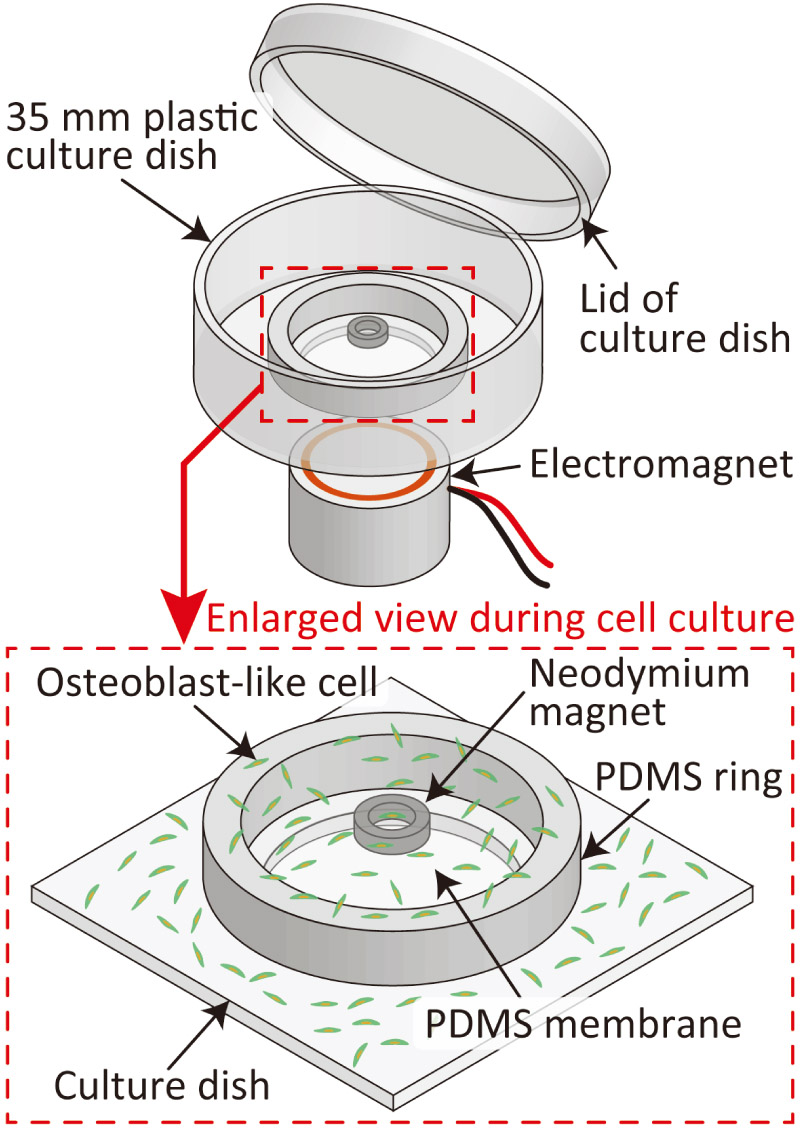
Magnetic cell-vibration device
- [1] F. Boccafoschi, M. Bosetti et al., “Dynamic fibroblast cultures: Response to mechanical stretching,” Cell Adhesion & Migration, Vol.1, No.3, pp. 124-128, 2007. https://doi.org/10.4161/cam.1.3.5144
- [2] K. Sato, T. Nakahara, and K. Minami, “Difference in the osteoblastic calcium signaling response between compression and stretching mechanical stimuli,” J. Robot. Mechatron., Vol.35, No.5, pp. 1135-1142, 2023. https://doi.org/10.20965/jrm.2023.p1135
- [3] T. Nakahara, S. Ono, and K. Minami, “Fabrication of a magnetically driven cell-stretching device for predefined cell alignment in vitro,” J. Robot. Mechatron., Vol.35, No.5, pp. 1143-1150, 2023. https://doi.org/10.20965/jrm.2023.p1143
- [4] S. Giulitti, A. Zambon et al., “Mechanotransduction through substrates engineering and microfluidic devices,” Current Opinion in Chemical Engineering, Vol.11, pp. 67-76, 2016. https://doi.org/10.1016/j.coche.2016.01.010
- [5] H. Gao, M. Zhai et al., “Low-level mechanical vibration enhances osteoblastogenesis via a canonical Wnt signaling-associated mechanism,” Molecular Medicine Reports, Vol.16, No.1, pp. 317-324, 2017. https://doi.org/10.3892/mmr.2017.6608
- [6] K. Sato and D. Oomori, “Development of vibration mechanical stimuli loading device for live cell fluorescence microscopy,” J. of Biomechanical Science and Engineering, Vol.17, No.2, Article No.21-00294, 2022. https://doi.org/10.1299/jbse.21-00294
- [7] S. García-López, R. E. Villanueva et al., “Micro-vibrations at 30 Hz on bone cells cultivated in vitro produce soluble factors for osteoclast inhibition and osteoblast activity,” Archives of Oral Biology, Vol.110, Article No.104594, 2020. https://doi.org/10.1016/j.archoralbio.2019.104594
- [8] Z. Fu, X. Huang et al., “Protective effects of low-magnitude high-frequency vibration on high glucose-induced osteoblast dysfunction and bone loss in diabetic rats,” J. of Orthopaedic Surgery and Research, Vol.16, Article No.650, 2021. https://doi.org/10.1186/s13018-021-02803-w
- [9] T. Shikata, T. Shiraishi et al., “Effects of amplitude and frequency of mechanical vibration stimulation on cultured osteoblasts,” J. of System Design and Dynamics, Vol.2, No.1, pp. 382-388, 2008. https://doi.org/10.1299/jsdd.2.382
- [10] D. Choi, T. Ishii et al., “Vertical vibration of mouse osteoblasts promotes cellular differentiation and cell cycle progression and induces aging in vitro,” Biomedicines, Vol.11, No.2, Article No.444, 2023. https://doi.org/10.3390/biomedicines11020444
- [11] Q. Guo, Y. Fu et al., “Amplitude dependence in extracellular vesicle release from cultured myotubes under vibration stimulation,” Biochemical and Biophysical Research Communications, Vol.797, Article No.153218, 2026. https://doi.org/10.1016/j.bbrc.2025.153218
- [12] W. W. Hou, Z. L. Zhu et al., “Involvement of wnt activation in the micromechanical vibration-enhanced osteogenic response of osteoblasts,” J. of Orthopaedic Science, Vol.16, No.5, pp. 598-605, 2011. https://doi.org/10.1007/s00776-011-0124-5
- [13] A. Matsushita, T. A. Kudo et al., “Frequency-regulated repeated micro-vibration promotes osteoblast differentiation through BMP signaling in MC3T3-E1 cells,” Life, Vol.15, No.4, Article No.588, 2025. https://doi.org/10.3390/life15040588
- [14] K.-i. Konno, T. Kosawada et al., “Development of three-dimensional micro vibration stage and its application to control device for cell culture,” J. of Biomechanical Science and Engineering, Vol.3, No.1, pp. 38-49, 2008. https://doi.org/10.1299/jbse.3.38
- [15] Y. Yamada, G. Umegaki et al., “Development of cell culture microdevice actuated by piezoelectric thin films for delivering mechanical vibratory stimuli to cells,” J. of Physics: Conf. Series, Vol.352, Article No.012027, 2012. https://doi.org/10.1088/1742-6596/352/1/012027
- [16] S. Benjakul, S. Jitpukdeebodintra et al., “Effects of low magnitude high frequency mechanical vibration combined with compressive force on human periodontal ligament cells in vitro,” European J. of Orthodontics, Vol.40, No.4, pp. 356-363, 2018. https://doi.org/10.1093/ejo/cjx062
- [17] K. Minami, T. Nakahara, and K. Sato, “Evaluation of the basic designs of a micro device that provides vibrational stimulation to cells,” J. Robot. Mechatron., Vol.35, No.5, pp. 1151-1157, 2023. https://doi.org/10.20965/jrm.2023.p1151
- [18] C. C. Shih, S. J. Lin et al., “The cytotoxicity of corrosion products of nitinol stent wire on cultured smooth muscle cells,” J. of Biomedial Materials Research, Vol.52, No.2, pp. 395-403, 2000. https://doi.org/10.1002/1097-4636(200011)52:2<395::aid-jbm21>3.0.co;2-b
- [19] S. Bhattacharya, A. Datta et al., “Studies on surface wettability of poly(dimethyl) siloxane (PDMS) and glass under oxygen-plasma treatment and correlation with bond strength,” J. of Microelectromechanical Systems, Vol.14, No.3, pp. 590-597, 2005. https://doi.org/10.1109/jmems.2005.844746
- [20] Y. Kanda, “Investigation of the freely available easy-to-use software ‘EZR’ for medical statistics,” Bone Marrow Transplantation, Vol.48, pp. 452-458, 2013. https://doi.org/10.1038/bmt.2012.244
- [21] M. Yashiro, M. Ohya et al., “Active vitamin D and vitamin D analogs stimulate fibroblast growth factor 23 production in osteocyte-like cells via the vitamin D receptor,” J. of Pharmaceutical and Biomedical Analysis, Vol.182, Article No.113139, 2020. https://doi.org/10.1016/j.jpba.2020.113139
- [22] T. Zhou, B. Gao et al., “Piezo1/2 mediate mechanotransduction essential for bone formation through concerted activation of NFAT-YAP1--catenin,” Elife, Vol.9, Article No.e52779, 2020. https://doi.org/10.7554/elife.52779
- [23] M. Sun, G. Chi et al., “Extracellular matrix stiffness controls osteogenic differentiation of mesenchymal stem cells mediated by integrin 5,” Stem Cell Research & Therapy, Vol.9, Article No.52, 2018. https://doi.org/10.1186/s13287-018-0798-0
- [24] P. S. Mathieu and E. G. Loboa, “Cytoskeletal and focal adhesion influences on mesenchymal stem cell shape, mechanical properties, and differentiation down osteogenic, adipogenic, and chondrogenic pathways,” Tissue Engineering Part B: Reviews, Vol.18, No.6, pp. 436-444, 2012. https://doi.org/10.1089/ten.teb.2012.0014
- [25] A. M. C. Barradas, H. A. M. Fernandes et al., “A calcium-induced signaling cascade leading to osteogenic differentiation of human bone marrow-derived mesenchymal stromal cells,” Biomaterials, Vol.33, No.11, pp. 3205-3215, 2012. https://doi.org/10.1016/j.biomaterials.2012.01.020
- [26] J.-M. Kim, Y.-S. Yang et al., “Biphasic regulation of osteoblast development via the ERK MAPK–mtor pathway,” Elife, Vol.11, Article No.e78069, 2022. https://doi.org/10.7554/elife.78069
This article is published under a Creative Commons Attribution-NoDerivatives 4.0 Internationa License.