
Paper:
Non-Enzymatic Electrochemical Detection of Glucose Using VO2-Based Tungsten Microneedles
Ganesh Kumar Mani*1,*2
 , Ponnusamy Dhivya*2,*3
, Madhanraj Ramu*4, Akshay Krishnakumar*2, Toshiaki Kanematsu*5, and Kazuyoshi Tsuchiya*2,*6
*1Department of Physics, Korea Advanced Institute of Science and Technology (KAIST)
291 Daehak-ro, Yuseong-gu, Daejeon 34141, Republic of Korea
*2Micro/Nano Technology Center, Tokai University
4-1-1 Kitakaname, Hiratsuka, Kanagawa 259-1292, Japan
*3Education Center for Emerging Energy Technology on Climate Change, Chungnam National University
99 Daehak-ro, Yuseong District, Daejeon 34134, Republic of Korea
*4Hindusthan College of Arts & Science (HICAS)
Avinashi Rd, behind Nava India, Udayampalayam, Coimbatore, Tamil Nadu 641028, India
*5Graduate School of Science and Technology, Tokai University
4-1-1 Kitakaname, Hiratsuka, Kanagawa 259-1292, Japan
*6Department of Mechanical Engineering, Tokai University
4-1-1 Kitakaname, Hiratsuka, Kanagawa 259-1292, Japan

Traditional glucose-detection methods, such as calorimetric, optical, and physical biosensors, are not suitable for real-time applications owing to their high maintenance, cost, and time requirements. This study aims to create a painless glucose detection system, employing a tungsten microneedle as a non-enzymatic electrochemical biosensing platform. It employs, for the first time, vanadium oxide (VO2)-based non-enzymatic tungsten microneedles as the working electrode for electrochemical detection. The correlation between glucose concentration and the microneedles’ reaction was evaluated by cyclic voltammetric and amperometric investigations. Furthermore, this work addresses the critical challenges that currently limit the practical implementation of embedded tissue sensing technologies, including weak signal propagation within biological tissues, long-term electrochemical stability, and biocompatibility. Thus, the proposed platform enables reliable and sustained transmission of biological information from living tissues.
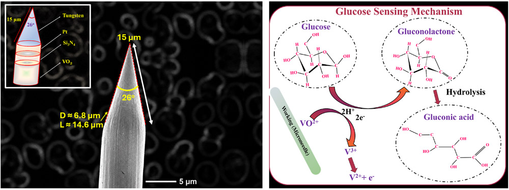
VO2 microneedle: structure and sensing
- [1] M. Blair, “Diabetes Mellitus Review,” Urol. Nurs., Vol.36, No.1, pp. 27-36, 2016.
- [2] A. B. Olokoba, O. A. Obateru, and L. B. Olokoba, “Type 2 diabetes mellitus: A review of current trends,” Oman Med. J., Vol.27, No.4, pp. 269-273, 2012. https://doi.org/10.5001/omj.2012.68
- [3] T. M. Dall, W. Yang, P. Halder, B. Pang, M. Massoudi, N. Wintfeld, A. P. Semilla, J. Franz, and P. F. Hogan, “The Economic Burden of Elevated Blood Glucose Levels in 2012: Diagnosed and Undiagnosed Diabetes, Gestational Diabetes Mellitus, and Prediabetes,” Diabetes Care, Vol.37, No.12, pp. 3172-3179, 2014. https://doi.org/10.2337/dc14-1036
- [4] K. B. Kim, W.-C. Lee, C.-H. Cho, D.-S. Park, S. J. Cho, and Y. B. Shim, “Continuous glucose monitoring using a microneedle array sensor coupled with a wireless signal transmitter,” Sensors Actuators, B Chem., Vol.281, pp. 14-21, 2019. https://doi.org/10.1016/j.snb.2018.10.081
- [5] S. S. Lund, P. Rossing, and A. A. Vaag, “Follow-up of intensive glucose control in type 2 diabetes,” N. Engl. J. Med., Vol.360, No.4, pp. 416-418, 2009. https://doi.org/10.1056/NEJMc082275
- [6] S. K. Vashist, “Continuous Glucose Monitoring Systems: A Review,” Diagnostics, Vol.3, No.4 pp. 385-412, 2013. https://doi.org/10.3390/diagnostics3040385
- [7] L. Hood, R. Balling, and C. Auffray, “Revolutionizing medicine in the 21st century through systems approaches,” Biotechnol. J., Vol.7, No.8, pp. 992-1001, 2012. https://doi.org/10.1002/biot.201100306
- [8] R. Esfandyarpour, H. Esfandyarpour, M. Javanmard, J. S. Harris, and R. W. Davis, “Microneedle biosensor: A method for direct label-free real time protein detection,” Sensors Actuators, B Chem., Vol.177, pp. 848-855, 2013. https://doi.org/10.1016/j.snb.2012.11.064
- [9] S. A. Ranamukhaarachchi, C. Padeste, M. Dübner, U. O. Häfeli, B. Stoeber, and V. J. Cadarso, “Integrated hollow microneedle-optofluidic biosensor for therapeutic drug monitoring in sub-nanoliter volumes,” Sci. Rep., Article No.29075, 2016. https://doi.org/10.1038/srep29075
- [10] J. Yang, X. Liu, Y. Fu, and Y. Song, “Recent advances of microneedles for biomedical applications: Drug delivery and beyond,” Acta Pharm. Sin. B. Vol.9, No.3, pp. 469-483, 2019. https://doi.org/10.1016/j.apsb.2019.03.007
- [11] L. Ventrelli, L. M. Strambini, and G. Barillaro, “Microneedles for Transdermal Biosensing: Current Picture and Future Direction,” Adv. Healthcare Mater., Vol.4, No.17, pp. 2606-2640, 2015. https://doi.org/10.1002/adhm.201500450
- [12] P. Bataillard, E. Steffgen, S. Haemmerli, A. Manz, and H. M. Widmer, “An integrated silicon thermopile as biosensor for the thermal monitoring of glucose, urea and penicillin,” Biosens. Bioelectron., Vol.8, No.2 pp. 89-98, 1993. https://doi.org/10.1016/0956-5663(93)80057-V
- [13] C. H. Schein, K. Kashiwagi, A. Fujisawa, and C. Weissmann, “Secretion of Mature IFN–2 and Accumulation of Uncleaved Precursor by Bacillus subtilis Transformed with a Hybrid -Amylase Signal Sequence-IFN-2 Gene,” Nat. Biotechnol., Vol.4, pp. 719-725, 1986. https://doi.org/10.1038/nbt0886-719
- [14] L. Doretti, D. Ferrara, P. Gattolin, and S. Lora, “Amperometric biosensor with physically immobilized glucose oxidase on a PVA cryogel membrane,” Talanta, Vol.44, No.5, pp. 859-866, 1997. https://doi.org/10.1016/S0039-9140(96)02130-3
- [15] T. Gan, Z. Shi, J. Sun, and Y. Liu, “Simple and novel electrochemical sensor for the determination of tetracycline based on iron/zinc cations–exchanged montmorillonite catalyst,” Talanta, Vol.121, pp. 187-193, 2014. https://doi.org/10.1016/j.talanta.2014.01.002
- [16] C. Yu, L. Wang, W. Li, C. Zhu, N. Bao, and H. Gu, “Detection of cellular H2O2 in living cells based on horseradish peroxidase at the interface of Au nanoparticles decorated graphene oxide,” Sensors Actuators, B Chem., Vol.211, pp. 17-24, 2015. https://doi.org/10.1016/j.snb.2015.01.064
- [17] Y. Li, Q. Lu, S. Wu, L. Wang, and X. Shi, “Hydrogen peroxide sensing using ultrathin platinum-coated gold nanoparticles with core@shell structure,” Biosens. Bioelectron., Vol.41, pp. 576-581, 2013. https://doi.org/10.1016/j.bios.2012.09.027
- [18] F. Meng, X. Yan, J. Liu, J. Gu, and Z. Zou, “Nanoporous gold as non-enzymatic sensor for hydrogen peroxide,” Electrochim. Acta., Vol.56, No.12, pp. 4657-4662, 2011. https://doi.org/10.1016/j.electacta.2011.02.105
- [19] A. Aherwar and M. Bahraminasab, “Biocompatibility evaluation and corrosion resistance of tungsten added Co-30Cr-4Mo-1Ni alloy,” Biomed. Mater. Eng., Vol.28, No.6, pp. 687-701, 2017. https://doi.org/10.3233/BME-171706
- [20] G. Yang, B. Wang, K. Tawfiq, H. Wei, S. Zhou, and G. Chen, “Electropolishing of surfaces: Theory and applications,” Surf. Eng., Vol.33, No.2, pp. 149-166, 2017. https://doi.org/10.1080/02670844.2016.1198452
- [21] L. Ramachandra Bhat, S. Vedantham, U. M. Krishnan, and J. B. B. Rayappan, “A non-enzymatic two step catalytic reduction of methylglyoxal by nanostructured V2O5 modified electrode,” Biosens. Bioelectron., Vol.103, pp. 143-150, 2018. https://doi.org/10.1016/j.bios.2017.12.036
- [22] K. Jayanth Babu, M. Sasya, N. Nesakumar, P. Shankar, M. B. Gumpu, B. L. Ramachandra, A. J. Kulandaisamy, and J. B. B. Rayappan, “Non-enzymatic detection of glucose in fruits using TiO-MnO hybrid nano interface,” Appl. Nanosci., Vol.7, pp. 309-316, 2017. https://doi.org/10.1007/s13204-017-0571-1
- [23] A. Mohamed Azharudeen, R. Karthiga, M. Rajarajan, and A. Suganthi, “Enhancement of electrochemical sensor for the determination of glucose based on mesoporous VO/PVA nanocomposites,” Surfaces and Interfaces, Vol.16 pp. 164-173, 2019. https://doi.org/10.1016/j.surfin.2019.05.005
- [24] S. T. R. Naqvi, B. Shirinfar, D. Hussain, S. Majeed, M. N. Ashiq, Y. Aslam, and N. Ahmed, “Electrochemical Sensing of Ascorbic Acid, Hydrogen Peroxide and Glucose by Bimetallic (Fe, Ni)–CNTs Composite Modified Electrode,” Electroanalysis, Vol.31, No.5, pp. 851-857, 2019. https://doi.org/10.1002/elan.201800768
- [25] A. Fatoni, A. N. Aziz, and M. D. Anggraeni, “Low-cost and real-time color detector developments for glucose biosensor,” Sens. Bio-Sens. Res., Vol.28, Article No.100325, 2020. https://doi.org/10.1016/j.sbsr.2020.100325
- [26] N. Mohamad Nor, N. S. Ridhuan, and K. Abdul Razak, “Progress of Enzymatic and Non-Enzymatic Electrochemical Glucose Biosensor Based on Nanomaterial-Modified Electrode,” Biosensors, Vol.12, No.12, Article No.1136, 2022. https://doi.org/10.3390/bios12121136
- [27] R. Ahmad, N. Tripathy, M.-S. Ahn, K. S. Bhat, T. Mahmoudi, Y. Wang, J.-Y. Yoo, D.-W. Kwon, H.-Y. Yang, and Y.-B. Hahn, “Highly efficient non-enzymatic glucose sensor based on CuO modified vertically-grown ZnO nanorods on electrode,” Sci. Rep., Vol.7, Article No.5715, 2017. https://doi.org/10.1038/s41598-017-06064-8
- [28] M. S. Reza, H. Song, Z. Islam, D. Kim, J. Go, and J. Y. Park, “A perfluorinated sulfonic acid-based double-layered membrane-coated polymeric microneedle glucose sensor with wide linear range for long-term stable monitoring,” Chem. Eng. J., Vol.529, Article No.172668, 2026. https://doi.org/10.1016/j.cej.2026.172668
- [29] Y.-J. Kim, S. R. Chinnadayyala, H. T. N. Le, and S. Cho, “Sensitive Electrochemical Non-Enzymatic Detection of Glucose Based on Wireless Data Transmission,” Sensors, Vol.22, No.7, Article No.2787, 2022. https://doi.org/10.3390/s22072787
This article is published under a Creative Commons Attribution-NoDerivatives 4.0 Internationa License.