
Paper:
Non-Enzymatic Electrochemical Detection of Glucose Using VO2-Based Tungsten Microneedles
Ganesh Kumar Mani*1,*2
 , Ponnusamy Dhivya*2,*3
, Madhanraj Ramu*4, Akshay Krishnakumar*2, Toshiaki Kanematsu*5, and Kazuyoshi Tsuchiya*2,*6
*1Department of Physics, Korea Advanced Institute of Science and Technology (KAIST)
291 Daehak-ro, Yuseong-gu, Daejeon 34141, Republic of Korea
*2Micro/Nano Technology Center, Tokai University
4-1-1 Kitakaname, Hiratsuka, Kanagawa 259-1292, Japan
*3Education Center for Emerging Energy Technology on Climate Change, Chungnam National University
99 Daehak-ro, Yuseong District, Daejeon 34134, Republic of Korea
*4Hindusthan College of Arts & Science (HICAS)
Avinashi Rd, behind Nava India, Udayampalayam, Coimbatore, Tamil Nadu 641028, India
*5Graduate School of Science and Technology, Tokai University
4-1-1 Kitakaname, Hiratsuka, Kanagawa 259-1292, Japan
*6Department of Mechanical Engineering, Tokai University
4-1-1 Kitakaname, Hiratsuka, Kanagawa 259-1292, Japan

Traditional glucose-detection methods, such as calorimetric, optical, and physical biosensors, are not suitable for real-time applications owing to their high maintenance, cost, and time requirements. This study aims to create a painless glucose detection system, employing a tungsten microneedle as a non-enzymatic electrochemical biosensing platform. It employs, for the first time, vanadium oxide (VO2)-based non-enzymatic tungsten microneedles as the working electrode for electrochemical detection. The correlation between glucose concentration and the microneedles’ reaction was evaluated by cyclic voltammetric and amperometric investigations. Furthermore, this work addresses the critical challenges that currently limit the practical implementation of embedded tissue sensing technologies, including weak signal propagation within biological tissues, long-term electrochemical stability, and biocompatibility. Thus, the proposed platform enables reliable and sustained transmission of biological information from living tissues.
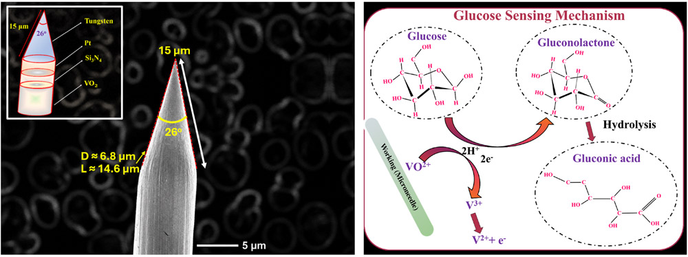
VO2 microneedle: structure and sensing
1. Introduction
Diabetes mellitus is a major cause of several health complications ranging from neurological disorders, as well as endocrine and cardiac diseases 1. People with Type 2 diabetes have both short- and long-term complications that may lead to premature death 2. Type 2 diabetes can even induce genetic and behavioral changes. Approximately 422 million people worldwide suffer from diabetic complications (World Health Organization, November 2019), which pose a high economic burden on developing countries 3. Rapid increase in blood-glucose levels causes diabetes, which in turn, causes various complications in the human body 4. Thus developing a reliable glucose monitoring system to eliminate such burgeoning health complications is cardinally crucial 5. Blood collection by pricking the finger is the most common method in glucose monitoring 6. As this process is painful and can lead to infections, such devices cannot be incorporated into a continuous monitoring system. Thus, developing a painless and continuous monitoring system, equipped in a proactive disease control environment, is necessary 7.
The advent of wearable health monitoring systems using stretchable and flexible electronics have paved the way for next-generation biosensors and drug delivery systems 8,9. Interstitial fluid (ISF) glucose concentration closely tracks blood glucose levels, making it a suitable target matrix for continuous non-invasive monitoring. Microneedles are thus well-positioned as sensing elements as they can introduce electrochemical transducers directly into biological tissue, enabling real-time information acquisition from within the tissue environment. They are consistent with the emerging paradigm of sensor introduction into cells and tissues for information transmission. In this regard, microneedles-based biomedical instruments have been used widely towards disease administration. Microneedles are minimally invasive, highly efficient, and versatile materials that have superior advantages of low cost, relative safety, and superior therapeutic effect 10. Additionally microneedles can rupture the skin’s toughest layer stratum corneum, which provides an easy access to the dermal and epidermal layers 9. Microneedles are widely used in cosmetic surgeries, drug delivery, and disease diagnostic systems 11. Therefore, this work focusses on developing tungsten microneedle-based biosensors towards diabetes mellitus diagnosis. Tungsten is a well-established biocompatible material used in implantable neural microelectrodes and surgical instruments, with demonstrated resistance to electrochemical corrosion in physiological saline environments.
Conventional detection strategies, such as calorimetric 12, optical 13, and physical 14 biosensors fall short in real-time applications with high maintenance, cost, and time consumption 15. Thus, recent researches have been focused on electrochemical detection of biological analytes using a metal oxide nanointerface, which can overcome these shortcomings. Enzymatic detection of biological analytes is a possible electrochemical strategy, by which efficient electrocatalytic activity could be attained 16. On the other hand, enzymatic detection suffers from serious disadvantages, such as high cost, chemical instability, and enzyme degradation 17,18. Thus, a non-enzymatic electrochemical detection approach of biological analytes was adopted herein due to its feasibility and long life.
Thus, a non-enzymatic electrochemical-based biosensing platform using a tungsten microneedle for a painless detection of glucose analyte was developed. Vanadium oxide (VO\(_{2}\))-based non-enzymatic tungsten microneedles (working electrode) were utilized for electrochemical detection. The response of the microneedles to glucose concentration was measured using cyclic voltammetric and amperometric studies. To the best of our knowledge this is the first study of its kind on non-enzymatic electrochemical detection of glucose using VO\(_{2}\)-based tungsten microneedles.
2. Materials and Methods
2.1. Materials
Tungsten microneedles with 300-μm diameter with 99.95% purity were procured from Nilaco Corporation, Japan. Silicon nitride (Si\(_{3}\)N\(_4\)) and VO\(_{2}\) target with 99.95% purity were purchased from Furuuchi Chemical Corporation, Japan. Ultrapure water from Milli-Q System (Millipore, USA) was used throughout the experiments. All purchased substrates and chemicals were used without any further purification.
2.2. Electropolishing and Surface Modification
Tungsten-based microneedles were manually cleaned with ethanol and water to remove the grease, slacks, and oxide layers. Progressively the microneedles were electropolished with potassium hydroxide (KOH) solution to clean the microneedle edges and deburr the surface. Electropolishing was performed with 2-M of KOH in a 100-mL beaker. A customized stirrer was added to the reaction beaker at a stirring rate of 300 rpm. A copper plate and tungsten microneedles, vibrated throughout the process, acted as an anode and a cathode, respectively. Subsequently, the microneedles were dipped in deionized water and ethanol to completely remove the remaining residues. The microneedle thus obtained was sharpened with electropolishing process.
The electroactive sensing material was coated on the surface of the tungsten thin films by a sputtering technique. An Si\(_3\)N\(_4\) layer was coated on the surface of the tungsten microneedle by radio frequency (RF) sputtering technique. The substrate, in which the microneedles were placed, was oriented perpendicular to the sputtering target. Additionally, the substrate was integrated with a motor (50 rpm) to obtain a uniform coating along the microneedle surface. Further platinum (Pt) was coated on the surface of the Si\(_3\)N\(_4\)-modified tungsten electrode to achieve an active surface in the working electrode. Subsequently VO\(_2\) was modified on the surface of the working electrode to enhance the redox reactions towards the glucose analyte. The detailed sputtering parameters are presented in Table 1.
| Factor | Condition | ||
| ECR | RF | ||
| Substrate | W | W/Si\(_3\)N\(_4\)/Pt | W |
| Target | Si\(_3\)N\(_4\) | V\(_2\)O\(_5\) | Pt |
| Sputtering time [min] | 40 | 120 | 20 |
| Ar gas flow rate [sccm] | 100 | 100 | 0.8 |
| Acceleration rate [eV] | 40 | 40 | 2500 |
| Pressure [Pa] | 3.0 | 2.0 | \(2.2 \times 10^{-2}\) |
2.3. Methods
The morphological of the fabricated microneedle was analyzed using a desktop scanning microscope (JCM-6000Plu, manufactured by JEOL Ltd.) and compositional analysis using a scanning electron microscope (FE-SEM) (JCM 6000, JEOL, Japan). Sensing measurements were carried out using a bi-potentiostat for RRDE-3A (Model 2325, ALS, Japan). Cyclic voltammetry studies and amperometric studies were carried out for electrochemical analysis of the VO\(_{2}\) microneedle. KCl saturated Ag/AgCl (013503, 0.5 mm diameter) and a platinum wire counter electrode (002233, 0.5 mm diameter) were also purchased from ALS Co., Ltd., Japan.
3. Results and Discussion
3.1. Microneedle Architecture and Characterization
Microneedle fabrication consists of four progressive layer modifications deposited using RF and electron cyclotron resonance (ECR) sputtering techniques.
Tungsten microneedle: Tungsten microneedles were preferred as the base for electrochemical detection of glucose. Tungsten has an unsurpassed strength and can be equipped in more delicate instruments. Tungsten wire is highly biocompatible and is electrochemical corrosion-resistant 19. Furthermore, the tungsten needles were electropolished to remove unwanted metallic workpieces and obtain a smooth surface. A smoothed microneedle was used for further modification 20.
Insulating layer: To inhibit tungsten participation in the redox reaction, Si\(_{3}\)N\(_4\), which is an insulating material with oxidation, shock, and mechanical fatigue resistance, was used as an insulation layer. Using a laser microscope, a complete deposition of Si\(_{3}\)N\(_{4}\) was observed on the surface of the microneedle.
Electrode surface layer: Pt was used as an active surface in the electrochemical detection. Pt is highly inert and does not participate in electrochemical redox reaction. Moreover, it is highly conductive, thereby providing a perpetual path for the flow of electrons. Platinum was used as a current collector for the working electrode from electroactive material during the redox reactions.
Redox interaction layer: Finally, VO\(_{2}\) was modified on the surface of the Pt microneedle electrode for glucose detection. VO\(_{2}\) has strong electrocatalytic and redox reaction properties that favor the electron transfer properties. The modified electroactive metal oxide has partially filled d-orbitals with high valance electrons, which triggers the redox reaction 21. Thus, VO\(_{2}\) was preferred as the nanointerface for glucose detection.
Surface morphology and elemental analysis: The surface morphology of the prepared tungsten microneedle was analyzed by FE-SEM to evaluate its tip geometry and dimensions. As shown in Fig. 1, the tapered tip exhibited an included angle of approximately 26° and a slanted surface length of approximately 15 μm. Based on these geometrical parameters, the corresponding tip diameter and axial length (perpendicular to the diameter) were estimated to be approximately 6.8 and 14.6 μm, respectively. To further contemplate the presence of VO\(_{2}\), elemental mapping and EDAX analysis were conducted. The surface of the microneedle had high concentrations of vanadium and oxygen, confirming the streamlined coating of the metal oxide nanointerface. The elemental composition of the microneedle surface was further examined by energy-dispersive X-ray spectroscopy (EDS). The EDS spectrum confirmed the presence of vanadium and oxygen; however, the resulting elemental percentages should not be directly interpreted as the stoichiometric composition of the VO\(_2\) coating. Because of the tapered multilayer structure and finite electron-beam interaction volume, the EDS signal might include contributions from the VO\(_2\) coating, underlying Si\(_3\)N\(_4\) insulation layer, and tungsten substrate, together with possible surface-adsorbed species.
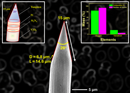
Fig. 1. FE-SEM micrograph of VO\(_2\)-based tungsten microneedle together with EDS elemental analysis. The upper-right inset presents the measured oxygen and vanadium elemental percentages obtained from the analyzed region. Because the EDS interaction volume may include the multilayer structure and underlying substrate, and EDS provides semi-quantitative elemental analysis, the displayed percentages should not be interpreted as the stoichiometric \(\mathrm{V}:\mathrm{O}\) ratio of the VO\(_2\) coating.
3.2. Biosensing Signatures
3.2.1. Choice of Electrolyte
NaCl–phosphate-buffered saline (PBS) buffer was chosen for the electrolyte for glucose detection. This electrolyte has a superior advantage over other electrolytes in reaction kinetics. Since NaCl is a strong electrolyte, the buffer immediately separates into constituent ions and helps in ion transfer. PBS solution consists of sodium, potassium, and phosphate ions, which can increase multiple reaction dynamics. Pt-modified tungsten microneedle was chosen to be the working electrode of the electrochemical setup. Therefore, the electrochemical potential swings of the microneedle must be between \(-\)0.9 and 0.9 V.
3.2.2. Electrochemical Characterization of Nanointerface
Cyclometric voltammetric (CV) analysis (Figs. 2(a) and (b)) was performed using an NaCl–PBS electrolyte both in the absence and presence of glucose concentration with voltage swing of \(-\)0.9\(\sim\)0.9 V. Scan rate studies of the VO\(_{2}\)-based tungsten microneedles were conducted in the presence of 10 mM of glucose concentration. The obtained graph confirms (Figs. 2(a) and (b)) that the obtained electrochemical reaction was a surface-controlled process. The scan rate of the electrochemical reaction was fixed as 0.05 Vs\(^{-1}\) for further analysis.
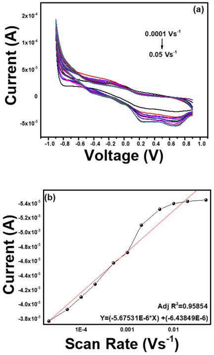
Fig. 2. (a) Cyclic voltammetric response for scan rates from 0.0001 to 0.05 Vs\(^{-1}\). (b) Linear regression plot with cathodic current vs. scan rate.
The CV graph response of the tungsten microneedles was analyzed in the absence and presence of the glucose analyte (Fig. 3(a)). No significant redox peak was observed from bare and VO\(_{2}\)-coated microneedles. However, with increase in glucose concentration (Fig. 3(b)), two distinctive cathodic peaks were observed. The peaks projected might be probably due to the reduction of glucose to gluconolactone in the presence of VO\(_{2}\) metal oxide 22.
Similarly, Mohamed Azharudeen et al. 23 developed PVA-based VO\(_{2}\), which hydrolyses gluconolactone to gluconic acid. From the past literature, it could also be observed that the lower reduction peak corresponds to the formation of gluconic acid by hydrolysis 24.
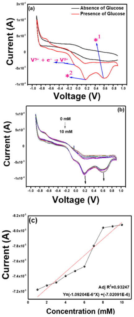
Fig. 3. (a) CV graph of microelectrode in absence and presence of glucose. Annotations denote oxidation peaks corresponding to the V\(^{2+}\)/V\(^{3+}\) (*1) and glucose oxidation-coupled current enhancement (*2), respectively. (b) Presence of 1–10 mM of glucose. Each color plot (light blue to dark blue) corresponds to different glucose concentrations in steps of 1 mM: 1–10 mM. (c) Linear calibration plot.
3.2.3. Increase in Concentration
The possible mechanism of the non-enzymatic glucose detection of the microneedles is presented in Fig. 4. The electrocatalytic oxidation of glucose to gluconolactone induces the reduction of VO\(^{2+}\) to form V\(^{3+}\) ions. The source of H\(^{+}\) and electrons for the electrochemical interaction was from the glucose. With increase in concentration, more glucose would be electrochemically oxidized, favoring the reduction of VO\(^{2+}\) ions. This could be validated by the slight increase in the shoulder peak around \(-\)0.03 V corresponding to the oxidation of V\(^{3+}\) ions.
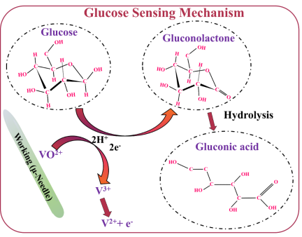
Fig. 4. Schematic of glucose-VO\(_2\) microneedle interaction.
3.2.4. Performance Characteristics
The prepared biosensor exhibited a sensitivity (Fig. 3(c)) of \(-\)1.092 μA mM\(^{-1}\) with a linear range of 1–10 mM. The limit of detection (LOD) and limit of quantification (LOQ) of the microneedle can be estimated from the formulae, \(3*\mathrm{SD}\) and \(10*\mathrm{SD}\), respectively. Thus, the LOD and LOQ of the microneedle were found to be 1.268 and 42 μM, respectively. The linear calibration curve showed an adjusted \(R^2\) value of 0.9324, indicating a strong linear relationship between glucose concentration and the measured current response. The adjusted \(R^2\) accounts for the number of predictors and observations in the regression model, thus giving a more conservative fit estimate than the standard \(R^2\). Unlike the standard \(R^2\), which does not decrease when predictors are added, the adjusted \(R^{2}\) can decrease if an added predictor fails to meaningfully improve the fit. Similarly, the obtained RSD value proved the repeatability and reproducibility of the microneedles.
For a broader perspective, Table 2 25,26,27,28,29 compares the proposed VO\(_2\)/Pt/Si\(_3\)N\(_4\) tungsten microneedle sensor with representative technologies across colorimetric/optical assays, conventional enzymatic electrochemical sensors, planar non-enzymatic nanomaterial electrodes, and microneedle-based enzymatic and non-enzymatic continuous monitoring platforms. Colorimetric and enzymatic sensors can achieve high selectivity and sensitivity, but are constrained by reagent or sample handling, enzyme stability, and finite operational lifetime. Planar non-enzymatic electrodes, meanwhile, achieve low detection limits and rapid response, but typically depend on external blood or serum sampling. Against this backdrop, the proposed tungsten microneedle sensor strikes a distinct balance of reagent-free operation, long-term stability, and minimally invasive tissue integration, though its raw sensitivity remains somewhat below that of the best-performing planar nanomaterial electrodes.
| Category | Platform | LOD | Linear range | Sensitivity | Method | Key limitation |
|
Colorimetric/ optical 25 |
Low-cost colorimetric enzymatic glucose biosensor | 0.14 mM | 0.1–2.5 mM | N/A (absorbance-based) |
Bench/point of care, sample extraction needed |
Reagent-dependent, single-point, not continuous |
| Conventional enzymatic electrochemical 26 |
GOx/PtNP/PAA-modified screen-printed carbon electrode |
7.6 μM | 0.02–2.3 mM |
\(\sim\)42.7 μA mM\(^{-1}\) cm\(^{-2}\) |
Finger-prick strip |
Enzyme instability, storage requirements, single-point |
| Non-enzymatic electrochemical 27 | CuO–ZnO nanorod/FTO electrode | 0.40 μM | 0.001–8.45 mM |
\(\sim\)2961.7 μA mM\(^{-1}\) cm\(^{-2}\) |
Bench electrode, requires blood/serum sample | Not minimally invasive, not implantable in current form |
| Microneedle-based (enzymatic, wearable Continuous glucose monitor) 28 |
Double-layer PSA membrane-coated polymeric microneedle |
1.8 μM |
up to 0–35 mM |
\(\sim\)18.46 μA mM\(^{-1}\) cm\(^{-2}\) |
Minimally invasive, wearable patch | Enzyme degradation limits long-term stability |
| Microneedle-based (non-enzymatic) 29 | Pt-black/Nafion wireless microneedle array | 268 μM | 1–30 mM |
\(\sim\)445.75 μA mM\(^{-1}\) cm\(^{-2}\) |
Minimally invasive, wireless-capable array |
Multi-needle array, single-needle non-enzymatic reports remain rare |
| This work |
VO\(_{\boldsymbol{2}}\)/Pt/Si\(_{\boldsymbol{3}}\)N\(_{\boldsymbol{4}}\) tungsten single microneedle |
1.268 μM |
1–10 mM (LOQ 42 μM) |
1.092 μA mM\(^{\boldsymbol{-1}}\) |
Minimally invasive, single microneedle, reagent-free | Lower raw sensitivity than best planar non-enzymatic electrodes, in vitro only |
4. Conclusions
A nonenzymatic glucose sensor was prepared by VO\(_{2}\)-based tungsten microneedles. The electroactive surface aided the electrocatalytic oxidation of glucose and reduction of VO\(^{2+}\) ions. The coating on the surface of the microneedle was rendered flawless so that none of the Si\(_{3}\)N\(_{4}\) participated in the electrochemical redox reactions. The results demonstrated that this technique can be integrated with wireless real-time continuous monitoring systems for glucose monitoring. From a practical implementation standpoint, the amperometric signal generated at the microneedle tip can be transmitted through the conductive tungsten shaft to an external or miniaturized readout circuit, thereby forming the sensing element of a complete tissue-to-transmission sensing chain.
Notably, the current linear range (1–10 mM) covers the normoglycemic and hypoglycemic thresholds; however, it does not extend to the upper end of hyperglycemic ISF concentrations (\(< 15\) mM), which can occur under poorly controlled diabetic conditions. Extending the linear range through surface area modification or electrode-geometry tuning represents an important future design direction toward full physiological coverage. The present measurements were also conducted in PBS, whose ionic strength and pH approximate those of ISF. Direct validation in artificial ISF, together with interference testing against endogenous species, such as ascorbic acid, uric acid, and acetaminophen, is planned as part of the next stage of sensor development. More broadly, while the present study establishes the electrochemical performance baseline under controlled in vitro conditions, electrochemical stability under biofouling conditions and long-term operational durability remain important subjects for future investigation. Integration with a compact potentiostat module and a Bluetooth low-energy wireless transmitter represents a feasible pathway toward a wearable, real-time continuous glucose monitoring platform. These efforts are currently underway and will be reported in a subsequent paper.
Acknowledgments
The authors wish to sincerely thank Micro/Nano Technology Center (MNTC), Tokai University (Shonan Campus), Japan, for their infrastructural and financial support, and Tokai Imaging Center for Advanced Research (TICAR), Tokai University (Shonan campus), Japan for helping with additional characterization techniques. Additionally, the authors are grateful to the Basic Science Research Program of the National Research Foundation of Korea (NRF), funded by the Ministry of Education (NRF-2022R1I1A1A01068997) and Japan Society for the Promotion of Science (JSPS KAKENHI 15K12560) for providing financial assistance to this study.
- [1] M. Blair, “Diabetes Mellitus Review,” Urol. Nurs., Vol.36, No.1, pp. 27-36, 2016.
- [2] A. B. Olokoba, O. A. Obateru, and L. B. Olokoba, “Type 2 diabetes mellitus: A review of current trends,” Oman Med. J., Vol.27, No.4, pp. 269-273, 2012. https://doi.org/10.5001/omj.2012.68
- [3] T. M. Dall, W. Yang, P. Halder, B. Pang, M. Massoudi, N. Wintfeld, A. P. Semilla, J. Franz, and P. F. Hogan, “The Economic Burden of Elevated Blood Glucose Levels in 2012: Diagnosed and Undiagnosed Diabetes, Gestational Diabetes Mellitus, and Prediabetes,” Diabetes Care, Vol.37, No.12, pp. 3172-3179, 2014. https://doi.org/10.2337/dc14-1036
- [4] K. B. Kim, W.-C. Lee, C.-H. Cho, D.-S. Park, S. J. Cho, and Y. B. Shim, “Continuous glucose monitoring using a microneedle array sensor coupled with a wireless signal transmitter,” Sensors Actuators, B Chem., Vol.281, pp. 14-21, 2019. https://doi.org/10.1016/j.snb.2018.10.081
- [5] S. S. Lund, P. Rossing, and A. A. Vaag, “Follow-up of intensive glucose control in type 2 diabetes,” N. Engl. J. Med., Vol.360, No.4, pp. 416-418, 2009. https://doi.org/10.1056/NEJMc082275
- [6] S. K. Vashist, “Continuous Glucose Monitoring Systems: A Review,” Diagnostics, Vol.3, No.4 pp. 385-412, 2013. https://doi.org/10.3390/diagnostics3040385
- [7] L. Hood, R. Balling, and C. Auffray, “Revolutionizing medicine in the 21st century through systems approaches,” Biotechnol. J., Vol.7, No.8, pp. 992-1001, 2012. https://doi.org/10.1002/biot.201100306
- [8] R. Esfandyarpour, H. Esfandyarpour, M. Javanmard, J. S. Harris, and R. W. Davis, “Microneedle biosensor: A method for direct label-free real time protein detection,” Sensors Actuators, B Chem., Vol.177, pp. 848-855, 2013. https://doi.org/10.1016/j.snb.2012.11.064
- [9] S. A. Ranamukhaarachchi, C. Padeste, M. Dübner, U. O. Häfeli, B. Stoeber, and V. J. Cadarso, “Integrated hollow microneedle-optofluidic biosensor for therapeutic drug monitoring in sub-nanoliter volumes,” Sci. Rep., Article No.29075, 2016. https://doi.org/10.1038/srep29075
- [10] J. Yang, X. Liu, Y. Fu, and Y. Song, “Recent advances of microneedles for biomedical applications: Drug delivery and beyond,” Acta Pharm. Sin. B. Vol.9, No.3, pp. 469-483, 2019. https://doi.org/10.1016/j.apsb.2019.03.007
- [11] L. Ventrelli, L. M. Strambini, and G. Barillaro, “Microneedles for Transdermal Biosensing: Current Picture and Future Direction,” Adv. Healthcare Mater., Vol.4, No.17, pp. 2606-2640, 2015. https://doi.org/10.1002/adhm.201500450
- [12] P. Bataillard, E. Steffgen, S. Haemmerli, A. Manz, and H. M. Widmer, “An integrated silicon thermopile as biosensor for the thermal monitoring of glucose, urea and penicillin,” Biosens. Bioelectron., Vol.8, No.2 pp. 89-98, 1993. https://doi.org/10.1016/0956-5663(93)80057-V
- [13] C. H. Schein, K. Kashiwagi, A. Fujisawa, and C. Weissmann, “Secretion of Mature IFN–2 and Accumulation of Uncleaved Precursor by Bacillus subtilis Transformed with a Hybrid -Amylase Signal Sequence-IFN-2 Gene,” Nat. Biotechnol., Vol.4, pp. 719-725, 1986. https://doi.org/10.1038/nbt0886-719
- [14] L. Doretti, D. Ferrara, P. Gattolin, and S. Lora, “Amperometric biosensor with physically immobilized glucose oxidase on a PVA cryogel membrane,” Talanta, Vol.44, No.5, pp. 859-866, 1997. https://doi.org/10.1016/S0039-9140(96)02130-3
- [15] T. Gan, Z. Shi, J. Sun, and Y. Liu, “Simple and novel electrochemical sensor for the determination of tetracycline based on iron/zinc cations–exchanged montmorillonite catalyst,” Talanta, Vol.121, pp. 187-193, 2014. https://doi.org/10.1016/j.talanta.2014.01.002
- [16] C. Yu, L. Wang, W. Li, C. Zhu, N. Bao, and H. Gu, “Detection of cellular H2O2 in living cells based on horseradish peroxidase at the interface of Au nanoparticles decorated graphene oxide,” Sensors Actuators, B Chem., Vol.211, pp. 17-24, 2015. https://doi.org/10.1016/j.snb.2015.01.064
- [17] Y. Li, Q. Lu, S. Wu, L. Wang, and X. Shi, “Hydrogen peroxide sensing using ultrathin platinum-coated gold nanoparticles with core@shell structure,” Biosens. Bioelectron., Vol.41, pp. 576-581, 2013. https://doi.org/10.1016/j.bios.2012.09.027
- [18] F. Meng, X. Yan, J. Liu, J. Gu, and Z. Zou, “Nanoporous gold as non-enzymatic sensor for hydrogen peroxide,” Electrochim. Acta., Vol.56, No.12, pp. 4657-4662, 2011. https://doi.org/10.1016/j.electacta.2011.02.105
- [19] A. Aherwar and M. Bahraminasab, “Biocompatibility evaluation and corrosion resistance of tungsten added Co-30Cr-4Mo-1Ni alloy,” Biomed. Mater. Eng., Vol.28, No.6, pp. 687-701, 2017. https://doi.org/10.3233/BME-171706
- [20] G. Yang, B. Wang, K. Tawfiq, H. Wei, S. Zhou, and G. Chen, “Electropolishing of surfaces: Theory and applications,” Surf. Eng., Vol.33, No.2, pp. 149-166, 2017. https://doi.org/10.1080/02670844.2016.1198452
- [21] L. Ramachandra Bhat, S. Vedantham, U. M. Krishnan, and J. B. B. Rayappan, “A non-enzymatic two step catalytic reduction of methylglyoxal by nanostructured V2O5 modified electrode,” Biosens. Bioelectron., Vol.103, pp. 143-150, 2018. https://doi.org/10.1016/j.bios.2017.12.036
- [22] K. Jayanth Babu, M. Sasya, N. Nesakumar, P. Shankar, M. B. Gumpu, B. L. Ramachandra, A. J. Kulandaisamy, and J. B. B. Rayappan, “Non-enzymatic detection of glucose in fruits using TiO-MnO hybrid nano interface,” Appl. Nanosci., Vol.7, pp. 309-316, 2017. https://doi.org/10.1007/s13204-017-0571-1
- [23] A. Mohamed Azharudeen, R. Karthiga, M. Rajarajan, and A. Suganthi, “Enhancement of electrochemical sensor for the determination of glucose based on mesoporous VO/PVA nanocomposites,” Surfaces and Interfaces, Vol.16 pp. 164-173, 2019. https://doi.org/10.1016/j.surfin.2019.05.005
- [24] S. T. R. Naqvi, B. Shirinfar, D. Hussain, S. Majeed, M. N. Ashiq, Y. Aslam, and N. Ahmed, “Electrochemical Sensing of Ascorbic Acid, Hydrogen Peroxide and Glucose by Bimetallic (Fe, Ni)–CNTs Composite Modified Electrode,” Electroanalysis, Vol.31, No.5, pp. 851-857, 2019. https://doi.org/10.1002/elan.201800768
- [25] A. Fatoni, A. N. Aziz, and M. D. Anggraeni, “Low-cost and real-time color detector developments for glucose biosensor,” Sens. Bio-Sens. Res., Vol.28, Article No.100325, 2020. https://doi.org/10.1016/j.sbsr.2020.100325
- [26] N. Mohamad Nor, N. S. Ridhuan, and K. Abdul Razak, “Progress of Enzymatic and Non-Enzymatic Electrochemical Glucose Biosensor Based on Nanomaterial-Modified Electrode,” Biosensors, Vol.12, No.12, Article No.1136, 2022. https://doi.org/10.3390/bios12121136
- [27] R. Ahmad, N. Tripathy, M.-S. Ahn, K. S. Bhat, T. Mahmoudi, Y. Wang, J.-Y. Yoo, D.-W. Kwon, H.-Y. Yang, and Y.-B. Hahn, “Highly efficient non-enzymatic glucose sensor based on CuO modified vertically-grown ZnO nanorods on electrode,” Sci. Rep., Vol.7, Article No.5715, 2017. https://doi.org/10.1038/s41598-017-06064-8
- [28] M. S. Reza, H. Song, Z. Islam, D. Kim, J. Go, and J. Y. Park, “A perfluorinated sulfonic acid-based double-layered membrane-coated polymeric microneedle glucose sensor with wide linear range for long-term stable monitoring,” Chem. Eng. J., Vol.529, Article No.172668, 2026. https://doi.org/10.1016/j.cej.2026.172668
- [29] Y.-J. Kim, S. R. Chinnadayyala, H. T. N. Le, and S. Cho, “Sensitive Electrochemical Non-Enzymatic Detection of Glucose Based on Wireless Data Transmission,” Sensors, Vol.22, No.7, Article No.2787, 2022. https://doi.org/10.3390/s22072787
This article is published under a Creative Commons Attribution-NoDerivatives 4.0 Internationa License.