
Paper:
Analysis of Intracellular DNA Sensor Dynamics Around DNA-Coated Microbeads Designed as Interfaces for Controlling Cellular Molecular Communication
Shouhei Kobayashi
 , Kenta Arai, Kayoko Ujiie, Noriko Fukuta, and Chie Mori
Bio-ICT Collaboration Center, Advanced ICT Research Institute, National Institute of Information and Communications Technology (NICT)
588-2 Iwaoka, Iwaoka-cho, Nishi-ku, Kobe, Hyogo 651-2492, Japan

As a model system for interface technologies to artificially control molecule-mediated communication in living cells (molecular communication), we developed a methodology in which biomolecule-coated polystyrene beads are internalized into cells, enabling measurement and control of cellular responses in the vicinity of the beads. We demonstrate that the cytosolic DNA sensor proteins barrier-to-autointegration factor and cyclic GMP-AMP synthase exhibit distinct behaviors around linear double-stranded DNA-coated polystyrene beads (DNA-beads, 2.8 µm in diameter) introduced into the cytoplasm. These findings highlight the importance of understanding and controlling the early events following cytoplasmic introduction of DNA-beads when such bead-based bio-artificial interfaces are used to regulate molecular communication in cells.
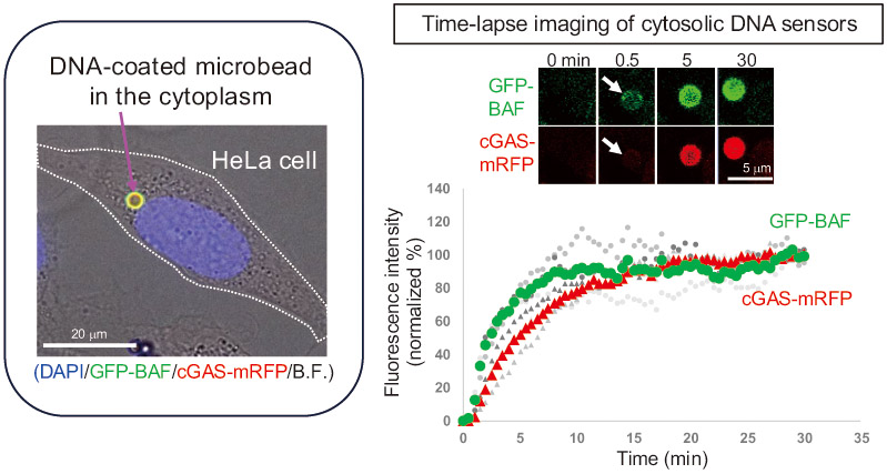
Biomolecule-coated microbeads to control molecular communication
- [1] T. Nakano, Y. Okaie, S. Kobayashi, T. Hara, Y. Hiraoka, and T. Haraguchi, “Methods and applications of mobile molecular communication,” Proc. of the IEEE, Vol.107, No.7, pp. 1442-1456, 2019. https://doi.org/10.1109/JPROC.2019.2917625
- [2] M. Egan, M. Kuscu, M. T. Barros, M. Booth, A. Llopis-Lorente, M. Magarini, D. P. Martins, M. Schäfer, and P. Stano, “Toward interdisciplinary synergies in molecular communications: Perspectives from synthetic biology, nanotechnology, communications engineering and philosophy of science,” Life, Vol.13, No.1, Article No.208, 2023. https://doi.org/10.3390/life13010208
- [3] J. Su, Y. Song, Z. Zhu, X. Huang, J. Fan, J. Qiao, and F. Mao, “Cell–cell communication: New insights and clinical implications,” Signal Transduction and Targeted Therapy, Vol.9, Article No.196, 2024. https://doi.org/10.1038/s41392-024-01888-z
- [4] S. Kobayashi, T. Kojidani, H. Osakada, A. Yamamoto, T. Yoshimori, Y. Hiraoka, and T. Haraguchi, “Artificial induction of autophagy around polystyrene beads in nonphagocytic cells,” Autophagy, Vol.6, No.1, pp. 36-45, 2010. https://doi.org/10.4161/auto.6.1.10324
- [5] S. Kobayashi, T. Koujin, T. Kojidani, H. Osakada, C. Mori, Y. Hiraoka, and T. Haraguchi, “BAF is a cytosolic DNA sensor that leads to exogenous DNA avoiding autophagy,” Proc. Natl. Acad. Sci. U.S.A., Vol.112, No.22, pp. 7027-7032, 2015. https://doi.org/10.1073/pnas.1501235112
- [6] Ş. Bilir, T. Kojidani, C. Mori, H. Osakada, S. Kobayashi, T. Koujin, Y. Hiraoka, and T. Haraguchi, “Roles of Nup133, Nup153 and membrane fenestrations in assembly of the nuclear pore complex at the end of mitosis,” Genes to Cells, Vol.24, No.5, pp. 338-353, 2019. https://doi.org/10.1111/gtc.12677
- [7] L. Sun, J. Wu, F. Du, X. Chen, and Z. J. Chen, “Cyclic GMP-AMP Synthase Is a Cytosolic DNA Sensor That Activates the Type I Interferon Pathway,” Science, Vol.339, No.6121, pp. 786-791, 2013. https://doi.org/10.1126/science.1232458
- [8] Q. Chen, L. Sun, and Z. J. Chen, “Regulation and Function of the cGAS–STING Pathway of Cytosolic DNA Sensing,” Nat. Immunol., Vol.17, No.10, pp. 1142-1149, 2016. https://doi.org/10.1038/ni.3558
- [9] M. S. Lee and R. Craigie, “A previously unidentified host protein protects retroviral DNA from autointegration,” Proc. Natl. Acad. Sci. U.S.A., Vol.95, No.4, pp. 1528-1533, 1998. https://doi.org/10.1073/pnas.95.4.1528
- [10] H. Chen and A. Engelman, “The barrier-to-autointegration protein is a host factor for HIV type 1 integration,” Proc. Natl. Acad. Sci. U.S.A., Vol.95, No.26, pp. 15270-15274, 1998. https://doi.org/10.1073/pnas.95.26.15270
- [11] M. Tsuchiya, H. Ogawa, T. Koujin, C. Mori, H. Osakada, S. Kobayashi, Y. Hiraoka, and T. Haraguchi, “p62/SQSTM1 promotes rapid ubiquitin conjugation to target proteins after endosome rupture during xenophagy,” FEBS Open Bio, Vol.8, No.3, pp. 470-480, 2018. https://doi.org/10.1002/2211-5463.12385
- [12] T. Haraguchi, T. Koujin, M. Segura-Totten, K. K. Lee, Y. Matsuoka, Y. Yoneda, K. L. Wilson, and Y. Hiraoka, “BAF is required for emerin assembly into the reforming nuclear envelope,” J. Cell Sci., Vol.114, No.24, pp. 4575-4585, 2001. https://doi.org/10.1242/jcs.114.24.4575
- [13] N. Lorén, J. Hagman, J. K. Jonasson, H. Deschout, D. Bernin, F. Cella-Zanacchi, A. Diaspro, J. G. McNally, M. Ameloot, N. Smisdom, M. Nydén, A.-M. Hermansson, M. Rudemo, and K. Braeckmans, “Fluorescence recovery after photobleaching in material and life sciences: Putting theory into practice,” Quarterly Reviews of Biophysics, Vol.48, No.3, pp. 323-387, 2015. https://doi.org/10.1017/S0033583515000013
- [14] B. Guey, M. Wischnewski, A. Decout, K. Makasheva, M. Kaynak, M. S. Sakar, B. Fierz, and A. Ablasser, “BAF restricts cGAS on nuclear DNA to prevent innate immune activation,” Science, Vol.369, No.6505, pp. 823-828, 2020. https://doi.org/10.1126/science.aaw6421
- [15] M. Du and Z. J. Chen, “DNA-Induced Liquid Phase Condensation of cGAS Activates Innate Immune Signaling,” Science, Vol.361, No.6403, pp. 704-709, 2018. https://doi.org/10.1126/science.aat1022
- [16] R. Zheng, R. Ghirlando, M. S. Lee, K. Mizuuchi, M. Krause, and R. Craigie, “Barrier-to-autointegration factor (BAF) bridges DNA in a discrete, higher-order nucleoprotein complex,” Proc. Natl. Acad. Sci. U.S.A., Vol.97, No.16, pp. 8997-9002, 2000. https://doi.org/10.1073/pnas.150240197
This article is published under a Creative Commons Attribution-NoDerivatives 4.0 Internationa License.