
Paper:
Delivery of Artificial Materials Into Adhesive Cells by Electromechanical Poration
Nobutoshi Ota*
 , Mayu Nakahigashi*, Taito Ikeuchi**
, Saki Kozawa**
, Kei Tateishi*, Yumi Ichinose*, Yuudai Aokusa*
, Yuma Minami*, Miho Tagawa**
, and Yoko Yamanishi*
*Department of Mechanical Engineering, Kyushu University
744 Motooka, Nishi-ku, Fukuoka, Fukuoka 819-0395, Japan
**Institute of Materials and Systems for Sustainability (IMaSS), Nagoya University
Furo-cho, Chikusa-ku, Nagoya, Aichi 464-8603, Japan

In order to transmit information from inside cells or living organisms, it is important to embed sensors or artificial objects inside cells or tissues rather than placing them on the surface of the tissue, to accurately extract internal information. However, sensors and artificial objects embedded in living tissues or cells have limitations because of their impact on cell viability and transfection efficiency. Electromechanical poration (EMP) is a promising method for introducing micrometer-scale materials into living cells. Unlike conventional electroporation, EMP combines high-voltage electrical stimulation with mechanical stimulation generated by the volume change of microbubbles at the electrode surface. These microbubbles create transient mechanical stress that facilitates membrane permeabilization while potentially reducing cellular damage. Using EMP, we investigated the delivery of polystyrene beads and silver-coated DNA-functionalized gold nanoparticle crystals (Au@Ag NPCs) into HeLa cells. Polystyrene beads up to 3 µm in diameter were successfully introduced, whereas delivery efficiency significantly decreased for beads larger than 6 µm, indicating a size-dependent limitation of cellular uptake. Importantly, cell viability remained high even 4 hours after bead delivery, regardless of bead size, indicating minimal cytotoxicity. In addition, Au@Ag NPCs (1–1.5 µm) were successfully delivered with a delivery rate of 0.5%, without inducing significant cytotoxicity over 1–4 days. These results demonstrated that EMP enabled efficient intracellular delivery of micrometer-scale materials while preserving cell viability, supporting its potential for intracellular sensing applications.
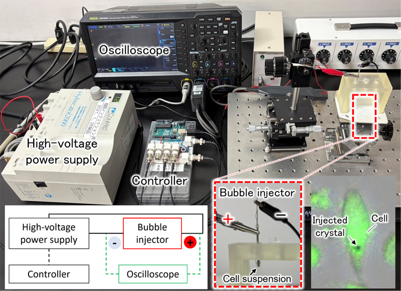
Intracellular crystal delivery by EMP
- [1] S. Modi, Swetha M. G., D. Goswami, G. D. Gupta, S. Mayor, and Y. Krishnan, “A DNA nanomachine that maps spatial and temporal pH changes inside living cells,” Nature Nanotech., Vol.4, pp. 325-330, 2009. https://doi.org/10.1038/nnano.2009.83
- [2] C. E. Talley, J. Jusinski, C. W. Hollars, S. M. Lane, and T. Huser, “Intracellular pH Sensors Based on Surface-Enhanced Raman Scattering,” Anal. Chem., Vol.76, No.23, pp. 7064-7068, 2004. https://doi.org/10.1021/ac049093j
- [3] D. F. Williams, “On the nature of biomaterials,” Biomaterials, Vol.30, No.30, pp. 5897-5909, 2009. https://doi.org/10.1016/j.biomaterials.2009.07.027
- [4] L. Dykman and N. Khlebtsov, “Gold nanoparticles in biomedical applications: Recent advances and perspectives,” Chem. Soc. Rev., Vol.41, No.6, pp. 2256-2282, 2012. https://doi.org/10.1039/c1cs15166e
- [5] M. B. Ross, C. A. Mirkin, and G. C. Schatz, “Optical Properties of One-, Two-, and Three-Dimensional Arrays of Plasmonic Nanostructures,” J. Phys. Chem. C, Vol.120, No.6, pp. 816-830, 2016. https://doi.org/10.1021/acs.jpcc.5b10800
- [6] T. Xu and Z. Geng, “Strategies to improve performances of LSPR biosensing: Structure, materials, and interface modification,” Biosensors and Bioelectronics, Vol.174, Article No.112850, 2021. https://doi.org/10.1016/j.bios.2020.112850
- [7] M. Yokomori, H. Suzuki, A. Nakamura, S. S. Sugano, and M. Tagawa, “DNA-functionalized colloidal crystals for macromolecular encapsulation,” Soft Matter, Vol.18, No.36, pp. 6954-6964, 2022. https://doi.org/10.1039/d2sm00949h
- [8] T. K. Kim and J. H. Eberwine, “Mammalian cell transfection: the present and the future,” Anal. Bioanal. Chem., Vol.397, No.8, pp. 3173-3178, 2010. https://doi.org/10.1007/s00216-010-3821-6
- [9] M. P. Stewart, A. Sharei, X. Ding, G. Sahay, R. Langer, and K. F. Jensen, “In vitro and ex vivo strategies for intracellular delivery,” Nature, Vol.538, pp. 183-192, 2016. https://doi.org/10.1038/nature19764
- [10] M. P. Stewart, R. Langer, and K. F. Jensen, “Intracellular Delivery by Membrane Disruption: Mechanisms, Strategies, and Concepts,” Chem. Rev., Vol.118, No.16, pp. 7409-7531, 2018. https://doi.org/10.1021/acs.chemrev.7b00678
- [11] P. E. Boukany, A. Morss, W.-c. Liao, B. Henslee, H. Jung, X. Zhang, B. Yu, X. Wang, Y. Wu, L. Li, K. Gao, X. Hu, X. Zhao, O. Hemminger, W. Lu, G. P. Lafyatis, and L. J. Lee, “Nanochannel electroporation delivers precise amounts of biomolecules into living cells,” Nature Nanotech., Vol.6, pp. 747-554, 2011. https://doi.org/10.1038/nnano.2011.164
- [12] A. Sharei, J. Zoldan, A. Adamo, W. Y. Sim, N. Cho, E. Jackson, S. Mao, S. Schneider, M.-J. Han, A. Lytton-Jean, P. A. Basto, S. Jhunjhunwala, J. Lee, D. A. Heller, J. W. Kang, G. C. Hartoularos, K.-S. Kim, D. G. Anderson, R. Langer, and K. F. Jensen, “A vector-free microfluidic platform for intracellular delivery,” PNAS, Vol.110, No.6, pp. 2082-2087, 2013. https://doi.org/10.1073/pnas.1218705110
- [13] W. Huang, S. Sakuma, N. Tottori, S. S. Sugano, and Y. Yamanishi, “Viscosity-aided electromechanical poration of cells for transfecting molecules,” Lab Chip, Vol.22, No.22, pp. 4276-4291, 2022. https://doi.org/10.1039/d2lc00628f
- [14] W. Huang, Y. Ma, N. Tottori, and Y. Yamanishi, “Enhancing suspended cell transfection by inducing localized distribution of the membrane actin cortex before exposure to electromechanical stimulation,” Biotechnol. Lett., Vol.45, pp. 1417-1430, 2023. https://doi.org/10.1007/s10529-023-03382-y
- [15] T. Ikeuchi, L. Zhang, S. Kozawa, S. Kojima, H. Niinomi, M. Kuwahara, K. Hiramatsu, and M. Tagawa, “Self-assembly of High-quality Single Crystals of DNA-functionalized Gold Nanoparticle Superlattices for In-vivo SERS Detection,” Proc. 15th Int. Conf. Metamaterials, Photonic Crystals and Plasmonics (META 2025), pp. 1840-1841, 2025.
- [16] A. B. Borle, “Kinetic Analyses of Calcium Movements in HeLa Cell Cultures: I. Calcium influx,” J. Gen. Physiol., Vol.53, No.1, pp. 43-56, 1969. https://doi.org/10.1085/jgp.53.1.43
- [17] M. Uzhytchak, B. Smolková, A, Frtús, A. Stupakov, M. Lunova, F. Scollo, M. Hof, P. Jurkiewicz, G. J. Sullivan, A. Dejneka, and O. Lunov, “Sensitivity of endogenous autofluorescence in HeLa cells to the application of external magnetic fields,” Sci. Rep., Vol.13, Article No.10818, 2023. https://doi.org/10.1038/s41598-023-38015-x
- [18] D. Guo, J. Li, G. Xie, Y. Wang, and J. Luo, “Elastic Properties of Polystyrene Nanospheres Evaluated with Atomic Force Microscopy: Size Effect and Error Analysis,” Langmuir, Vol.30, No.24, pp. 7206-7212, 2014. https://doi.org/10.1021/la501485e
- [19] J. Bush, C.-H. Hu, and R. Veneziano, “Mechanical Properties of DNA Hydrogels: Towards Highly Programmable Biomaterials,” Applied Sciences, Vol.11, No.4, Article No.1885, 2021. https://doi.org/10.3390/app11041885
- [20] Y. Wang, Y. Zhang, Q. Zhang, X. Li, Q. Yan, and Y. Zhu, “Mechanical properties modulation and biological applications of DNA hydrogels,” Advanced Sensor and Energy Materials, Vol.3, No.3, Article No.100113, 2024. https://doi.org/10.1016/j.asems.2024.100113
This article is published under a Creative Commons Attribution-NoDerivatives 4.0 Internationa License.