
Paper:
Delivery of Artificial Materials Into Adhesive Cells by Electromechanical Poration
Nobutoshi Ota*
 , Mayu Nakahigashi*, Taito Ikeuchi**
, Saki Kozawa**
, Kei Tateishi*, Yumi Ichinose*, Yuudai Aokusa*
, Yuma Minami*, Miho Tagawa**
, and Yoko Yamanishi*
*Department of Mechanical Engineering, Kyushu University
744 Motooka, Nishi-ku, Fukuoka, Fukuoka 819-0395, Japan
**Institute of Materials and Systems for Sustainability (IMaSS), Nagoya University
Furo-cho, Chikusa-ku, Nagoya, Aichi 464-8603, Japan

In order to transmit information from inside cells or living organisms, it is important to embed sensors or artificial objects inside cells or tissues rather than placing them on the surface of the tissue, to accurately extract internal information. However, sensors and artificial objects embedded in living tissues or cells have limitations because of their impact on cell viability and transfection efficiency. Electromechanical poration (EMP) is a promising method for introducing micrometer-scale materials into living cells. Unlike conventional electroporation, EMP combines high-voltage electrical stimulation with mechanical stimulation generated by the volume change of microbubbles at the electrode surface. These microbubbles create transient mechanical stress that facilitates membrane permeabilization while potentially reducing cellular damage. Using EMP, we investigated the delivery of polystyrene beads and silver-coated DNA-functionalized gold nanoparticle crystals (Au@Ag NPCs) into HeLa cells. Polystyrene beads up to 3 µm in diameter were successfully introduced, whereas delivery efficiency significantly decreased for beads larger than 6 µm, indicating a size-dependent limitation of cellular uptake. Importantly, cell viability remained high even 4 hours after bead delivery, regardless of bead size, indicating minimal cytotoxicity. In addition, Au@Ag NPCs (1–1.5 µm) were successfully delivered with a delivery rate of 0.5%, without inducing significant cytotoxicity over 1–4 days. These results demonstrated that EMP enabled efficient intracellular delivery of micrometer-scale materials while preserving cell viability, supporting its potential for intracellular sensing applications.
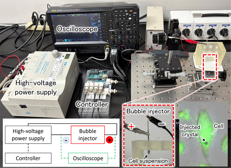
Intracellular crystal delivery by EMP
1. Introduction
Unlike surface-equipped sensors, which measure information appearing on or accessible from the body surface, implantable sensors enable the measurement of information inside the body, such as within cells, blood, and interstitial fluid. In particular, intracellular information cannot be obtained using conventional approaches for in vivo measurements based on blood or body fluid sampling, creating a demand for sensors that can operate inside cells (Fig. 1). Hence, it is preferable to construct intracellular sensors using biomolecule-based or biocompatible materials to ensure safe operation of a sensor within a cell 1,2.
Biocompatible materials include inert polymers and metals 3. Among these materials, gold nanoparticles (AuNPs) exhibit high biocompatibility and size-dependent optical properties. By functionalizing AuNPs with sensor molecules such as aptamers, AuNPs can therefore be utilized as sensors 4.
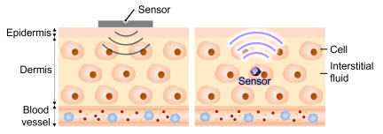
Fig. 1. Images of biosensors. (Left) A sensor placed on the body surface and (right) a sensor injected beneath the epidermis.
AuNPs are also expected to enhance their sensor performance when aligned at appropriate distances 5,6, which is possibly controlled by DNA attached to the gold surface 7. Hybridization of the DNA strands on modified AuNP surfaces form crosslinked structures, which promote the self-assembly of AuNPs. This DNA-mediated crosslinking maintains a constant distance between gold nanoparticles to form nanoparticle colloidal crystals (NPCs).
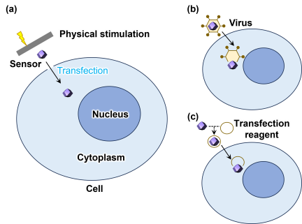
Fig. 2. Methods for delivery of artificial materials into cells.
When introducing microparticles such as NPCs into cells, delivery methods can be broadly classified into three categories: physical, chemical, and biological (Fig. 2) 8,9. Although chemical and biological approaches, such as lipid/polymer-based carriers and viral vectors, are widely used, their applicability to large or structurally complex cargos can be limited by endosomal entrapment, cargo degradation during intracellular trafficking, cytotoxicity or immunogenicity, and constraints on cargo size and surface properties 9,10. Physical methods, including microinjection, electroporation, particle bombardment, and microfluidic membrane deformation rely on direct membrane perturbation by mechanical, electrical, or ballistic forces. Each physical method has its own limitations, such as the low throughput of microinjection or the possible loss of viability and nonuniform permeabilization associated with bulk electroporation. Nevertheless, physical delivery methods offer several advantages, including applicability to a wide range of cell types and cargo materials, precise control of physical parameters for material delivery, and the absence of a requirement to encapsulate a delivered material in a carrying agent such as a lipid membrane 11,12.
In this study, we employed electromechanical poration (EMP), a physical method of material delivery. EMP offers the same advantages as other physical delivery approaches. EMP uses both electrical and mechanical stimulation. Electrical stimulation permeabilizes the cell membrane. Mechanical stimulation arising from oscillatory changes in microbubble volume transiently perturbs the cytoskeleton and facilitates membrane permeabilization. This synergistic effect enables intracellular delivery of materials with low applied energy, thereby reducing cellular damage and improving cell viability 13,14. In the following sections, we discuss the delivery of artificial materials, polystyrene beads and silver-coated gold nanoparticle crystals (Au@Ag NPCs), into cells using EMP. Au@Ag NPCs were produced using NPCs synthesized via DNA self-assembly as precursors, followed by formation of a core–shell structure around the nanoparticles through a silver reduction reaction. Silver coating is expected to reduce gaps between nanoparticles and enhance surface-enhanced Raman scattering, thereby enabling highly sensitive detection of trace molecules, even within cells 15.
2. EMP
2.1. Concept of EMP
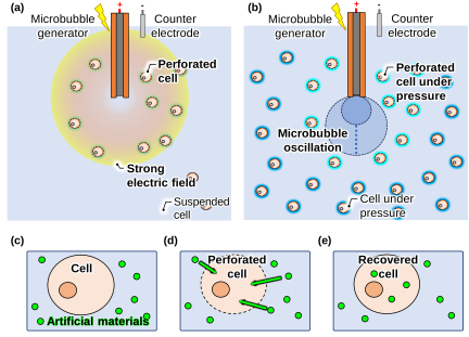
Fig. 3. Illustration of EMP and delivery of artificial materials into a cell. (a) Electrical stimulation for perforating cell membranes during EMP. (b) Mechanical stimulation for monomerizing the cytoskeleton of cells during EMP. (c–e) Delivery of artificial particles into a cell by EMP. (c) Before EMP, (d) during EMP, and (e) after EMP.
EMP induces membrane perforation by combining electrical and mechanical stimuli. EMP generates pressure fluctuations through changes in the microbubble volume. This pressure fluctuation transiently depolymerizes the cytoskeleton that supports the cell membrane. As a result, the cell membrane of EMP-treated cells becomes more capable of maintaining pores compared to untreated cells, thereby facilitating material delivery into the cells (Figs. 3(a) and (b)) 13.
When EMP is applied to cells, (1) pores are formed on the cell membrane; (2) external materials are taken up into the cytoplasm; and (3) the cell recovers its membrane, which in turn closes the pores and retains the materials inside the cell. This method can be used not only for genetic materials such as plasmids, but also for artificial microparticles (Figs. 3(c)–(e)). By introducing artificial materials into cells using this approach, it is possible to evaluate their compatibility at the cellular level.
2.2. Instrumentation for EMP
EMP was performed using a bubble injector, as shown in Fig. 4. The injector in this study had a core–shell structure, consisting of a tungsten electrode of 400-μm diameter and a glass capillary to insulate the electrode. When the injector is immersed in an aqueous solution, such as a cell suspension, and a high-voltage pulse is applied, a strong electric field is generated near the tip of the electrode. Simultaneously, the intense electric field at the electrode tip induces the formation of a microbubble. This microbubble changes its volume in response to the applied electric pulses and intervals (Fig. 3(b)). This oscillation of microbubble volume generates pressure fluctuations in the liquid, thereby altering the state of the cytoskeleton 14.
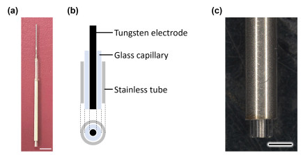
Fig. 4. A bubble injector. (a) An image of a bubble injector. Tungsten electrode and glass capillary worked as a microbubble generator. Stainless tube worked as a counter electrode. Scale bar: 5 mm. (b) Illustration of a bubble injector. (c) A microscopic image of the tip of a bubble injector. Scale bar: 1 mm.
Figure 5 shows the entire arrangement of the bubble injection system. The cathode of the power supply was connected to the bubble injector and the anode was connected to a stainless-steel counter electrode attached to the outer shell of the bubble injector. The voltage and current applied to the bubble injector were monitored by using an oscilloscope. A high-voltage power supply (Hyfrecator 2000, CONMED, Largo, FL, USA) was used as the pulse source to control the electrical power during EMP. The number of electrical pulses was controlled using a microcontroller (Uno-R3, Arduino, Monza, Italy).
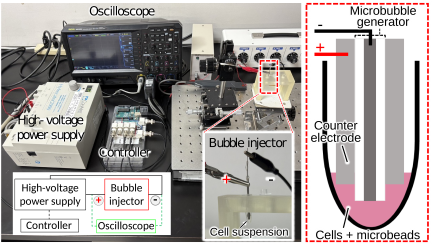
Fig. 5. Setup for EMP. EMP was produced by a bubble injector, consisting of a microbubble generator, a counter electrode, and a power supply. The controller, connected to the power supply, controlled the number of electrical pulses.
2.3. Material Delivery to Adherent Cells
To investigate the delivery of artificial materials by EMP, HeLa cells, a type of adherent cells, were used as the target of material delivery. A total of \(1.5 \times 10^6\) HeLa cells were suspended in 4.5 μL of culture medium (OptiMEM, Thermo Fisher Scientific, Waltham, MA, USA) to prepare a cell suspension. Immediately before EMP treatment, the suspension was mixed by tapping to ensure that both electrical and mechanical stimuli reached as many cells as possible.
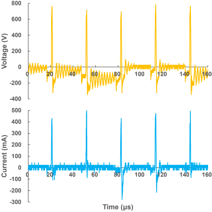
Fig. 6. Profiles of voltage and current during EMP with applied electric power of 12 W.
During EMP treatment, the pulse generator was operated in bipolar mode, and an electrical power of 12 W was applied. Each pulse cycle consisted of a 1.1-μs pulse followed by a 30-μs interval. One set comprised 1,000 pulse cycles, and 20 sets were applied in a single EMP treatment, corresponding to 20,000 electrical pulses delivered to the cell suspension. A 2-s pause was introduced between sets. These EMP parameters were selected based on our previous studies 13,14, in which energy input of 1.1-μs pulse width with 30-μs intervals generated microbubbles on the order of 100 μm and effectively induced membrane permeabilization in adherent cells without markedly reducing cell viability.
During EMP treatment, the voltage and current waveforms were continuously monitored using an oscilloscope. Representative voltage and current waveforms obtained during the normal EMP operation are shown in Fig. 6.
Fluorescent polystyrene microbeads (2, 3, 6, and 10 μm in diameter; Fluoresbrite, Polysciences, Warrington, PA, USA) and NPCs 7 were used as artificial materials for EMP-mediated delivery. After EMP treatment, the treated cell suspension was diluted into a culture medium, OptiMEM, containing 10% (v/v) fetal bovine serum (Biosera, Cholet, France), at a ratio of \(1:99\) (v/v) and incubated at 37°C in a 5% CO\(_2\) incubator.
The material-loaded cells were stained for evaluation using fluorescence microscopy. Propidium iodide (PI) (Dojindo, Kumamoto, Japan) was used to stain dead cells. Hoechst 33342 (Dojindo) was used to stain cell nuclei, and 5-chloromethylfluorescein diacetate (CMFDA) (Thermo Fisher Scientific, Waltham, MA, USA) was used for cytoplasmic staining.
3. Results and Discussion
3.1. Bubble Formation During EMP
The generation of microbubbles responsible for the mechanical stimulus during EMP is shown at the macroscopic level in Fig. 7. Upon applying electrical pulses during EMP, microbubble formation was accompanied by light emission. The generated bubbles were ejected downward from the electrode surface to the liquid phase. Subsequently, the bubbles rose toward the liquid surface owing to buoyancy. Microscopic images of bubble formation were obtained using a high-speed camera (HPV-X2, Shimadzu, Kyoto, Japan) (Fig. 8). The application of multiple electrical pulses generates small microbubbles on the electrode surface. The continuous application of pulses increased the bubble volume, leading to the formation of an integrated microbubble. This microbubble subsequently collapsed and was ejected from the electrode surface while generating a microjet. This cycle of microbubble formation and ejection occurred multiple times within one set of applied pulses (1,000 pulses).
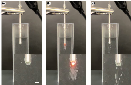
Fig. 7. Bubble formation and irradiation induced by electric pulses. (a) Before electric pulses were applied. (b) Microbubble generation and irradiation from the microbubbles by applying electric pulses. (c) Microbubble discharge. Scale bar: 100 μm.
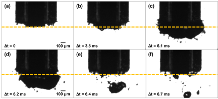
Fig. 8. Bubble formation on the electrode surface of a bubble injector by applying electric pulses. (a) The beginning of applying electric pulses. (b) Formation of small bubbles. (c) Integration of small bubbles. (d) Bubble shrinkage. (e) Bubble collapse. (f) Bubble discharge.
3.2. Results of Bead Injection
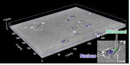
Fig. 9. A bead-injected cell observed under a confocal microscope. Green: green fluorescent microbeads; blue: nuclei of cells stained by Hoechst 33342. A magnified image in \(xy\) plane of the cell enclosed by a dashed line is shown on the lower right corner.
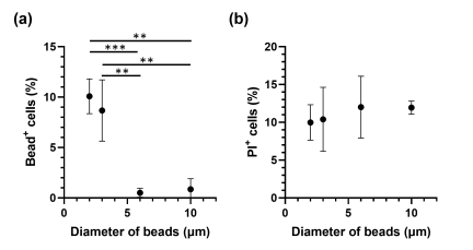
Fig. 10. Injection of fluorescent beads into HeLa cells. (a) The rate of successful bead delivery into HeLa cells (bead\(^{+}\) cells) at various bead diameters. (b) The rate of cell death at 24 h after delivery of beads into HeLa cells at various bead diameters. \(^{**}\): \(p< 0.01\); \(^{***}\): \(p<0.001\).
Fluorescent polystyrene beads were delivered to HeLa cells using EMP. After bead delivery, cells were cultured for 24 h. Before observation under a fluorescence microscope, the culture medium containing free polystyrene beads was aspirated, and the cells were washed with PBS to remove beads that were not taken up by the cells. Washed HeLa cells were observed using fluorescence microscopy. Cell nuclei were stained with Hoechst 33342 to identify HeLa cells using blue fluorescence. Bright-field images were used to determine cell boundaries, and cells containing green fluorescent polystyrene beads within the cell contour were counted. To confirm that the beads were internalized, rather than attached to the cell surface, confocal microscopy was used to obtain cross-sectional images along the \(z\)-axis (Fig. 9).
The correlation between bead diameter and delivery efficiency is shown in Fig. 10. Delivery efficiency was calculated as the number of bead-containing cells divided by the total number of cells. Bead-containing cells were defined as those in which both polystyrene beads with green fluorescence and nuclei with blue fluorescence were observed within the cell boundary. The total number of cells was determined based on the number of nuclei.
Beads with diameters up to 3 μm exhibited high delivery efficiency into HeLa cells, whereas beads with diameters exceeding 6 μm showed a significant decrease in delivery efficiency. The diameter of non-adherent HeLa cells during EMP treatment was estimated to be 17 μm 16,17. Assuming HeLa cells were spherical, a membrane opening sufficient for a 3-μm particle would correspond to 0.78% of the cell surface area, whereas that for a 6-μm particle would correspond to 3.1% of the cell surface area. These results suggest that HeLa cells readily take up objects approximately up to 0.8% of their surface area. The selected EMP conditions may have supported the uptake of micrometer-scale particles through the combined effects of electrical and mechanical stimulation. Electrical stimulation can increase membrane permeability, whereas mechanical stimulation generated by oscillatory changes in microbubble volume may transiently perturb the cytoskeleton and facilitate membrane deformation. Such transient membrane deformation would be beneficial for the entry of particles to approximately 3 μm in diameter. In contrast, particles exceeding 6 μm would require larger membrane openings and greater membrane deformation, which may explain the marked decrease in delivery efficiency.
Cell viability was also evaluated 24 h after bead delivery (Figs. 10(a) and (b)). Viability was calculated based on the number of dead cells relative to the total number of cells; dead cells were defined as red fluorescent (PI\(^{+}\)) cells; 24-h cell viability was comparable regardless of bead size. This suggests that the polystyrene microparticles did not exhibit cytotoxicity toward HeLa cells within the range of tested bead sizes.
3.3. Results of Silver-Coated DNA-Functionalized Gold Nanoparticle Crystals (Au@Ag NPC)
Au@Ag NPCs were delivered into HeLa cells using EMP under the same conditions as those used for polystyrene bead delivery. Based on the bead delivery results, NPC diameter was adjusted to approximately 1–1.5 μm to facilitate uptake. Aggregated Au@Ag NPCs exceeding 6 μm in diameter were excluded from counting, as they were expected to exhibit low delivery efficiency.
After NPC delivery, the cells were cultured for 1–4 d. Prior to fluorescence observation, the culture medium containing free Au@Ag NPCs was aspirated, and the cells were washed with PBS to remove Au@Ag NPCs that were not taken up by the cells. The washed cells were observed using fluorescence microscopy (Fig. 11). The cytoplasm was stained with CMFDA to show green fluorescence, and cells containing non-fluorescent Au@Ag NPCs within the cytoplasm were counted. Bright-field images of the same samples were used to confirm the presence of NPCs in the cells.
After 24 h of culture, NPC-containing cells accounted for 0.50% of the total cell population. Of these, 61% was attributed to EMP-mediated delivery, whereas the remaining 39% was considered to result from spontaneous cellular uptake, which occurred to cells with no EMP treatment.
The lower delivery efficiency of Au@Ag NPCs compared to that of polystyrene beads may be attributed to differences in mechanical stiffness. Polystyrene beads have a Young’s modulus in the GPa range 18, whereas Au@Ag NPCs are crosslinked by DNA strands between nanoparticles and are therefore expected to have lower mechanical stiffness 19,20. During microbubble collapse, rigid polystyrene beads may transfer momentum more effectively to the cell membrane, whereas softer Au@Ag NPCs may undergo deformation. Such a deformation could dissipate a fraction of the impact energy and reduce the force transmitted to the membrane, resulting in a lower delivery efficiency. The optimization of the EMP parameters for Au@Ag NPC delivery requires further investigation. Nevertheless, because NPC delivery via EMP was confirmed despite the difference in stiffness, the cytotoxicity of Au@Ag NPCs was evaluated.
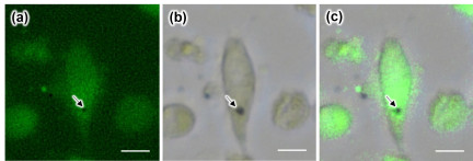
Fig. 11. An Au@Ag NPC-injected cell observed under a microscope. (a) An Au@Ag NPC in a cell in which cytoplasm emitted green fluorescence of CMFDA. (b) An Au@Ag NPC in a cell observed in bright field. (c) A merged image of (a) and (b). Arrows indicate an Au@Ag NPC in a cell. Scale bar: 10 μm.
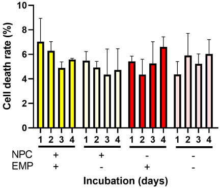
Fig. 12. Number of cell deaths influenced by injection of Au@Ag NPCs. The number of dead cells in each Au@Ag NPC/EMP condition was counted at 1–4 d of incubation. Yellow: Au@Ag NPC\(^{+}\)/EMP\(^{+}\); white: Au@Ag NPC\(^{+}\)/EMP\(^{-}\); red: Au@Ag NPC-/EMP\(^{+}\); pink: Au@Ag NPC\(^{-}\)/EMP\(^{-}\). NPC: Au@Ag NPC-treated condition; EMP: EMP with 12 W pulses applied.
The number of dead cells after Au@Ag NPC delivery is shown in Fig. 12. Because Au@Ag NPCs remained in the culture environment after treatment, this experiment enabled evaluation of potential acute toxicity of Au@Ag NPCs. HeLa cells were divided into four groups based on the presence or absence of Au@Ag NPC and EMP. No significant differences were observed in the number of dead cells among the four groups. In addition, no substantial change in cell death was observed over 1–4 d in any group. These results indicate that the presence of Au@Ag NPCs in the culture environment did not induce acute cytotoxicity in HeLa cells. It has also been demonstrated that EMP treatment stimulate cells within a recoverable range. Thus, EMP-mediated Au@Ag NPC delivery, which combines a low-cytotoxicity material with a mild delivery method, resulted in minimal acute cytotoxicity.
4. Conclusions
Microbubbles can be generated simultaneously with an electric field by using a bubble injector in combination with high-voltage pulses. EMP is based on this phenomenon, and artificial materials can be introduced into cells by applying electrical stimulation from high-voltage pulses and mechanical stimulation derived from volume changes in microbubbles. In this study, polystyrene beads and Au@Ag NPCs were successfully introduced into HeLa cells. Polystyrene beads with diameters of up to 3 μm exhibited a delivery efficiency of approximately 10%, and HeLa cells showed viability of approximately 90% after bead delivery, regardless of bead diameter.
Au@Ag NPCs exhibited a delivery efficiency of 0.5%, and no significant change in cell viability was observed over 1–4 d, regardless of Au@Ag NPC delivery or EMP treatment. These results indicate that Au@Ag NPCs in the culture environment did not exert acute cytotoxic effects on HeLa cells. In addition, no significant increase in the number of dead cells was observed when HeLa cells were subjected to EMP treatment alone, suggesting that cellular damage caused by EMP treatment was limited. Together with the successful delivery of microparticles and Au@Ag NPCs larger than 1 μm in diameter, these results demonstrate that EMP enables the delivery of μm-scale artificial materials into cells without causing excessive cellular damage.
The delivery of Au@Ag NPCs by EMP suggests that not only submicrometer artificial sensors, such as AuNPs, but also larger artificial sensors, such as Au@Ag NPCs, which generate stronger signals, can be delivered to cells. Cells incorporating such sensors are expected to function as implantable sensors, as they can be used not only to measure intracellular information, but also to reflect the dynamics of surrounding cells and tissues when introduced into living organisms.
Acknowledgments
This research project was funded by JSPS KAKENHI (JP22H00198, 25H00410, 25H00832), JST CREST (JPMJCR19S6) and JST Moonshot R&D (JPMJMS2217-3-1). The authors thank Yuki Okugawa (the Center for Advanced Technical and Educational Supports, Faculty of Agriculture, Kyushu University) for her technical assistance on analysis by confocal microscopy.
- [1] S. Modi, Swetha M. G., D. Goswami, G. D. Gupta, S. Mayor, and Y. Krishnan, “A DNA nanomachine that maps spatial and temporal pH changes inside living cells,” Nature Nanotech., Vol.4, pp. 325-330, 2009. https://doi.org/10.1038/nnano.2009.83
- [2] C. E. Talley, J. Jusinski, C. W. Hollars, S. M. Lane, and T. Huser, “Intracellular pH Sensors Based on Surface-Enhanced Raman Scattering,” Anal. Chem., Vol.76, No.23, pp. 7064-7068, 2004. https://doi.org/10.1021/ac049093j
- [3] D. F. Williams, “On the nature of biomaterials,” Biomaterials, Vol.30, No.30, pp. 5897-5909, 2009. https://doi.org/10.1016/j.biomaterials.2009.07.027
- [4] L. Dykman and N. Khlebtsov, “Gold nanoparticles in biomedical applications: Recent advances and perspectives,” Chem. Soc. Rev., Vol.41, No.6, pp. 2256-2282, 2012. https://doi.org/10.1039/c1cs15166e
- [5] M. B. Ross, C. A. Mirkin, and G. C. Schatz, “Optical Properties of One-, Two-, and Three-Dimensional Arrays of Plasmonic Nanostructures,” J. Phys. Chem. C, Vol.120, No.6, pp. 816-830, 2016. https://doi.org/10.1021/acs.jpcc.5b10800
- [6] T. Xu and Z. Geng, “Strategies to improve performances of LSPR biosensing: Structure, materials, and interface modification,” Biosensors and Bioelectronics, Vol.174, Article No.112850, 2021. https://doi.org/10.1016/j.bios.2020.112850
- [7] M. Yokomori, H. Suzuki, A. Nakamura, S. S. Sugano, and M. Tagawa, “DNA-functionalized colloidal crystals for macromolecular encapsulation,” Soft Matter, Vol.18, No.36, pp. 6954-6964, 2022. https://doi.org/10.1039/d2sm00949h
- [8] T. K. Kim and J. H. Eberwine, “Mammalian cell transfection: the present and the future,” Anal. Bioanal. Chem., Vol.397, No.8, pp. 3173-3178, 2010. https://doi.org/10.1007/s00216-010-3821-6
- [9] M. P. Stewart, A. Sharei, X. Ding, G. Sahay, R. Langer, and K. F. Jensen, “In vitro and ex vivo strategies for intracellular delivery,” Nature, Vol.538, pp. 183-192, 2016. https://doi.org/10.1038/nature19764
- [10] M. P. Stewart, R. Langer, and K. F. Jensen, “Intracellular Delivery by Membrane Disruption: Mechanisms, Strategies, and Concepts,” Chem. Rev., Vol.118, No.16, pp. 7409-7531, 2018. https://doi.org/10.1021/acs.chemrev.7b00678
- [11] P. E. Boukany, A. Morss, W.-c. Liao, B. Henslee, H. Jung, X. Zhang, B. Yu, X. Wang, Y. Wu, L. Li, K. Gao, X. Hu, X. Zhao, O. Hemminger, W. Lu, G. P. Lafyatis, and L. J. Lee, “Nanochannel electroporation delivers precise amounts of biomolecules into living cells,” Nature Nanotech., Vol.6, pp. 747-554, 2011. https://doi.org/10.1038/nnano.2011.164
- [12] A. Sharei, J. Zoldan, A. Adamo, W. Y. Sim, N. Cho, E. Jackson, S. Mao, S. Schneider, M.-J. Han, A. Lytton-Jean, P. A. Basto, S. Jhunjhunwala, J. Lee, D. A. Heller, J. W. Kang, G. C. Hartoularos, K.-S. Kim, D. G. Anderson, R. Langer, and K. F. Jensen, “A vector-free microfluidic platform for intracellular delivery,” PNAS, Vol.110, No.6, pp. 2082-2087, 2013. https://doi.org/10.1073/pnas.1218705110
- [13] W. Huang, S. Sakuma, N. Tottori, S. S. Sugano, and Y. Yamanishi, “Viscosity-aided electromechanical poration of cells for transfecting molecules,” Lab Chip, Vol.22, No.22, pp. 4276-4291, 2022. https://doi.org/10.1039/d2lc00628f
- [14] W. Huang, Y. Ma, N. Tottori, and Y. Yamanishi, “Enhancing suspended cell transfection by inducing localized distribution of the membrane actin cortex before exposure to electromechanical stimulation,” Biotechnol. Lett., Vol.45, pp. 1417-1430, 2023. https://doi.org/10.1007/s10529-023-03382-y
- [15] T. Ikeuchi, L. Zhang, S. Kozawa, S. Kojima, H. Niinomi, M. Kuwahara, K. Hiramatsu, and M. Tagawa, “Self-assembly of High-quality Single Crystals of DNA-functionalized Gold Nanoparticle Superlattices for In-vivo SERS Detection,” Proc. 15th Int. Conf. Metamaterials, Photonic Crystals and Plasmonics (META 2025), pp. 1840-1841, 2025.
- [16] A. B. Borle, “Kinetic Analyses of Calcium Movements in HeLa Cell Cultures: I. Calcium influx,” J. Gen. Physiol., Vol.53, No.1, pp. 43-56, 1969. https://doi.org/10.1085/jgp.53.1.43
- [17] M. Uzhytchak, B. Smolková, A, Frtús, A. Stupakov, M. Lunova, F. Scollo, M. Hof, P. Jurkiewicz, G. J. Sullivan, A. Dejneka, and O. Lunov, “Sensitivity of endogenous autofluorescence in HeLa cells to the application of external magnetic fields,” Sci. Rep., Vol.13, Article No.10818, 2023. https://doi.org/10.1038/s41598-023-38015-x
- [18] D. Guo, J. Li, G. Xie, Y. Wang, and J. Luo, “Elastic Properties of Polystyrene Nanospheres Evaluated with Atomic Force Microscopy: Size Effect and Error Analysis,” Langmuir, Vol.30, No.24, pp. 7206-7212, 2014. https://doi.org/10.1021/la501485e
- [19] J. Bush, C.-H. Hu, and R. Veneziano, “Mechanical Properties of DNA Hydrogels: Towards Highly Programmable Biomaterials,” Applied Sciences, Vol.11, No.4, Article No.1885, 2021. https://doi.org/10.3390/app11041885
- [20] Y. Wang, Y. Zhang, Q. Zhang, X. Li, Q. Yan, and Y. Zhu, “Mechanical properties modulation and biological applications of DNA hydrogels,” Advanced Sensor and Energy Materials, Vol.3, No.3, Article No.100113, 2024. https://doi.org/10.1016/j.asems.2024.100113
This article is published under a Creative Commons Attribution-NoDerivatives 4.0 Internationa License.