
Paper:
Production of Single-Cell-Laden Alginate Microgels Through Seamless Separation and In-Flow Gelation Using Deterministic Lateral Displacement
Hiroki Fukunaga, Naotomo Tottori†
 , Shinya Sakuma
, and Yoko Yamanishi
Department of Mechanical Engineering, Faculty of Engineering, Kyushu University
744 Motooka, Nishi-ku, Fukuoka, Fukuoka 819-0395, Japan
†Corresponding author

Cell-laden alginate microgels have been extensively studied for three-dimensional cell culture, tissue engineering, and cell-based screening because they provide microscale compartments that support cell culture in a defined microenvironment. In particular, single-cell-laden alginate microgels have attracted increasing interest as platforms for studying cellular heterogeneity. These applications require efficient and scalable production of single-cell-laden alginate microgels. Droplet microfluidics has therefore emerged as a promising method for producing alginate microgels with high uniformity and throughput. However, conventional passive encapsulation is governed by Poisson statistics, resulting in a large fraction of empty alginate microgels and only a limited fraction containing a single cell. Consequently, additional pre- or post-processing steps, such as cell pre-coating or off-chip purification, are often required, complicating the workflow and reducing process continuity. In this study, we propose a microfluidic device that integrates continuous separation of cell-encapsulated droplets with ionic gelation in a deterministic lateral displacement (DLD) micropillar array. This integrated design allows for the seamless production of single-cell-laden alginate microgels with improved purity. As a proof of concept, we demonstrated the continuous production of single-cell-laden alginate microgels using NIH3T3-3-4 as a model cell line, with a purity of 64% and a separation efficiency of 53%. These results show the potential of DLD-based integration for the streamlined and scalable production of single-cell-laden alginate microgels for single-cell analysis and related advanced biomedical applications.
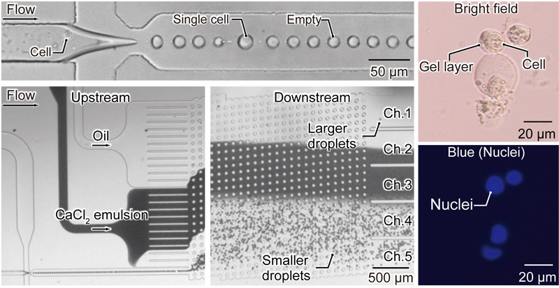
On-chip separation and in-flow gelation
- [1] M. Y. Wang, H. L. Tao, J. Li, X. Y. Chen, Y. C. Gu, R. Tang, and X. Q. Liu, “The cell-loaded alginate microspheres in cell culture and disease treatment,” Smart Mater. Med., Vol.6, Issue 3, pp. 387-405, 2025. https://doi.org/10.1016/j.smaim.2025.11.002
- [2] S.-M. Kang, J.-H. Lee, Y. S. Huh, and S. Takayama, “Alginate microencapsulation for three-dimensional in vitro cell culture,” ACS Biomater. Sci. Eng., Vol.7, Issue 7, pp. 2864-2879, 2021. https://doi.org/10.1021/acsbiomaterials.0c00457
- [3] B. Namgung, K. Ravi, P. P. Vikraman, S. Sengupta, and H. L. Jang, “Engineered cell-laden alginate microparticles for 3D culture,” Biochem. Soc. Trans., Vol.49, Issue 2, pp. 761-773, 2021. https://doi.org/10.1042/BST20200673
- [4] S. Utech, R. Prodanovic, A. S. Mao, R. Ostafe, D. J. Mooney, and D. A. Weitz, “Microfluidic generation of monodisperse, structurally homogeneous alginate microgels for cell encapsulation and 3D cell culture,” Adv. Healthc. Mater., Vol.4, Issue 11, pp. 1628-1633, 2015. https://doi.org/10.1002/adhm.201500021
- [5] X. Qin, Z. Gan, H. Liu, T. Tao, J. He, X. Li, D. Shang, X. Li, F. Xie, and J. Qin, “A pump-free strategy for the controllable generation of alginate microgels as cellular microcarriers,” ACS Biomater. Sci. Eng., Vol.10, Issue 6, pp. 3958-3967, 2024. https://doi.org/10.1021/acsbiomaterials.4c00375
- [6] C. An, W. Liu, Y. Zhang, B. Pang, H. Liu, Y. Zhang, H. Zhang, L. Zhang, H. Liao, C. Ren, and H. Wang, “Continuous microfluidic encapsulation of single mesenchymal stem cells using alginate microgels as injectable fillers for bone regeneration,” Acta Biomater., Vol.111, pp. 181-196, 2020. https://doi.org/10.1016/j.actbio.2020.05.024
- [7] H. Soleymani, M. M. Moghaddam, H. Naderi-Manesh, and R. A. Taheri, “Single-layer graphene oxide nanosheets induce proliferation and osteogenesis of single-cell hBMSCs encapsulated in alginate microgels,” Sci. Rep., Vol.14, Article No.25272, 2024. https://doi.org/10.1038/s41598-024-76957-y
- [8] V. Iansante, A. Dhawan, F. Masmoudi, C. A. Lee, R. Fernandez-Dacosta, S. Walker, E. Fitzpatrick, R. R. Mitry, and C. Filippi, “A new high throughput screening platform for cell encapsulation in alginate hydrogel shows improved hepatocyte functions by mesenchymal stromal cells co-encapsulation,” Front. Med., Vol.5, Article No.216, 2018. https://doi.org/10.3389/fmed.2018.00216
- [9] Z. Zhao, X. Feng, H. Wu, S. Chen, C. Ma, Z. Guan, L. Lei, K. Tang, X. Chen, Y. Dong, and Y. Tang, “Construction of a lung cancer 3D culture model based on alginate/gelatin micro-beads for drug evaluation,” Transl. Lung Cancer Res., Vol.13, No.10, pp. 2698-2712, 2024. https://doi.org/10.21037/tlcr-24-490
- [10] Y. Wang, Z. Shen, H. Wang, Z. Song, D. Yu, G. Li, X. Liu, and W. Liu, “Progress in research on metal ion crosslinking alginate-based gels,” Gels, Vol.11, Issue 1, Article No.16, 2025. https://doi.org/10.3390/gels11010016
- [11] M. Pires-Santos, S. Nadine, and J. F. Mano, “Unveiling the potential of single-cell encapsulation in biomedical applications: Current advances and future perspectives,” Small Sci., Vol.4, Issue 5, Article No.2300332, 2024. https://doi.org/10.1002/smsc.202300332
- [12] B. M. Tiemeijer and J. Tel, “Hydrogels for single-cell microgel production: Recent advances and applications,” Front. Bioeng. Biotechnol., Vol.10, Article No.891461, 2022. https://doi.org/10.3389/fbioe.2022.891461
- [13] R. Dubay, J. N. Urban, and E. M. Darling, “Single-cell microgels for diagnostics and therapeutics,” Adv. Funct. Mater., Vol.31, Issue 44, Article No.2009946, 2021. https://doi.org/10.1002/adfm.202009946
- [14] A. S. Mao, J.-W. Shin, S. Utech, H. Wang, O. Uzun, W. Li, M. Cooper, Y. Hu, L. Zhang, D. A. Weitz, and D. J. Mooney, “Deterministic encapsulation of single cells in thin tunable microgels for niche modelling and therapeutic delivery,” Nat. Mater., Vol.16, pp. 236-243, 2017. https://doi.org/10.1038/nmat4781
- [15] T. Kamperman, S. Henke, A. van den Berg, S. R. Shin, A. Tamayol, A. Khademhosseini, M. Karperien, and J. Leijten, “Single cell microgel based modular bioinks for uncoupled cellular micro- and macroenvironments,” Adv. Healthc. Mater., Vol.6, Issue 3, Article No.1600913, 2017. https://doi.org/10.1002/adhm.201600913
- [16] L. R. Huang, E. C. Cox, R. H. Austin, and J. C. Sturm, “Continuous particle separation through deterministic lateral displacement,” Science, Vol.304, Issue 5673, pp. 987-990, 2004. https://doi.org/10.1126/science.1094567
- [17] N. Tottori, Y. Tang, Y. Kanno, and T. Nisisako, “Continuous production and extraction of monodisperse alginate microgels via deterministic lateral displacement,” Soft Matter, Vol.22, Issue 7, pp. 1483-1493, 2026. https://doi.org/10.1039/d5sm00757g
- [18] T. Jing, R. Ramji, M. E. Warkiani, J. Han, C. T. Lim, and C. H. Chen, “Jetting microfluidics with size-sorting capability for single-cell protease detection,” Biosens. Bioelectron., Vol.66, pp. 19-23, 2015. https://doi.org/10.1016/j.bios.2014.11.001
- [19] N. Tottori, T. Hatsuzawa, and T. Nisisako, “Separation of main and satellite droplets in a deterministic lateral displacement microfluidic device,” RSC Adv., Vol.7, Issue 56, pp. 35516-35524, 2017. https://doi.org/10.1039/c7ra05852g
- [20] N. Tottori and T. Nisisako, “High-throughput production of satellite-free droplets through a parallelized microfluidic deterministic lateral displacement device,” Sens. Actuators B: Chem., Vol.260, pp. 918-926, 2018. https://doi.org/10.1016/j.snb.2018.01.112
- [21] H. Fukunaga, N. Tottori, T. Tsubouchi, T. Kumazaki, S. Sakuma, and Y. Yamanishi, “Spatially constrained encapsulation of cells in microdroplets enables continuous cell fusion within a microfluidic flow environment,” Sens. Actuators B: Chem., Vol.458, Article No.139779, 2026. https://doi.org/10.1016/j.snb.2026.139779
- [22] J. A. Davis, “Microfluidic Separation of Blood Components through Deterministic Lateral Displacement,” Doctoral thesis, Princeton University, 2008.
- [23] Y. Liu, N. Tottori, and T. Nisisako, “Microfluidic synthesis of highly spherical calcium alginate hydrogels based on external gelation using an emulsion reactant,” Sens. Actuators B: Chem., Vol.283, pp. 802-809, 2019. https://doi.org/10.1016/j.snb.2018.12.101
- [24] S. I. R. Lane, J. Butement, J. Harrington, T. Underwood, J. Shrimpton, and J. West, “Perpetual sedimentation for the continuous delivery of particulate suspensions,” Lab Chip, Vol.19, Issue 22, pp. 3771-3775, 2019. https://doi.org/10.1039/c9lc00774a
- [25] D. Holmes, G. Whyte, J. Bailey, N. Vergara-Irigaray, A. Ekpenyong, J. Guck, and T. Duke, “Separation of blood cells with differing deformability using deterministic lateral displacement,” Interface Focus, Vol.4, Issue 6, Article No.20140011, 2014. https://doi.org/10.1098/rsfs.2014.0011
- [26] S. Ye, X. Shao, Z. Yu, and W. Yu, “Effects of the particle deformability on the critical separation diameter in the deterministic lateral displacement device,” J. Fluid Mech., Vol.743, pp. 60-74, 2014. https://doi.org/10.1017/jfm.2014.22
- [27] T. Krüger, D. Holmes, and P. V. Coveney, “Deformability-based red blood cell separation in deterministic lateral displacement devices—A simulation study,” Biomicrofluidics, Vol.8, Issue 5, Article No.054114, 2014. https://doi.org/10.1063/1.4897913
- [28] N. Tottori and T. Nisisako, “Tunable deterministic lateral displacement of particles flowing through thermo-responsive hydrogel micropillar arrays,” Sci. Rep., Vol.13, Article No.4994, 2023. https://doi.org/10.1038/s41598-023-32233-z
- [29] Z. Jiang, Y. Kanno, and T. Nisisako, “Tunable cell separation using a thermo-responsive deterministic lateral displacement device,” Lab Chip, Vol.25, Issue 24, pp. 6454-6464, 2025. https://doi.org/10.1039/d5lc00783f
- [30] M. Aghaamoo, A. Aghilinejad, X. Chen, and J. Xu, “On the design of deterministic dielectrophoresis for continuous separation of circulating tumor cells from peripheral blood cells,” Electrophoresis, Vol.40, Issue 10, pp. 1486-1493, 2019. https://doi.org/10.1002/elps.201800459
- [31] J. P. Beech, P. Jönsson, and J. O. Tegenfeldt, “Tipping the balance of deterministic lateral displacement devices using dielectrophoresis,” Lab Chip, Vol.9, Issue 18, pp. 2698-2706, 2009. https://doi.org/10.1039/b823275j
- [32] T. S. H. Tran, B. D. Ho, J. P. Beech, and J. O. Tegenfeldt, “Open channel deterministic lateral displacement for particle and cell sorting,” Lab Chip, Vol.17, Issue 21, pp. 3592-3600, 2017. https://doi.org/10.1039/c7lc00707h
- [33] J. P. Beech, K. Keim, B. D. Ho, C. Guiducci, and J. O. Tegenfeldt, “Active posts in deterministic lateral displacement devices,” Adv. Mater. Technol., Vol.4, Issue 9, Article No.1900339, 2019. https://doi.org/10.1002/admt.201900339
- [34] V. Calero, P. Garcia-Sanchez, C. Honrado, A. Ramos, and H. Morgan, “AC electrokinetic biased deterministic lateral displacement for tunable particle separation,” Lab Chip, Vol.19, Issue 8, pp. 1386-1396, 2019. https://doi.org/10.1039/c8lc01416g
- [35] B. M. Dincau, A. Aghilinejad, X. Chen, S. Y. Moon, and J.-H. Kim, “Vortex-free high-Reynolds deterministic lateral displacement (DLD) via airfoil pillars,” Microfluid. Nanofluidics, Vol.22, Article No.137, 2018. https://doi.org/10.1007/s10404-018-2160-3
- [36] B. M. Dincau, A. Aghilinejad, T. Hammersley, X. Chen, and J.-H. Kim, “Deterministic lateral displacement (DLD) in the high Reynolds number regime: High-throughput and dynamic separation characteristics,” Microfluid. Nanofluidics, Vol.22, Article No.59, 2018. https://doi.org/10.1007/s10404-018-2078-9
- [37] H. Fukunaga, N. Tottori, S. Sakuma, T. Hayakawa, and Y. Yamanishi, “Tunable particle separation using vibration induced flow in deterministic lateral displacement micropillar arrays,” 2025 23rd Int. Conf. Solid-State Sensors, Actuators Microsystems, pp. 227-230, 2025. https://doi.org/10.1109/transducers61432.2025.11111463
- [38] H. Huang and X. He, “Interfacial tension based on-chip extraction of microparticles confined in microfluidic stokes flows,” Appl. Phys. Lett., Vol.105, Issue 14, Article No.143704, 2014. https://doi.org/10.1063/1.4898040
- [39] Y. Deng, N. Zhang, L. Zhao, X. Yu, X. Ji, W. Liu, S. Guo, K. Liu, and X.-Z. Zhao, “Rapid purification of cell encapsulated hydrogel beads from oil phase to aqueous phase in a microfluidic device,” Lab Chip, Vol.11, Issue 23, pp. 4117-4121, 2011. https://doi.org/10.1039/c1lc20494g
- [40] T. Hu and A. C. Y. Lo, “Collagen–alginate composite hydrogel: Application in tissue engineering and biomedical sciences,” Polymers, Vol.13, Issue 11, Article No.1852, 2021. https://doi.org/10.3390/polym13111852
- [41] K. Y. Lee and D. J. Mooney, “Alginate: Properties and biomedical applications,” Prog. Polym. Sci., Vol.37, Issue 1, pp. 106-126, 2012. https://doi.org/10.1016/j.progpolymsci.2011.06.003
- [42] J. A. Rowley and D. J. Mooney, “Alginate type and RGD density control myoblast phenotype,” J. Biomed. Mater. Res., Vol.60, Issue 2, pp. 217-223, 2002. https://doi.org/10.1002/jbm.1287
- [43] B. Sarker, R. Singh, R. Silva, J. A. Roether, J. Kaschta, R. Detsch, D. W. Schubert, I. Cicha, and A. R. Boccaccini, “Evaluation of Fibroblasts Adhesion and Proliferation on Alginate-Gelatin Crosslinked Hydrogel,” PLoS One, Vol.9, No.9, Article No.e107952, 2014. https://doi.org/10.1371/journal.pone.0107952
- [44] W. Bouhlel, J. Kui, J. Bibette, and N. Bremond, “Encapsulation of cells in a collagen matrix surrounded by an alginate hydrogel shell for 3D cell culture,” ACS Biomater. Sci. Eng., Vol.8, Issue 6, pp. 2700-2708, 2022. https://doi.org/10.1021/acsbiomaterials.1c01486
- [45] W. Zhang, S. Zhao, W. Rao, J. Snyder, J. K. Choi, J. Wang, I. A. Khan, N. B. Saleh, P. J. Mohler, J. Yu, T. J. Hund, C. Tang, and X. He, “A novel core–shell microcapsule for encapsulation and 3D culture of embryonic stem cells,” J. Mater. Chem. B, Vol.1, Issue 7, pp. 1002-1009, 2013. https://doi.org/10.1039/c2tb00058j
- [46] H. Zhang, E. Tumarkin, R. Peerani, Z. Nie, R. M. A. Sullan, G. C. Walker, and E. Kumacheva, “Microfluidic production of biopolymer microcapsules with controlled morphology,” J. Am. Chem. Soc., Vol.128, Issue 37, pp. 12205-12210, 2006. https://doi.org/10.1021/ja0635682
- [47] P. Agarwal, J. K. Choi, H. Huang, S. Zhao, J. Dumbleton, J. Li, and X. He, “A biomimetic core–shell platform for miniaturized 3D cell and tissue engineering,” Part. Part. Syst. Charact., Vol.32, Issue 8, pp. 809-816, 2015. https://doi.org/10.1002/ppsc.201500025
- [48] H. Aoki, Y. Masahiro, M. Shimizu, Y. Hongoh, M. Ohkuma, and Y. Yamagata, “Agarose gel microcapsules enable easy-to-prepare, picolitre-scale, single-cell genomics, yielding high-coverage genome sequences,” Sci. Rep., Vol.12, Article No.17014, 2022. https://doi.org/10.1038/s41598-022-20923-z
- [49] M. Chau, M. Abolhasani, H. Thérien-Aubin, Y. Li, Y. Wang, D. Velasco, E. Tumarkin, A. Ramachandran, and E. Kumacheva, “Microfluidic generation of composite biopolymer microgels with tunable compositions and mechanical properties,” Biomacromolecules, Vol.15, Issue 7, pp. 2419-2425, 2014. https://doi.org/10.1021/bm5002813
This article is published under a Creative Commons Attribution-NoDerivatives 4.0 Internationa License.