
Paper:
Production of Single-Cell-Laden Alginate Microgels Through Seamless Separation and In-Flow Gelation Using Deterministic Lateral Displacement
Hiroki Fukunaga, Naotomo Tottori†
 , Shinya Sakuma
, and Yoko Yamanishi
Department of Mechanical Engineering, Faculty of Engineering, Kyushu University
744 Motooka, Nishi-ku, Fukuoka, Fukuoka 819-0395, Japan
†Corresponding author

Cell-laden alginate microgels have been extensively studied for three-dimensional cell culture, tissue engineering, and cell-based screening because they provide microscale compartments that support cell culture in a defined microenvironment. In particular, single-cell-laden alginate microgels have attracted increasing interest as platforms for studying cellular heterogeneity. These applications require efficient and scalable production of single-cell-laden alginate microgels. Droplet microfluidics has therefore emerged as a promising method for producing alginate microgels with high uniformity and throughput. However, conventional passive encapsulation is governed by Poisson statistics, resulting in a large fraction of empty alginate microgels and only a limited fraction containing a single cell. Consequently, additional pre- or post-processing steps, such as cell pre-coating or off-chip purification, are often required, complicating the workflow and reducing process continuity. In this study, we propose a microfluidic device that integrates continuous separation of cell-encapsulated droplets with ionic gelation in a deterministic lateral displacement (DLD) micropillar array. This integrated design allows for the seamless production of single-cell-laden alginate microgels with improved purity. As a proof of concept, we demonstrated the continuous production of single-cell-laden alginate microgels using NIH3T3-3-4 as a model cell line, with a purity of 64% and a separation efficiency of 53%. These results show the potential of DLD-based integration for the streamlined and scalable production of single-cell-laden alginate microgels for single-cell analysis and related advanced biomedical applications.
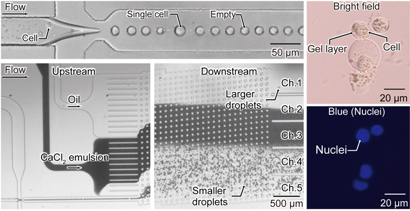
On-chip separation and in-flow gelation
1. Introduction
Cell-laden alginate microgels have attracted considerable attention as microscale three-dimensional (3D) culture units 1,2,3,4 because alginate is biocompatible and highly permeable to nutrients and metabolites, providing a suitable microenvironment for 3D cell culture. Because of these properties, alginate hydrogels have been extensively studied for 3D cell culture 4,5, tissue engineering 6,7, and cell-based screening applications 8,9. Conventionally, alginate hydrogels are produced by first generating droplets of an alginate precursor solution, which are subsequently solidified through ionic crosslinking, most commonly using calcium ions (Ca\(^{2+}\)) 4,10.
Droplet microfluidics is particularly attractive for producing monodisperse alginate microgels and has therefore been widely used for this purpose 3,4. Recently, single-cell-laden alginate microgels have become increasingly important as microscale compartments for studying cellular heterogeneity and constructing well-defined cellular microenvironments 11,12,13. However, in conventional passive droplet generation, the number of cells encapsulated in each droplet is largely governed by Poisson statistics, resulting in a substantial fraction of empty alginate microgels and a limited fraction of single-cell-laden alginate microgels 11. Therefore, additional pre- or post-processing steps are often required, such as cell precoating strategies to enrich cell-laden alginate microgels 14 or off-chip purification processes 15. These additional operations complicate the workflow and reduce process continuity.
Deterministic lateral displacement (DLD) is a passive microfluidic technique that uses micropillar arrays to continuously separate suspended particles based on their physical properties 16. A recent study demonstrated that DLD can be combined with size-based purification and in-flow ionic gelation of alginate precursor droplets by guiding them through a Ca\(^{2+}\)-containing water-in-oil (W/O) emulsion (CaCl\(_2\) emulsion) and oil-to-water transfer 17. Although this method allows for continuous on-chip gelation and extraction of alginate microgels, it does not address the selective enrichment of single-cell-laden alginate microgels, which is fundamentally limited by Poisson statistics during droplet generation. To the best of our knowledge, no previous studies have integrated the selective separation of single-cell-encapsulated droplets (single-cell droplets) from droplets without cells (empty droplets) with subsequent gelation in a continuous microfluidic workflow.
In this study, we propose a microfluidic device that integrates size-based separation of cell-encapsulated droplets from empty droplets with in-flow ionic gelation within a DLD micropillar array. By selectively directing single-cell droplets along deterministic trajectories while excluding empty droplets, the proposed device allows for the continuous production of single-cell-laden alginate microgels with improved purity in a seamless microfluidic workflow. This method provides a foundation for the integration of single-cell-laden alginate microgel production with downstream analysis, screening, and other advanced biomedical applications. Moreover, by allowing for controlled handling of cell-associated artificial microenvironments at the single-cell level, the present platform contributes to the development of systems capable of stably integrating functional materials or sensing components with living cells and tissues.
2. Materials and Methods
2.1. Design of a Microfluidic Device
The designed microfluidic device consisted of two functional modules (Fig. 1(a)): (i) a microfluidic droplet generator (MFDG) that produces droplets that encapsulate cells using a cross-junction geometry (Fig. 1(b)) and (ii) a DLD module that uses a micropillar array for size-based droplet separation 17,18,19,20,21 (Fig. 1(c)). The MFDG was configured to generate W/O droplets, with 50-μm-wide inlet channels and a 75-μm-wide downstream outlet channel. A 30-μm-wide constricted region was introduced downstream of the junction to stabilize droplet breakup. Each microchannel maintained a consistent height of 25 μm. The DLD array geometry was selected with the aim of separating larger single-cell droplets from smaller empty droplets. Droplets larger than the critical diameter (\(D_\mathrm{c})\) were laterally displaced toward the left sidewall, whereas smaller droplets remained near the channel centerline. \(D_\mathrm{c}\) was estimated using the conventional DLD design relation 22 as follows:
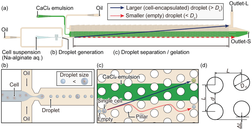
Fig. 1. Droplet microfluidic device for producing single-cell-laden alginate microgels through seamless separation and in-flow ionic gelation using DLD micropillar arrays. (a) Schematic overview of the microfluidic device, (b) generation of droplets encapsulating single cells, and (c) separation of empty droplets and ionic gelation of precursor droplets containing cells. (d) Geometric parameters of the unit cell within the DLD micropillar array: pillar diameter (\(D_\mathrm{p}\)) \(=45\) μm, array periodicity (\(L\)) \(=90\) μm, displacement ratio (\(\Delta L\)/\(L\)) \(=0.1\), and gap width (\(d=L -D_\mathrm{p}\)) \(=45\) μm.
2.2. Device Fabrication
Microfluidic channels with micropillars were fabricated from poly(dimethylsiloxane) (PDMS) using standard soft lithography techniques. A silicon wafer was first coated with a negative photoresist (SU-8 3025, Nippon Kayaku, Tokyo, Japan) by spin-coating at 2500 rpm to obtain a resist layer with approximately 25 μm thickness. After soft baking at 95°C for 30 min, the resist layer was patterned with ultraviolet exposure through a film photomask defining the microchannel layout, followed by post-exposure baking at 90°C for 15 min. The exposed wafer was developed in PM thinner (Tokyo Ohka Kogyo, Kanagawa, Japan) to generate a relief mold containing channels and micropillar features.
To facilitate demolding, the mold surface was treated with vapor-phase silanization using chlorotrimethylsilane (Tokyo Chemical Industry, Tokyo, Japan). The PDMS prepolymer and curing agent (Toray, Tokyo, Japan) were mixed in a mass ratio of \(10:1\) and degassed under vacuum for 60 min to remove trapped air. The mixture was poured onto the mold, degassed again for 60 min, and thermally cured at 90°C for 60 min. After curing, the PDMS replica was carefully peeled from the mold.
Access ports for fluidic connections were created using a biopsy punch (Kai Industries, Gifu, Japan) with diameters of approximately 1.0 mm for the cell suspension inlet, oil inlets, collection outlet, and waste outlet. The PDMS layer was irreversibly bonded to the glass substrates following air plasma activation (CUTE-MPR, FEMTO SCIENCE, Hwaseong, Korea), and the assembled device was subsequently heated at 120°C for 30 min to reinforce the bonding.
To impart hydrophobicity to the channel walls, the microchannels were flushed with fluorinated oil (HFE-7500, Fluorochem, Hadfield, UK) containing 0.5 wt% trichloro (3,3,4,4,5,5,6,6,7,7,8,8,8-tridecafluorooctyl) silane (FUJIFILM Wako Pure Chemical, Osaka, Japan) and incubated for 5 min. The treatment solution was then removed, and the device was dried under vacuum in a desiccator for 30 min, followed by a final heat treatment at 120°C for 30 min.
2.3. Cell Preparation and Culture
NIH3T3-3-4 mouse fibroblasts (RCB1862; RIKEN BRC, Japan) were cultured under standard conditions, as described in previous studies 21. To perform fluorescence imaging, the nuclei were stained with Hoechst 33342. Prior to encapsulation, cells were resuspended at \(3\times 10^6\) cells/mL in a 5 wt% aqueous sodium alginate solution (ULV-L3G, KIMICA Corporation, Tokyo, Japan) prepared in the presence of 200 mM mannitol.
2.4. Sample Preparation for Droplet Generation
W/O droplets were generated using corn oil (FUJIFILM Wako Pure Chemical, Osaka, Japan) supplemented with 2 wt% surfactant (SY-Glyster CRS-75, Sakamoto Yakuhin Kogyo, Osaka, Japan) as the continuous phase and an aqueous sodium alginate solution containing cells (NIH3T3-3-4) as the dispersed phase. For in-flow ionic gelation, a CaCl\(_2\) emulsion was prepared by dissolving calcium chloride (CaCl\(_2\); 30 wt%; FUJIFILM Wako Pure Chemical, Osaka, Japan) in ultrapure water, followed by emulsification in corn oil with 0.5 wt% surfactant (SY-Glyster CRS-75). Emulsification was performed using a homogenizer (T10 basic ULTRA-TURRAX, IKA, Germany) at a water-to-oil volume ratio of \(1:3\) 23. Corn oil supplemented with 1 wt% surfactant (SY-Glyster CRS-75) was used as an additional oil phase throughout the experiments.
2.5. Device Setup and Procedures
To operate the microfluidic platform for single-cell-laden alginate microgel production (Fig. 2), the continuous phase (corn oil), CaCl\(_2\) emulsion, and additional oil phase (corn oil) were loaded into 15-mL centrifuge tubes, while the dispersed phase (cell-containing alginate solution) was placed in a 2.0-mL microtube. These reservoirs were connected to the microfluidic device using PEEK tubing (with an inner diameter of 0.254 mm and outer diameter of 0.79 mm) and PTFE tubing (with an inner diameter of 0.5 mm and outer diameter of 1.5 mm), together with inline flow sensors (Flow Unit M, Fluigent, Le Kremlin-Bicêtre, France). Positive pressure was applied to each reservoir using pressure controllers (Flow EZ 7000 mbar; Fluigent, Le Kremlin-Bicêtre, France) to control the fluid flow. The flow rates of the continuous phase, dispersed phase, CaCl\(_2\) emulsion, and additional oil phase were continuously monitored and regulated using feedback control to maintain the prescribed operating conditions. The flow behavior within the microchannels was observed and recorded using an inverted microscope (ECLIPSE Ts2; Nikon, Tokyo, Japan) equipped with a high-speed camera (FASTCAM Nova S9; Photron, Tokyo, Japan). The collected droplets were subsequently examined with an inverted microscope (ECLIPSE Ti2U; Nikon, Tokyo, Japan) and imaged using a digital camera (WRAYCAM-VEX830; WRAYMER, Osaka, Japan).
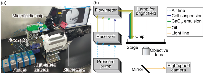
Fig. 2. System configuration for producing single-cell-laden alginate microgels. (a) Photograph of the experimental setup of the droplet microfluidic system. (b) Schematic overview of the microfluidic platform.
2.6. Measurement and Image Analysis
The viscosities of the oil and CaCl\(_2\) emulsion phases were measured at room temperature (25°C) using a rotational viscometer (ViscoQC 300-L, Anton Paar, Graz, Austria).
Quantitative image analyses were performed using MATLAB R2024b (MathWorks, MA, USA) and ImageJ (National Institutes of Health, MD, USA). MATLAB was used to extract droplet diameters from high-speed video recordings, whereas ImageJ was used for static microscopy images. The velocities of the droplet and cell were evaluated using time-resolved image data acquired at defined temporal and spatial resolutions. A stage micrometer was used for spatial calibration, and the conversion factors from pixels to microns were derived from the known dimensions of the camera pixels and magnification of the objective. The velocities were then estimated from the temporal evolution of droplet or cell positions within a defined region of interest, using centroid tracking as applicable.
The separation performance was evaluated by manually counting the number of droplets containing cells in each relevant DLD outlet channel based on the recorded videos. Droplets collected at the collection outlet were manually inspected to quantify the fraction of cell-laden droplets. Fluorescence microscopy was used to verify the successful encapsulation and gelation of cells within the alginate microgels.
2.7. Recovery of Cell-Laden Alginate Microgels
For off-chip observations, the cell-laden alginate microgels collected from the microfluidic device were transferred from the oil phase to an aqueous medium. The culture medium was first dispensed into a 15 mL centrifuge tube, after which the collected alginate microgels suspended in oil were gently layered onto the aqueous phase. The tube was briefly centrifuged to promote migration of the alginate microgels into the lower aqueous phase. Following centrifugation, the upper oil layer was carefully aspirated, and the aqueous phase containing the recovered cell-laden alginate microgels was transferred to a clean microtube for fluorescence microscopy.
2.8. Statistical Analysis
Quantitative data were presented as mean \(\pm\) standard deviation (SD), along with sample size. Statistical analyses of droplet diameter measurements, comparing empty droplets to single-cell droplets, were performed under a representative flow condition used in subsequent experiments. Welch’s \(t\)-test was used, with the significance level set at \(p<0.05\). Performance metrics based on proportions, including purity and separation efficiency, were reported as point estimates with the absolute number of analyzed droplets. Proportion-based comparisons were performed using Fisher’s exact test. Specifically, \(2\times 2\) contingency tables were constructed to compare the single-cell fraction between the generated droplet population and channel-1 (Ch. 1), and between Ch. 1 and channel-2 (Ch. 2). A \(p\)-value of less than 0.05 was considered statistically significant.
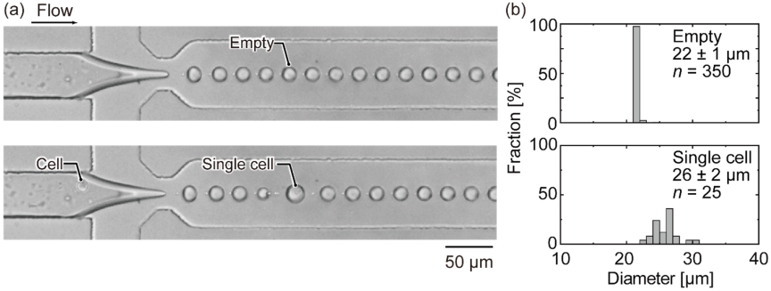
Fig. 3. Cell encapsulation in droplets generated by the MFDG. (a) High-speed recordings acquired at 15000 fps illustrate the formation of empty droplets and single-cell droplets at \(Q_\mathrm{c}=3.3\) μL/min and \(Q_\mathrm{d}=0.3\) μL/min. (b) Distributions of droplet diameters for empty droplets and single-cell droplets. The mean diameters (\(\pm\) SD) were 22\(\pm\)1 μm (\(n=350\)) for empty droplets and 26\(\pm\)2 μm (\(n=25\)) for single-cell droplets. Single-cell droplets exhibited significantly larger diameters under these representative operating conditions (Welch’s \(t\)-test, \(p<0.05\)).
3. Results
3.1. Cell Encapsulation
Cell encapsulation performance was evaluated by introducing NIH3T3-3-4 cells into the MFDG. To determine \(D_\mathrm{c}\) required for downstream DLD separation, the diameters of empty and cell-containing droplets were measured. Adjusting \(Q_\mathrm{c}\) and \(Q_\mathrm{d}\) to 3.3 and 0.3 μL/min, respectively, resulted in uniformly sized empty droplets with a mean diameter of approximately 22 μm at a generation rate of \(1.3\times 10^3\) droplets/s. Under these conditions, droplets formed from the tip of the dispersed-phase thread, with breakup occurring at a downstream constriction located immediately after the cross-junction, where the thread narrowed below the channel width (Fig. 3(a)). When the cells were introduced into the cross-junction, they became encapsulated within the droplets, increasing their diameter (Fig. 3(a)). The effective in-channel cell concentration at the droplet-generation junction was estimated to be \(9.5\times 10^5\) cells/mL, which was lower than the prepared cell concentration of \(3\times 10^6\) cells/mL. Single-cell droplets had a mean diameter of 26\(\pm\)2 μm (mean \(\pm\) SD) and were produced at approximately 3 droplets/s (Fig. 3(b)). The droplet velocities at the cross-junction, determined from the frame-to-frame centroid displacement in the high-speed recordings, were approximately 90.0\(\pm\)1.0 mm/s. In comparison, the velocity of cells flowing in the dispersed phase was measured to be 5.0\(\pm\)0.7 mm/s.
For comparison, the expected fractions of empty, single-cell, and multicell droplets were estimated using Poisson statistics as follows:
3.2. Separation and Gelation of Single-Cell Droplets
The size-based separation performance of the DLD module was evaluated under identical flow conditions, with all inlet flow rates maintained constant, as shown in Fig. 4. Droplets with diameters larger than \(D_\mathrm{c}\) underwent lateral displacement along the inclination of the micropillar array, moving toward the left sidewall relative to the flow direction. In contrast, droplets smaller than \(D_\mathrm{c}\) remained on their initial streamlines and were transported along the primary flow direction (Fig. 4). Consequently, single-cell droplets, with a mean diameter of 25\(\pm\)3 μm, were primarily recovered from Ch. 1, while empty droplets, with a diameter of 22\(\pm\)1 μm, were directed into Ch. 2, which served as the waste outlet (Figs. 5(a)–(c)).
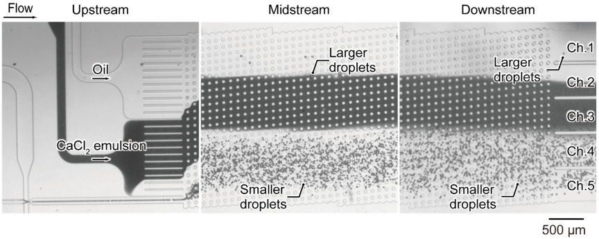
Fig. 4. Size-selective separation and in-flow gelation of droplets in a DLD micropillar array. High-speed imaging at 1000 fps illustrates droplets generated at the cross-junction, their entry into the micropillar array, and subsequent transport through the separation and gelation region. Experiments were conducted at flow rates of 3.3 μL/min (continuous phase; oil), 0.3 μL/min (dispersed phase; cell suspension), 0.7 μL/min (CaCl\(_2\) emulsion), and 1.8 μL/min (additional oil).
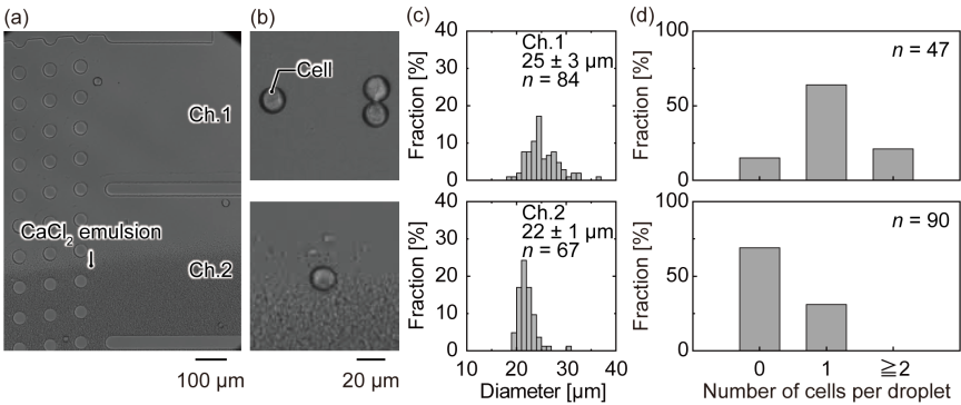
Fig. 5. Droplets processed through a DLD micropillar array. (a) High-speed imaging at 1000 fps illustrates droplet transport through the DLD separator toward the outlet channels (Ch. 1 and Ch. 2). (b) Magnified views of droplets recovered from Ch. 1 and Ch. 2, together with (c) the corresponding droplet diameter distributions. The mean droplet diameters (\(\pm\) SD) were 25\(\pm\)3 μm (\(n=84\)) for Ch. 1 and 22\(\pm\)1 μm (\(n=67\)) for Ch. 2. (d) Distribution of empty, single-cell, and multi-cell droplets observed at Ch. 1 and Ch. 2. Single-cell droplets collected from Ch. 1 showed a purity of 64% (\(30/47\)) and a separation efficiency of 53% (\(30/57\)). Fisher’s exact test showed that the single-cell fraction was significantly higher in Ch. 1 than before separation (\(p<0.0001\)) and significantly higher in Ch. 1 than in Ch. 2 (\(p=2.23\times 10^{-4}\)).
The separation outcome was assessed by evaluating the purity (\(P_\mathrm{p}\)) and separation efficiency (\(P_\mathrm{s}\)) of single-cell droplets recovered from Ch. 1, based on the analysis of high-speed imaging data. The purity was calculated as follows:
In comparison, the expected fraction of single-cell droplets predicted by Poisson statistics (Eq. (2)) was 1.8% under the experimental condition (\(\lambda=0.02\); cell concentration \(=3 \, \times \, 10^6\) cells/mL; droplet diameter \(=22\) μm). Even at the theoretical optimum (\(\lambda=1\)), the predicted fraction of single-cell droplets was 36.8%, which was lower than the experimentally achieved single-cell fraction after separation (64% in Ch. 1).
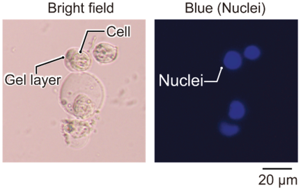
Fig. 6. Bright-field and fluorescence images of cell-laden alginate microgels collected after exiting the microchannel. The recovered alginate microgels were imaged after phase exchange from oil to culture medium.
High-speed image sequence analysis showed that the droplets traveled through the DLD separation region at a mean velocity of 4.2\(\pm\)0.8 mm/s. During transit through the micropillar array, the separated droplets passed through a Ca\(^{2+}\)-containing gelation stream (CaCl\(_2\) emulsion) under stable laminar flow conditions, allowing in-flow ionic crosslinking of the alginate phase. Under the present conditions, the droplet residence time within the gelation region was estimated to be on the order of 1–2 s. To assess the potential influence of multiphase flow, the viscosities of the oil phase and CaCl\(_2\) emulsion phase were measured to be 72 and 157 mPa\({\cdot}\)s, respectively. Although a viscosity difference existed, there was no obvious irregular droplet motion or suppression of lateral displacement near the oil-emulsion interface. The diameters of single-cell droplets were measured before and after passage through the CaCl\(_2\) emulsion stream. Following gelation, the average diameter decreased from 26 to 22 μm, indicating shrinkage during the gelation process.
After collection from the device and subsequent off-chip handling, the alginate microgels were transferred from the oil phase to the culture medium and examined using bright-field and fluorescence microscopy (Fig. 6). Microscopic observations showed an alginate gel layer surrounding the individual encapsulated cells, indicating that ionic crosslinking occurred during passage through the DLD micropillar array.
4. Discussion
We demonstrated the separation of single-cell droplets and their in-flow gelation into alginate microgels using a DLD micropillar array, allowing selective enrichment of single-cell-laden microgels within a continuous microfluidic workflow. Although this study provides a proof-of-concept for seamless separation and gelation, several challenges remain unaddressed.
Under the present experimental conditions (cell concentration of \(3 \, \times \, 10^6\) cells/mL and a droplet diameter of 22 μm), the generation rate of single-cell droplets was approximately 3 droplets/s, which was substantially lower than the Poisson-based expectation of approximately 20 droplets/s. To investigate the cause of this discrepancy, the concentration of the cells delivered to the droplet generation junction was evaluated. Upstream measurements indicated that the effective local cell concentration was reduced to \(9.5 \, \times \, 10^5\) cells/mL, suggesting nonuniform cell delivery, potentially owing to sedimentation or partial blockage in the sample reservoir, tubing, or inlet port. Even with this effective in-channel concentration, the Poisson distribution predicted a single-cell droplet generation rate of 7 droplets/s, which was higher than the experimentally observed rate (3 droplets/s). This discrepancy was even more pronounced for multicell droplets. According to the effective in-channel concentration, the Poisson distribution predicted a multicell droplet generation rate of approximately 0.02 droplets/s. In contrast, the experimentally observed multicell droplet generation rate was approximately 1.6 droplets/s. This result indicates that multicell droplets were generated much more frequently than predicted by the ideal Poisson model. These results suggest that this discrepancy cannot be explained solely by the reduced effective cell concentration. The cell encapsulation behavior most likely deviated from the Poisson prediction because of nonuniform cell distribution, such as cell aggregation or local concentration fluctuations prior to encapsulation. These effects may be mitigated by using cell introduction strategies that suppress particle depletion prior to entering the microchannel; for example, by rotating the syringe reservoir to maintain a homogeneous suspension 24.
The second limitation concerns the purity and separation efficiency of recovered single-cell droplets. As shown in Fig. 5(d), the single-cell collection channel contained not only single-cell droplets but also empty droplets and droplets containing two or more cells, reducing the apparent single-cell purity. Because \(D_\mathrm{c}\) was set to separate single-cell droplets from smaller empty droplets, droplets containing two or more cells, whose diameters exceeded those of single-cell droplets, were also directed to the same outlet. In addition to this design constraint, partial overlap between the size distributions of empty, single-cell, and multicell droplets reduces separation specificity. This overlap can be attributed to biological variability in cell size and fluctuations in droplet formation, which can generate relatively large empty droplets. Therefore, the current purity of 64% and separation efficiency of 53% should be regarded as proof-of-concept values rather than optimal performance. For direct downstream use without additional purification, such as cell culture, imaging, or cell-based assays, a purity of approximately 90% or higher is desirable, as reported for fluorescence-activated microgel-sorting methods 15. Although the current purity was lower than that of optimized high-purity sorting methods, it was within the range reported for some passive enrichment methods 14. To improve separation efficiency, future designs should focus on reducing the loss of single-cell droplets to non-target outlets by optimizing \(D_\mathrm{c}\), outlet geometry, and lateral position and width of the droplet stream. To improve purity, contamination by empty and multicell droplets must be reduced, primarily by minimizing the overlap in size distributions. In particular, hierarchical separation sections that direct empty, single-cell, and multi-cell droplets to different outlets may be effective when a sufficient size contrast exists. In addition, improving droplet size uniformity and reducing cell aggregation prior to encapsulation would minimize overlap among these droplet populations and improve single-cell specificity.
In the present study, only a single set of flow conditions was examined; therefore, the influence of the flow conditions on the size of the recovered single-cell-laden alginate microgels was not systematically investigated. However, previous studies have shown that increasing droplet size reduces the size difference between empty and single-cell droplets 21. Such a reduction in size contrast, particularly near \(D_\mathrm{c}\), is expected to decrease the purity of single-cell-laden alginate microgels through DLD-based separation. This suggests a tradeoff between recovering larger single-cell-laden alginate microgels and maintaining high purity.
In addition to the droplet size at generation, flow conditions within the DLD array can affect separation performance. In particular, the flow rates of the additional oil and CaCl\(_2\) emulsion affect the lateral position and width of the droplet stream entering the micropillar array. This can alter the initial droplet position and outlet distribution. Although these parameters were not systematically varied in the present study, their influence is reflected in the observed droplet trajectories under the selected operating conditions. Moreover, higher flow rates may increase the shear stress acting on droplets during their passage through the pillar gaps, resulting in droplet deformation and altered lateral displacement behavior. Because the estimated Reynolds number in the current device was low (\(\textit{Re}=0.004\)), inertial migration was expected to be negligible; however, deformation-related effects may still influence droplet migration behavior near \(D_\mathrm{c}\) owing to changes in effective droplet size. Thus, Eq. (1) should be considered as a first-order design guideline for deformable droplets because it is derived under the assumption of rigid particles and does not explicitly account for flow-dependent deformation effects. Future studies should evaluate how droplet deformation (as characterized by the capillary number) affects migration behavior relative to \(D_\mathrm{c}\) under varying flow rate conditions.
Partial gelation during transit through the DLD array may alter droplet deformability, influencing migration behavior near the critical diameter and affecting separation performance 25,26,27. In addition, gelation-induced shrinkage can reduce the effective droplet size during or after passage through the DLD array. In the present study, the average diameter of single-cell droplets decreased from 26 μm before passage through the CaCl\(_2\) emulsion stream to 22 μm after gelation, indicating shrinkage during the gelation process. This size reduction may decrease the effective droplet size relative to the designed \(D_\mathrm{c}\), causing some single-cell droplets that would otherwise be laterally displaced toward Ch. 1 to instead follow streamlines toward non-target outlets.
Future studies should systematically evaluate how initial droplet size, flow-rate-dependent droplet deformation, changes in deformability caused by partial gelation, and gelation-induced shrinkage affect droplet migration behavior relative to \(D_\mathrm{c}\) and separation performance. To accommodate such changes in droplet size and deformability, dynamic tuning of DLD separation characteristics, such as on-the-fly modulation of the effective \(D_\mathrm{c}\) using responsive micropillars 28,29, external fields 30,31,32,33,34, or flow-property control 35,36,37, may provide a promising strategy to adapt separation behavior to variations in droplet size and material properties, as demonstrated in recent DLD-based platforms.
Additionally, in this study, the recovered alginate microgels were transferred from oil to the culture medium using bulk centrifugation. To allow for the rapid utilization of the produced cell-laden alginate microgels in downstream applications, phase exchange should ideally be integrated into the microfluidic device. This may be achieved by incorporating an in-array solution transfer method 17 or integrating a continuous-flow oil-to-aqueous extraction unit 38,39.
Cell viability after recovery was not quantitatively evaluated in this study because the number of recovered cell-laden alginate microgels was limited under the experimental conditions. Tottori et al. reported a post-processing cell viability of approximately 80% for MCF-7 cells encapsulated in alginate microgels produced by droplet-based gelation and extraction 17. Although this suggests that alginate droplet-based microgel formation may be compatible with cell handling, cell viability may depend strongly on device-specific conditions, including gelation chemistry, crosslinking exposure time, oil/surfactant systems, and recovery processes. Therefore, a direct viability evaluation under the present device conditions will be required in future studies.
This study focused exclusively on alginate microgels, with no investigations into other materials or material combinations. For applications involving adherent cells, additional material design is required because alginate alone generally lacks cell-adhesive functionality 40,41. One method to overcome this limitation is to introduce cell-adhesive functionality through material design, for example, by incorporating cell-adhesive motifs such as RGD peptides 42, or by blending alginate with complementary biopolymers such as gelatin 43 or collagen 44. In addition to material composition, the temporal exposure of droplets to the crosslinking agent is an additional design parameter. According to Tottori et al. 17, this exposure time is expected to be tunable by geometric and flow-related factors, including the inclination angle of the DLD array, width of the reactant stream, and continuous-phase flow rate. Controlling these parameters would allow for the systematic regulation of microgel morphology, ranging from homogeneous gels to core-shell structures with a liquid core 45 and adjustable shell thickness 46, providing a route to tailor the mechanical properties for specific applications. Furthermore, the use of multi-stream laminar co-flow configurations offers opportunities to produce hybrid or composite microgels combining alginate with other biopolymers 47,48,49.
Finally, although the current study represents a proof-of-concept demonstration, parallelization of droplet generators and DLD arrays 20 could provide a straightforward route toward higher throughput and scalable production of single-cell-laden alginate microgels. Overall, the current study demonstrates an integrated microfluidic framework that combines size-based droplet separation with in-flow ionic gelation, highlighting the potential of engineered hydrogel systems for advanced single-cell-laden alginate microgel production.
5. Conclusion
We proposed an integrated droplet microfluidic method for generating single-cell-laden alginate microgels by coupling size-based separation with in-flow ionic gelation in DLD micropillar arrays. This platform enabled continuous in-flow crosslinking and selective separation of cell-laden alginate microgels from empty droplets, thereby eliminating the need for additional pre- or post-processing steps for single-cell enrichment that are commonly required in conventional methods. Although the achieved purity (64%) and separation efficiency (53%) represent proof-of-concept values, the results confirmed the feasibility of producing single-cell-laden alginate microgels in an integrated and continuous workflow. With further refinements in device design and integration with downstream single-cell analysis platforms, this method is expected to support the systematic investigation of single-cell behavior and heterogeneity in advanced biomedical applications. The current platform provides a foundational technology for constructing cell-associated systems that support the stable positioning of functional materials or sensing components in close proximity to individual cells, potentially contributing to future strategies for information transmission from living cells and tissues.
Acknowledgments
This work was supported by JSPS KAKENHI (Grant Number JP22K14547) and partially supported by JST Moonshot R&D Program (JPMJMS2217-5-1).
- [1] M. Y. Wang, H. L. Tao, J. Li, X. Y. Chen, Y. C. Gu, R. Tang, and X. Q. Liu, “The cell-loaded alginate microspheres in cell culture and disease treatment,” Smart Mater. Med., Vol.6, Issue 3, pp. 387-405, 2025. https://doi.org/10.1016/j.smaim.2025.11.002
- [2] S.-M. Kang, J.-H. Lee, Y. S. Huh, and S. Takayama, “Alginate microencapsulation for three-dimensional in vitro cell culture,” ACS Biomater. Sci. Eng., Vol.7, Issue 7, pp. 2864-2879, 2021. https://doi.org/10.1021/acsbiomaterials.0c00457
- [3] B. Namgung, K. Ravi, P. P. Vikraman, S. Sengupta, and H. L. Jang, “Engineered cell-laden alginate microparticles for 3D culture,” Biochem. Soc. Trans., Vol.49, Issue 2, pp. 761-773, 2021. https://doi.org/10.1042/BST20200673
- [4] S. Utech, R. Prodanovic, A. S. Mao, R. Ostafe, D. J. Mooney, and D. A. Weitz, “Microfluidic generation of monodisperse, structurally homogeneous alginate microgels for cell encapsulation and 3D cell culture,” Adv. Healthc. Mater., Vol.4, Issue 11, pp. 1628-1633, 2015. https://doi.org/10.1002/adhm.201500021
- [5] X. Qin, Z. Gan, H. Liu, T. Tao, J. He, X. Li, D. Shang, X. Li, F. Xie, and J. Qin, “A pump-free strategy for the controllable generation of alginate microgels as cellular microcarriers,” ACS Biomater. Sci. Eng., Vol.10, Issue 6, pp. 3958-3967, 2024. https://doi.org/10.1021/acsbiomaterials.4c00375
- [6] C. An, W. Liu, Y. Zhang, B. Pang, H. Liu, Y. Zhang, H. Zhang, L. Zhang, H. Liao, C. Ren, and H. Wang, “Continuous microfluidic encapsulation of single mesenchymal stem cells using alginate microgels as injectable fillers for bone regeneration,” Acta Biomater., Vol.111, pp. 181-196, 2020. https://doi.org/10.1016/j.actbio.2020.05.024
- [7] H. Soleymani, M. M. Moghaddam, H. Naderi-Manesh, and R. A. Taheri, “Single-layer graphene oxide nanosheets induce proliferation and osteogenesis of single-cell hBMSCs encapsulated in alginate microgels,” Sci. Rep., Vol.14, Article No.25272, 2024. https://doi.org/10.1038/s41598-024-76957-y
- [8] V. Iansante, A. Dhawan, F. Masmoudi, C. A. Lee, R. Fernandez-Dacosta, S. Walker, E. Fitzpatrick, R. R. Mitry, and C. Filippi, “A new high throughput screening platform for cell encapsulation in alginate hydrogel shows improved hepatocyte functions by mesenchymal stromal cells co-encapsulation,” Front. Med., Vol.5, Article No.216, 2018. https://doi.org/10.3389/fmed.2018.00216
- [9] Z. Zhao, X. Feng, H. Wu, S. Chen, C. Ma, Z. Guan, L. Lei, K. Tang, X. Chen, Y. Dong, and Y. Tang, “Construction of a lung cancer 3D culture model based on alginate/gelatin micro-beads for drug evaluation,” Transl. Lung Cancer Res., Vol.13, No.10, pp. 2698-2712, 2024. https://doi.org/10.21037/tlcr-24-490
- [10] Y. Wang, Z. Shen, H. Wang, Z. Song, D. Yu, G. Li, X. Liu, and W. Liu, “Progress in research on metal ion crosslinking alginate-based gels,” Gels, Vol.11, Issue 1, Article No.16, 2025. https://doi.org/10.3390/gels11010016
- [11] M. Pires-Santos, S. Nadine, and J. F. Mano, “Unveiling the potential of single-cell encapsulation in biomedical applications: Current advances and future perspectives,” Small Sci., Vol.4, Issue 5, Article No.2300332, 2024. https://doi.org/10.1002/smsc.202300332
- [12] B. M. Tiemeijer and J. Tel, “Hydrogels for single-cell microgel production: Recent advances and applications,” Front. Bioeng. Biotechnol., Vol.10, Article No.891461, 2022. https://doi.org/10.3389/fbioe.2022.891461
- [13] R. Dubay, J. N. Urban, and E. M. Darling, “Single-cell microgels for diagnostics and therapeutics,” Adv. Funct. Mater., Vol.31, Issue 44, Article No.2009946, 2021. https://doi.org/10.1002/adfm.202009946
- [14] A. S. Mao, J.-W. Shin, S. Utech, H. Wang, O. Uzun, W. Li, M. Cooper, Y. Hu, L. Zhang, D. A. Weitz, and D. J. Mooney, “Deterministic encapsulation of single cells in thin tunable microgels for niche modelling and therapeutic delivery,” Nat. Mater., Vol.16, pp. 236-243, 2017. https://doi.org/10.1038/nmat4781
- [15] T. Kamperman, S. Henke, A. van den Berg, S. R. Shin, A. Tamayol, A. Khademhosseini, M. Karperien, and J. Leijten, “Single cell microgel based modular bioinks for uncoupled cellular micro- and macroenvironments,” Adv. Healthc. Mater., Vol.6, Issue 3, Article No.1600913, 2017. https://doi.org/10.1002/adhm.201600913
- [16] L. R. Huang, E. C. Cox, R. H. Austin, and J. C. Sturm, “Continuous particle separation through deterministic lateral displacement,” Science, Vol.304, Issue 5673, pp. 987-990, 2004. https://doi.org/10.1126/science.1094567
- [17] N. Tottori, Y. Tang, Y. Kanno, and T. Nisisako, “Continuous production and extraction of monodisperse alginate microgels via deterministic lateral displacement,” Soft Matter, Vol.22, Issue 7, pp. 1483-1493, 2026. https://doi.org/10.1039/d5sm00757g
- [18] T. Jing, R. Ramji, M. E. Warkiani, J. Han, C. T. Lim, and C. H. Chen, “Jetting microfluidics with size-sorting capability for single-cell protease detection,” Biosens. Bioelectron., Vol.66, pp. 19-23, 2015. https://doi.org/10.1016/j.bios.2014.11.001
- [19] N. Tottori, T. Hatsuzawa, and T. Nisisako, “Separation of main and satellite droplets in a deterministic lateral displacement microfluidic device,” RSC Adv., Vol.7, Issue 56, pp. 35516-35524, 2017. https://doi.org/10.1039/c7ra05852g
- [20] N. Tottori and T. Nisisako, “High-throughput production of satellite-free droplets through a parallelized microfluidic deterministic lateral displacement device,” Sens. Actuators B: Chem., Vol.260, pp. 918-926, 2018. https://doi.org/10.1016/j.snb.2018.01.112
- [21] H. Fukunaga, N. Tottori, T. Tsubouchi, T. Kumazaki, S. Sakuma, and Y. Yamanishi, “Spatially constrained encapsulation of cells in microdroplets enables continuous cell fusion within a microfluidic flow environment,” Sens. Actuators B: Chem., Vol.458, Article No.139779, 2026. https://doi.org/10.1016/j.snb.2026.139779
- [22] J. A. Davis, “Microfluidic Separation of Blood Components through Deterministic Lateral Displacement,” Doctoral thesis, Princeton University, 2008.
- [23] Y. Liu, N. Tottori, and T. Nisisako, “Microfluidic synthesis of highly spherical calcium alginate hydrogels based on external gelation using an emulsion reactant,” Sens. Actuators B: Chem., Vol.283, pp. 802-809, 2019. https://doi.org/10.1016/j.snb.2018.12.101
- [24] S. I. R. Lane, J. Butement, J. Harrington, T. Underwood, J. Shrimpton, and J. West, “Perpetual sedimentation for the continuous delivery of particulate suspensions,” Lab Chip, Vol.19, Issue 22, pp. 3771-3775, 2019. https://doi.org/10.1039/c9lc00774a
- [25] D. Holmes, G. Whyte, J. Bailey, N. Vergara-Irigaray, A. Ekpenyong, J. Guck, and T. Duke, “Separation of blood cells with differing deformability using deterministic lateral displacement,” Interface Focus, Vol.4, Issue 6, Article No.20140011, 2014. https://doi.org/10.1098/rsfs.2014.0011
- [26] S. Ye, X. Shao, Z. Yu, and W. Yu, “Effects of the particle deformability on the critical separation diameter in the deterministic lateral displacement device,” J. Fluid Mech., Vol.743, pp. 60-74, 2014. https://doi.org/10.1017/jfm.2014.22
- [27] T. Krüger, D. Holmes, and P. V. Coveney, “Deformability-based red blood cell separation in deterministic lateral displacement devices—A simulation study,” Biomicrofluidics, Vol.8, Issue 5, Article No.054114, 2014. https://doi.org/10.1063/1.4897913
- [28] N. Tottori and T. Nisisako, “Tunable deterministic lateral displacement of particles flowing through thermo-responsive hydrogel micropillar arrays,” Sci. Rep., Vol.13, Article No.4994, 2023. https://doi.org/10.1038/s41598-023-32233-z
- [29] Z. Jiang, Y. Kanno, and T. Nisisako, “Tunable cell separation using a thermo-responsive deterministic lateral displacement device,” Lab Chip, Vol.25, Issue 24, pp. 6454-6464, 2025. https://doi.org/10.1039/d5lc00783f
- [30] M. Aghaamoo, A. Aghilinejad, X. Chen, and J. Xu, “On the design of deterministic dielectrophoresis for continuous separation of circulating tumor cells from peripheral blood cells,” Electrophoresis, Vol.40, Issue 10, pp. 1486-1493, 2019. https://doi.org/10.1002/elps.201800459
- [31] J. P. Beech, P. Jönsson, and J. O. Tegenfeldt, “Tipping the balance of deterministic lateral displacement devices using dielectrophoresis,” Lab Chip, Vol.9, Issue 18, pp. 2698-2706, 2009. https://doi.org/10.1039/b823275j
- [32] T. S. H. Tran, B. D. Ho, J. P. Beech, and J. O. Tegenfeldt, “Open channel deterministic lateral displacement for particle and cell sorting,” Lab Chip, Vol.17, Issue 21, pp. 3592-3600, 2017. https://doi.org/10.1039/c7lc00707h
- [33] J. P. Beech, K. Keim, B. D. Ho, C. Guiducci, and J. O. Tegenfeldt, “Active posts in deterministic lateral displacement devices,” Adv. Mater. Technol., Vol.4, Issue 9, Article No.1900339, 2019. https://doi.org/10.1002/admt.201900339
- [34] V. Calero, P. Garcia-Sanchez, C. Honrado, A. Ramos, and H. Morgan, “AC electrokinetic biased deterministic lateral displacement for tunable particle separation,” Lab Chip, Vol.19, Issue 8, pp. 1386-1396, 2019. https://doi.org/10.1039/c8lc01416g
- [35] B. M. Dincau, A. Aghilinejad, X. Chen, S. Y. Moon, and J.-H. Kim, “Vortex-free high-Reynolds deterministic lateral displacement (DLD) via airfoil pillars,” Microfluid. Nanofluidics, Vol.22, Article No.137, 2018. https://doi.org/10.1007/s10404-018-2160-3
- [36] B. M. Dincau, A. Aghilinejad, T. Hammersley, X. Chen, and J.-H. Kim, “Deterministic lateral displacement (DLD) in the high Reynolds number regime: High-throughput and dynamic separation characteristics,” Microfluid. Nanofluidics, Vol.22, Article No.59, 2018. https://doi.org/10.1007/s10404-018-2078-9
- [37] H. Fukunaga, N. Tottori, S. Sakuma, T. Hayakawa, and Y. Yamanishi, “Tunable particle separation using vibration induced flow in deterministic lateral displacement micropillar arrays,” 2025 23rd Int. Conf. Solid-State Sensors, Actuators Microsystems, pp. 227-230, 2025. https://doi.org/10.1109/transducers61432.2025.11111463
- [38] H. Huang and X. He, “Interfacial tension based on-chip extraction of microparticles confined in microfluidic stokes flows,” Appl. Phys. Lett., Vol.105, Issue 14, Article No.143704, 2014. https://doi.org/10.1063/1.4898040
- [39] Y. Deng, N. Zhang, L. Zhao, X. Yu, X. Ji, W. Liu, S. Guo, K. Liu, and X.-Z. Zhao, “Rapid purification of cell encapsulated hydrogel beads from oil phase to aqueous phase in a microfluidic device,” Lab Chip, Vol.11, Issue 23, pp. 4117-4121, 2011. https://doi.org/10.1039/c1lc20494g
- [40] T. Hu and A. C. Y. Lo, “Collagen–alginate composite hydrogel: Application in tissue engineering and biomedical sciences,” Polymers, Vol.13, Issue 11, Article No.1852, 2021. https://doi.org/10.3390/polym13111852
- [41] K. Y. Lee and D. J. Mooney, “Alginate: Properties and biomedical applications,” Prog. Polym. Sci., Vol.37, Issue 1, pp. 106-126, 2012. https://doi.org/10.1016/j.progpolymsci.2011.06.003
- [42] J. A. Rowley and D. J. Mooney, “Alginate type and RGD density control myoblast phenotype,” J. Biomed. Mater. Res., Vol.60, Issue 2, pp. 217-223, 2002. https://doi.org/10.1002/jbm.1287
- [43] B. Sarker, R. Singh, R. Silva, J. A. Roether, J. Kaschta, R. Detsch, D. W. Schubert, I. Cicha, and A. R. Boccaccini, “Evaluation of Fibroblasts Adhesion and Proliferation on Alginate-Gelatin Crosslinked Hydrogel,” PLoS One, Vol.9, No.9, Article No.e107952, 2014. https://doi.org/10.1371/journal.pone.0107952
- [44] W. Bouhlel, J. Kui, J. Bibette, and N. Bremond, “Encapsulation of cells in a collagen matrix surrounded by an alginate hydrogel shell for 3D cell culture,” ACS Biomater. Sci. Eng., Vol.8, Issue 6, pp. 2700-2708, 2022. https://doi.org/10.1021/acsbiomaterials.1c01486
- [45] W. Zhang, S. Zhao, W. Rao, J. Snyder, J. K. Choi, J. Wang, I. A. Khan, N. B. Saleh, P. J. Mohler, J. Yu, T. J. Hund, C. Tang, and X. He, “A novel core–shell microcapsule for encapsulation and 3D culture of embryonic stem cells,” J. Mater. Chem. B, Vol.1, Issue 7, pp. 1002-1009, 2013. https://doi.org/10.1039/c2tb00058j
- [46] H. Zhang, E. Tumarkin, R. Peerani, Z. Nie, R. M. A. Sullan, G. C. Walker, and E. Kumacheva, “Microfluidic production of biopolymer microcapsules with controlled morphology,” J. Am. Chem. Soc., Vol.128, Issue 37, pp. 12205-12210, 2006. https://doi.org/10.1021/ja0635682
- [47] P. Agarwal, J. K. Choi, H. Huang, S. Zhao, J. Dumbleton, J. Li, and X. He, “A biomimetic core–shell platform for miniaturized 3D cell and tissue engineering,” Part. Part. Syst. Charact., Vol.32, Issue 8, pp. 809-816, 2015. https://doi.org/10.1002/ppsc.201500025
- [48] H. Aoki, Y. Masahiro, M. Shimizu, Y. Hongoh, M. Ohkuma, and Y. Yamagata, “Agarose gel microcapsules enable easy-to-prepare, picolitre-scale, single-cell genomics, yielding high-coverage genome sequences,” Sci. Rep., Vol.12, Article No.17014, 2022. https://doi.org/10.1038/s41598-022-20923-z
- [49] M. Chau, M. Abolhasani, H. Thérien-Aubin, Y. Li, Y. Wang, D. Velasco, E. Tumarkin, A. Ramachandran, and E. Kumacheva, “Microfluidic generation of composite biopolymer microgels with tunable compositions and mechanical properties,” Biomacromolecules, Vol.15, Issue 7, pp. 2419-2425, 2014. https://doi.org/10.1021/bm5002813
This article is published under a Creative Commons Attribution-NoDerivatives 4.0 Internationa License.