
Paper:
Method for Evaluating Cell Deformability Using Forced Flow Cessation in Microchannels
Mitsuhiro Horade*,†
 , Kotaro Tsujimoto*, and Hiroaki Ito**
*Faculty of Science and Engineering, Setsunan University
17-8 Ikeda-nakamachi, Neyagawa, Osaka 572-8508, Japan
†Corresponding author
**Chiba University
1-33 Yayoicho, Inage-ku, Chiba, Chiba 263-8522, Japan

The characteristics of red blood cells (RBCs) strongly correlate with disease, making measurement techniques valuable for diagnostics. This study focused on RBC deformability and developed an image-based evaluation method using a microfluidic device that applies stress by forcibly stopping flow. Unlike biochemical analyses, such as polymerase chain reaction, which provide averaged data, this method enables single-cell mechanical property assessment. It is also simple, requiring minimal specialized equipment and manual operation. RBCs were confirmed to deform upon forced stopping. We discussed deformation behavior and load estimation, confirming that wall shear stress, not inertial force, dominates deformation. Normal RBCs deformed by 31.5%, whereas adenosine triphosphate (ATP)-depleted RBCs, modeling diseased states, deformed by only 11.1%. The difference was statistically significant (p=0.025). We validated reproducibility by quantifying variability from manual syringe operation, showing consistent deformation measurements across operators. Sensitivity analyses confirmed that assumptions in shear stress estimation, such as gap size and viscosity, had minimal impact. The deformation metric was defined and shown to be robust against image processing variations. Statistical rigor was improved by adding effect sizes, confidence intervals, and power analysis. We discussed the biological relevance and limitations of the ATP depletion model, noting the need for validation with pathological samples. Finally, we highlighted the method’s contribution to intracellular sensor research, positioning mechanical deformability assessment as complementary to molecular intracellular sensing. These results suggest the potential of the method for evaluating blood diseases and related conditions.
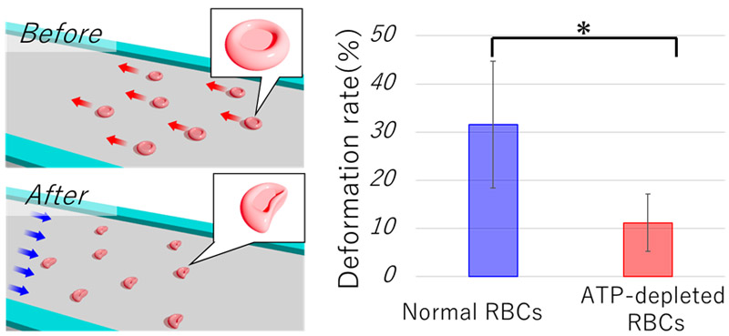
Comparison of RBC images before and after deformation
- [1] M. Battistelli et al., “Skeletal muscle cell behavior after physical agent treatments,” Curr. Pharm. Des., Vol.21, No.25, pp. 3665-3672, 2015. https://doi.org/10.2174/1381612821666150122123412
- [2] K.-A. Hansson and E. Eftestøl, “Scaling of nuclear numbers and their spatial arrangement in skeletal muscle cell size regulation,” Mol. Biol. Cell, Vol.34, No.8, Article No.pe3, 2023. https://doi.org/10.1091/mbc.E22-09-0424
- [3] A. Jögi, M. Vaapil, M. Johansson, and S. Påhlman, “Cancer cell differentiation heterogeneity and aggressive behavior in solid tumors,” Upsala J. Med. Sci., Vol.117, No.2, pp. 217-224, 2012. https://doi.org/10.3109/03009734.2012.659294
- [4] C. A. Moxon, G. E. Grau, and A. G. Craig, “Malaria: Modification of the red blood cell and consequences in the human host,” Br. J. Haematol., Vol.154, No.6, pp. 670-679, 2011. https://doi.org/10.1111/j.1365-2141.2011.08755.x
- [5] J. Pernow, A. Mahdi, J. Yang, and Z. Zhou, “Red blood cell dysfunction: A new player in cardiovascular disease,” Cardiovasc. Res., Vol.115, No.11, pp. 1596-1605, 2019. https://doi.org/10.1093/cvr/cvz156
- [6] H. H. Patel, H. R. Patel, and J. M. Higgins, “Modulation of red blood cell population dynamics is a fundamental homeostatic response to disease,” Am. J. Hematol., Vol.90, No.5, pp. 422-428, 2015. https://doi.org/10.1002/ajh.23982
- [7] B. George-Gay and K. Parker, “Understanding the complete blood count with differential,” J. Perianesth. Nurs., Vol.18, No.2, pp. 96-117, 2003. https://doi.org/10.1053/jpan.2003.50013
- [8] S. Sen et al., “Early clinical complete blood count changes in severe burn injuries,” Burns, Vol.45, No.1, pp. 97-102, 2019. https://doi.org/10.1016/j.burns.2018.09.004
- [9] L. Agnello et al., “The value of a complete blood count (CBC) for sepsis diagnosis and prognosis,” Diagnostics, Vol.11, No.10, Article No.1881, 2021. https://doi.org/10.3390/diagnostics11101881
- [10] M. Aghaei et al., “Evaluation of complete blood count parameters in patients with diabetes mellitus: A systematic review,” Health Sci. Rep., Vol.8, No.2, Article No.e70488, 2025. https://doi.org/10.1002/hsr2.70488
- [11] K. Wisanuvej et al., “Comparison between blood hemoglobin concentration determined by point-of-care device and complete blood count in adult patients with dengue,” PLOS Negl. Trop. Dis., Vol.15, No.8, Article No.e0009692, 2021. https://doi.org/10.1371/journal.pntd.0009692
- [12] G. Hao et al., “Association of blood glucose and lipid levels with complete blood count indices to establish a regression model,” Biomed. Rep., Vol.6, No.3, pp. 339-345, 2017. https://doi.org/10.3892/br.2017.852
- [13] R. Poonperm et al., “scRepli-RamDA-seq: A multi-omics technology enabling the analysis of gene expression dynamics during S-phase,” Nat. Commun., Vol.16, No.1, Article No.10902, 2025. https://doi.org/10.1038/s41467-025-64688-1
- [14] S. V. Avery, “Microbial cell individuality and the underlying sources of heterogeneity,” Nat. Rev. Microbiol., Vol.4, No.8, pp. 577-587, 2006. https://doi.org/10.1038/nrmicro1460
- [15] J. Zhao, “Cell individuality: A basic multicellular phenomenon and its role in the pathogenesis of disease,” Med. Hypotheses, Vol.44, No.5, pp. 400-402, 1995. https://doi.org/10.1016/0306-9877(95)90267-8
- [16] M. B. Ginzberg, R. Kafri, and M. Kirschner, “On being the right (cell) size,” Science, Vol.348, No.6236, Article No.1245075, 2015. https://doi.org/10.1126/science.1245075
- [17] M. Horade, C.-H. D. Tsai, and M. Kaneko, “On-chip cell incubator for simultaneous observation of culture with and without periodic hydrostatic pressure,” Micromachines, Vol.10, No.2, Article No.133, 2019. https://doi.org/10.3390/mi10020133
- [18] Asadullah et al., “Combined heterogeneity in cell size and deformability promotes cancer invasiveness,” J. Cell Sci., Vol.134, No.7, Article No.jcs250225, 2021. https://doi.org/10.1242/jcs.250225
- [19] X. He, H. Kimura, and T. Fujii, “A high-throughput device for patterned differentiation of embryoid bodies,” J. Robot. Mechatron., Vol.25, No.4, pp. 623-630, 2013. https://doi.org/10.20965/jrm.2013.p0623
- [20] C. W. Shields IV, C. D. Reyes, and G. P. López, “Microfluidic cell sorting: A review of the advances in the separation of cells from debulking to rare cell isolation,” Lab Chip, Vol.15, No.5, pp. 1230-1249, 2015. https://doi.org/10.1039/c4lc01246a
- [21] F. Tang et al., “mRNA-Seq whole-transcriptome analysis of a single cell,” Nat. Methods, Vol.6, No.5, pp. 377-382, 2009. https://doi.org/10.1038/nmeth.1315
- [22] E. Z. Macosko et al., “Highly parallel genome-wide expression profiling of individual cells using nanoliter droplets,” Cell, Vol.161, No.5, pp. 1202-1214, 2015. https://doi.org/10.1016/j.cell.2015.05.002
- [23] E. Danese, G. Lippi, and M. Montagnana, “Red blood cell distribution width and cardiovascular diseases,” J. Thorac. Dis., Vol.7, No.10, pp. E402-E411, 2015. https://doi.org/10.3978/j.issn.2072-1439.2015.10.04
- [24] M. Tonelli et al., “Relation between red blood cell distribution width and cardiovascular event rate in people with coronary disease,” Circulation, Vol.117, No.2, pp. 163-168, 2008. https://doi.org/10.1161/CIRCULATIONAHA.107.727545
- [25] G. L. Thompson and D. Kavanagh, “Diagnosis and treatment of thrombotic microangiopathy,” Int. J. Lab. Hematol., Vol.44, No.S1, pp. 101-113, 2022. https://doi.org/10.1111/ijlh.13954
- [26] K. Inoue, D. Nishi, T. Takubo, T. Tanikawa, and T. Arai, “Performance evaluation of teleoperation for manipulating micro objects using two-fingered micro hand,” J. Robot. Mechatron., Vol.19, No.5, pp. 577-584, 2007. https://doi.org/10.20965/jrm.2007.p0577
- [27] Y. Tanaka, “Double-arm optical tweezer system for precise and dexterous handling of micro-objects in 3D workspace,” Opt. Lasers Eng., Vol.111, pp. 65-70, 2018. https://doi.org/10.1016/j.optlaseng.2018.07.019
- [28] S. Sakuma and F. Arai, “Cellular force measurement using a nanometric-probe-integrated microfluidic chip with a displacement reduction mechanism,” J. Robot. Mechatron., Vol.25, No.2, pp. 277-284, 2013. https://doi.org/10.20965/jrm.2013.p0277
- [29] Y. Yamanishi et al., “Design and fabrication of all-in-one unified microfluidic chip for automation of embryonic cell manipulation,” J. Robot. Mechatron., Vol.22, No.3, pp. 371-379, 2010. https://doi.org/10.20965/jrm.2010.p0371
- [30] A. Raj and A. K. Sen, “Entry and passage behavior of biological cells in a constricted compliant microchannel,” RSC Adv., Vol.8, No.37, pp. 20884-20893, 2018. https://doi.org/10.1039/C8RA02763C
- [31] J. Chen, X. Zou, D. C. Spencer, and H. Morgan, “Single-cell electro-mechanical shear flow deformability cytometry,” Microsyst. Nanoeng., Vol.10, No.1, Article No.173, 2024. https://doi.org/10.1038/s41378-024-00810-5
- [32] T. Yang, F. Bragheri, and P. Minzioni, “A comprehensive review of optical stretcher for cell mechanical characterization at single-cell level,” Micromachines, Vol.7, No.5, Article No.90, 2016. https://doi.org/10.3390/mi7050090
- [33] H. T. K. Tse et al., “Quantitative diagnosis of malignant pleural effusions by single-cell mechanophenotyping,” Sci. Transl. Med., Vol.5, No.212, Article No.212ra163, 2013. https://doi.org/10.1126/scitranslmed.3006559
- [34] S. Sakuma et al., “High resolution cell positioning based on a flow reduction mechanism for enhancing deformability mapping,” Micromachines, Vol.5, No.4, pp. 1188-1201, 2014. https://doi.org/10.3390/mi5041188
- [35] M. L. Racine and F. A. Dinenno, “Reduced deformability contributes to impaired deoxygenation-induced ATP release from red blood cells of older adult humans,” J. Physiol., Vol.597, No.17, pp. 4503-4519, 2019. https://doi.org/10.1113/JP278338
- [36] Y. Li, J. Zhou, I. Burkovskiy, P. Yeung, and C. Lehmann, “ATP in red blood cells as biomarker for sepsis in humans,” Med. Hypotheses, Vol.124, pp. 84-86, 2019. https://doi.org/10.1016/j.mehy.2019.02.014
- [37] D. S. Parekh, W. A. Eaton, and S. L. Thein, “Recent developments in the use of pyruvate kinase activators as a new approach for treating sickle cell disease,” Blood, Vol.143, No.10, pp. 866-871, 2024. https://doi.org/10.1182/blood.2023021167
- [38] D. H. Bonfanti et al., “ATP-dependent potassium channels and type 2 diabetes mellitus,” Clin. Biochem., Vol.48, Nos.7-8, pp. 476-482, 2015. https://doi.org/10.1016/j.clinbiochem.2014.12.026
- [39] G. Ramdani and G. Langsley, “ATP, an extracellular signaling molecule in red blood cells: A messenger for malaria?,” Biomed. J., Vol.37, No.5, pp. 284-292, 2014. https://doi.org/10.4103/2319-4170.132910
- [40] X. Zhang, E. Agborbesong, and X. Li, “The role of mitochondria in acute kidney injury and chronic kidney disease and its therapeutic potential,” Int. J. Mol. Sci., Vol.22, No.20, Article No.11253, 2021. https://doi.org/10.3390/ijms222011253
- [41] H. Bruus, “Theoretical microfluidics,” Oxford University Press, 2007. https://doi.org/10.1093/oso/9780199235087.001.0001
- [42] A. Link, I. L. Pardo, B. Porr, and T. Franke, “AI based image analysis of red blood cells in oscillating microchannels,” RSC Adv., Vol.13, No.41, pp. 28576-28582, 2023. https://doi.org/10.1039/d3ra04644c
- [43] A. R. Pries, D. Neuhaus, and P. Gaehtgens, “Blood viscosity in tube flow: Dependence on diameter and hematocrit,” Am. J. Physiol. Heart Circ. Physiol., Vol.263, No.6, pp. H1770-H1778, 1992. https://doi.org/10.1152/ajpheart.1992.263.6.H1770
- [44] M. Horade, M. Moriga, and S. Murakami, “Cell size and deformation measurement using constrictions integrated into a microfluidic device,” Phys. Scr., Vol.99, No.7, Article No.075007, 2024. https://doi.org/10.1088/1402-4896/ad5060
- [45] K. Mizoue, K. Teramura, C.-H. D. Tsai, and M. Kaneko, “Transfer function of macro-micro manipulation on a PDMS microfluidic chip,” Micromachines, Vol.8, No.3, Article No.80, 2017. https://doi.org/10.3390/mi8030080
- [46] M. Puig-de-Morales-Marinkovic, K. T. Turner, J. P. Butler, J. J. Fredberg, and S. Suresh, “Viscoelasticity of the human red blood cell,” Am. J. Physiol. Cell Physiol., Vol.293, No.2, pp. C597-C605, 2007. https://doi.org/10.1152/ajpcell.00562.2006
- [47] M. Liang et al., “Pathophysiological associations and measurement techniques of red blood cell deformability,” Biosensors, Vol.15, No.9, Article No.566, 2025. https://doi.org/10.3390/bios15090566
- [48] G. Tomaiuolo, “Biomechanical properties of red blood cells in health and disease towards microfluidics,” Biomicrofluidics, Vol.8, No.5, Article No.051501, 2014. https://doi.org/10.1063/1.4895755
This article is published under a Creative Commons Attribution-NoDerivatives 4.0 Internationa License.