
Paper:
Electrical Impedance Spectroscopy-Based Characterization of Cell Spheroids Using Microelectrode Arrays
Takahiro Himuro*, Kenichi Yoshikawa**, Yusuke Yoshida**, Hisashi Hisatomi**
 , and Yoji Saito**
*National Institute of Technology, Kure College
2-2-11 Agaminami, Kure, Hiroshima 737-8506, Japan
**Graduate School of Science and Engineering, Seikei University
3-3-1 Kichijoji-Kitamachi, Musashino, Tokyo 180-8633, Japan

Cell spheroids are three-dimensional in vitro models that, compared with conventional two-dimensional cultures, more accurately reproduce the physiological properties of multicellular tissues. In this paper, we present a label-free method for evaluating cell spheroids using electrical impedance spectroscopy (EIS) with microelectrode arrays. A thin-film microelectrode array was fabricated on a glass substrate and integrated with a polydimethylsiloxane chamber to enable stable placement of spheroids on the electrodes. Impedance was measured over a frequency range of 1 kHz to 5 MHz using an alternating current voltage of 0.1 V. HEK293T spheroids exhibited impedance values that differed by approximately one order of magnitude from those of the culture medium. Equivalent circuit analysis allowed estimation of extracellular and intracellular resistances. Impedance also increased with spheroid size, reflecting differences in the number of cells contained within the spheroids. Cancer cell spheroids (DLD-1 and HepG2) showed electrical characteristics distinct from those of HEK293T spheroids, indicating sensitivity to cell type. Surfactant-induced membrane disruption resulted in a decrease in membrane capacitance, corresponding to loss of cell viability. These results demonstrate that EIS enables noninvasive, quantitative evaluation of cell spheroids, with potential applications in drug screening and biomedical diagnostics.
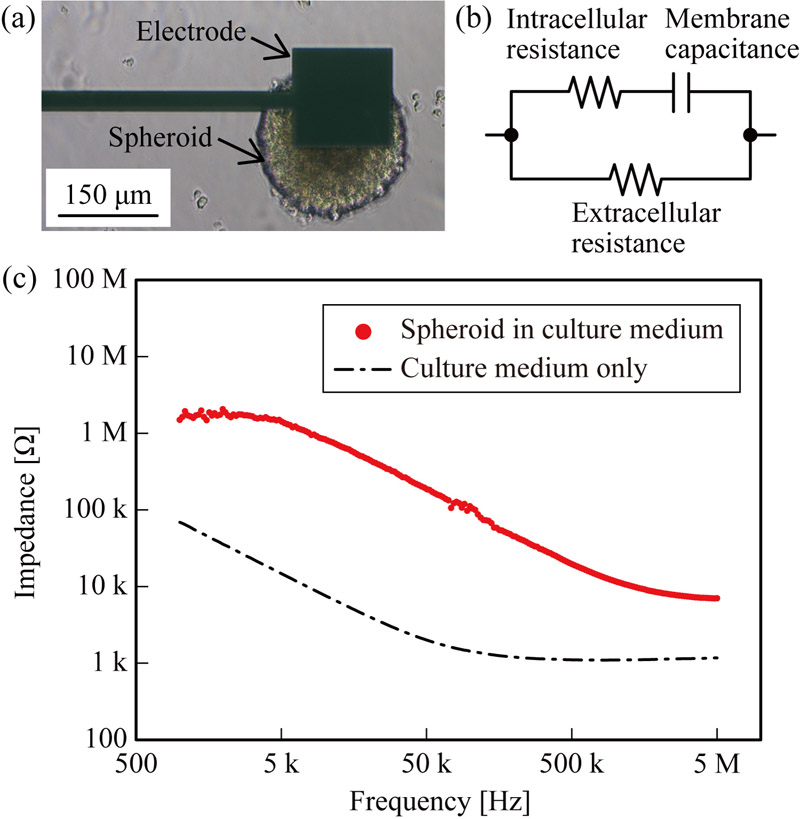
Impedance characterization of a cell spheroid
- [1] R. Langer and J. P. Vacanti, “Tissue Engineering,” Science, Vol.260, Issue 5110, pp. 920-926, 1993. https://doi.org/10.1126/science.8493529
- [2] K. Takahashi and S. Yamanaka, “Induction of Pluripotent Stem Cells from Mouse Embryonic and Adult Fibroblast Cultures by Defined Factors,” Cell, Vol.126, Issue 4, pp. 663-676, 2006. https://doi.org/10.1016/j.cell.2006.07.024
- [3] R. Nasiri, A. Sankaranthi, and G. Pratx, “Organ-on-a-chip systems for modeling tumor and normal tissue microenvironments in radiotherapy research,” Trends in Biotechnology, Vol.44, Issue 2, pp. 333-350, 2026. https://doi.org/10.1016/j.tibtech.2025.07.002
- [4] A. M. Bratt-Leal, R. L. Carpenedo, and T. C. McDevitt, “Engineering the embryoid body microenvironment to direct embryonic stem cell differentiation,” Biotechnology Progress, Vol.25, Issue 1, pp. 43-51, 2009. https://doi.org/10.1002/btpr.139
- [5] S. F. Abu-Absi, J. R. Friend, L. K. Hansen, and W.-S. Hu, “Structural Polarity and Functional Bile Canaliculi in Rat Hepatocyte Spheroids,” Experimental Cell Research, Vol.274, Issue 1, pp. 56-67, 2002. https://doi.org/10.1006/excr.2001.5467
- [6] C. S. Ong, X. Zhou, J. Han, C. Y. Huang, A. Nashed, S. Khatri, G. Mattson, T. Fukunishi, H. Zhang, and N. Hibino, “In vivo therapeutic applications of cell spheroids,” Biotechnology Advances, Vol.36, Issue 2, pp. 494-505, 2018. https://doi.org/10.1016/j.biotechadv.2018.02.003
- [7] S. Sant and P. A. Johnston, “The production of 3D tumor spheroids for cancer drug discovery,” Drug Discovery Today: Technologies, Vol.23, pp. 27-36, 2017. https://doi.org/10.1016/j.ddtec.2017.03.002
- [8] G. H. Underhill and S. R. Khetani, “Bioengineered Liver Models for Drug Testing and Cell Differentiation Studies,” Cellular and Molecular Gastroenterology and Hepatology, Vol.5, Issue 3, pp. 426-439, 2018. https://doi.org/10.1016/j.jcmgh.2017.11.012
- [9] H. Mihara, M. Kugawa, K. Sayo, F. Tao, M. Shinohara, M. Nishikawa, Y. Sakai, T. Akama, and N. Kojima, “Improved Oxygen Supply to Multicellular Spheroids Using a Gas-permeable Plate and Embedded Hydrogel Beads,” Cells, Vol.8, Issue 6, Article No.525, 2019. https://doi.org/10.3390/cells8060525
- [10] G. Mai, N. T. Huy, P. Morel, J. Mei, A. Andres, D. Bosco, R. Baertschiger, C. Toso, T. Berney, P. Majno, G. Mentha, D. Trono, and L. H. Buhler, “Treatment of fulminant liver failure by transplantation of microencapsulated primary or immortalized xenogeneic hepatocytes,” Xenotransplantation, Vol.12, Issue 6, pp. 457-464, 2005. https://doi.org/10.1111/j.1399-3089.2005.00248.x
- [11] S. Pengnam, S. Plianwong, P. Patrojanasophon, W. Radchatawedchakoon, B. Yingyongnarongkul, P. Opanasopit, and P. Charoensuksai, “Synergistic Effect of Doxorubicin and siRNA-Mediated Silencing of Mcl-1 Using Cationic Niosomes against 3D MCF-7 Spheroids,” Pharmaceutics, Vol.13, Issue 4, Article No.550, 2021. https://doi.org/10.3390/pharmaceutics13040550
- [12] L. F. E. Huerta-Nuñez, G. Gutierrez-Iglesias, A. Martinez-Cuazitl, M. M. Mata-Miranda, V. D. Alvarez-Jiménez, V. Sánchez-Monroy, A. Golberg, and C. A. González-Díaz, “A biosensor capable of identifying low quantities of breast cancer cells by electrical impedance spectroscopy,” Scientific Reports, Vol.9, Article No.6419, 2019. https://doi.org/10.1038/s41598-019-42776-9
- [13] Y. Hiraoka, T. Shimi, and T. Haraguchi, “Multispectral Imaging Fluorescence Microscopy for Living Cells,” Cell Structure and Function, Vol.27, Issue 5, pp. 367-374, 2002. https://doi.org/10.1247/csf.27.367
- [14] B. Eker, R. Meissner, A. Bertsch, K. Mehta, and P. Renaud, “Label-Free Recognition of Drug Resistance via Impedimetric Screening of Breast Cancer Cells,” PLOS ONE, Vol.8, Issue 3, Article No.e57423, 2013. https://doi.org/10.1371/journal.pone.0057423
- [15] A. Filipiak-Duliban, K. Brodaczewska, A. Kajdasz, and C. Kieda, “Spheroid Culture Differentially Affects Cancer Cell Sensitivity to Drugs in Melanoma and RCC Models,” Int. J. of Molecular Sciences, Vol.23, Issue 3, Article No.1166, 2022. https://doi.org/10.3390/ijms23031166
- [16] T. Himuro and Y. Saito, “Assessment of Deoxyribonuclease Activity Using DNA Molecules Immobilized Between Microelectrodes,” J. of Electronic Materials, Vol.50, No.2, pp. 537-542, 2021. https://doi.org/10.1007/s11664-020-08596-8
- [17] T. Himuro, S. Tsukamoto, and Y. Saito, “Fabrication of a Deoxyribonuclease Sensor Based on the Electrical Characteristics of DNA Molecules,” ACS Omega, Vol.7, Issue 27, pp. 23549-23554, 2022. https://doi.org/10.1021/acsomega.2c02069
- [18] Y. Wang, H. Wu, Y. Geng, Z. Zhang, J. Fu, J. Ouyang, and Z. Zhu, “Equivalent Circuit Modeling and Analysis for Microfluidic Electrical Impedance Monitoring of Single-Cell Growth,” Biosensors, Vol.15, Issue 2, Article No.113, 2025. https://doi.org/10.3390/bios15020113
- [19] F. Zhang, T. Jin, Q. Hu, and P. He, “Distinguishing skin cancer cells and normal cells using electrical impedance spectroscopy,” J. of Electroanalytical Chemistry, Vol.823, pp. 531-536, 2018. https://doi.org/10.1016/j.jelechem.2018.06.021
- [20] K. Iuchi, K. Oya, K. Hosoya, K. Sasaki, Y. Sakurada, T. Nakano, and H. Hisatomi, “Different morphologies of human embryonic kidney 293T cells in various types of culture dishes,” Cytotechnology, Vol.72, pp. 131-140, 2020. https://doi.org/10.1007/s10616-019-00363-w
- [21] C.-W. Huang, Y.-C. Lin, C.-H. Hung, H.-M. Chen, J.-T. Lin, C.-J. Wang, and S.-H. Kao, “Adenine inhibits the invasive potential of DLD-1 human colorectal cancer cell via the AMPK/FAK axis,” Pharmaceuticals, Vol.14, Issue 9, Article No.860, 2021. https://doi.org/10.3390/ph14090860
- [22] R. Negoro, M. Tasaka, S. Deguchi, K. Takayama, and T. Fujita, “Generation of HepG2 Cells with High Expression of Multiple Drug-Metabolizing Enzymes for Drug Discovery Research Using a PITCh System,” Cells, Vol.11, Issue 10, Article No.1677, 2022. https://doi.org/10.3390/cells11101677
- [23] M. Inbar, “Fluidity of membrane lipids: A single cell analysis of mouse normal lymphocytes and malignant lymphoma cells,” FEBS Letters, Vol.67, Issue 2, pp. 180-185, 1976. https://doi.org/10.1016/0014-5793(76)80361-4
- [24] E. Di Gregorio, S. Israel, M. Staelens, G. Tankel, K. Shankar, and J. A. Tuszyński, “The distinguishing electrical properties of cancer cells,” Physics of Life Reviews, Vol.43, pp. 139-188, 2022. https://doi.org/10.1016/j.plrev.2022.09.003
This article is published under a Creative Commons Attribution-NoDerivatives 4.0 Internationa License.