
Review:
How and Why Do We Measure Cellular Heat?–Significance and History—
Naoki Inomata

Department of Robotics, Graduate School of Engineering, Tohoku University
6-6-01 aza-Aoba, Aramaki, Aoba-ku, Sendai, Miyagi 980-8579, Japan

Temperature is a fundamental physical quantity that governs chemical reaction rates, molecular diffusion, and protein stability, and is therefore intimately linked to biological function. In cells, heat is generated through a variety of nonequilibrium processes, including metabolism and mechanical activity. Cellular thermometry is increasingly recognized not only as a method for measuring temperature but also as a framework for investigating intracellular energy conversion, heat dissipation, and the spatial localization of cellular function. This review examines the significance of thermal measurement in cell-related research and traces the historical development of the field, from bulk calorimetry of cell populations to single-cell and subcellular measurements enabled by optical and microfabricated sensors. We summarize the major methodological platforms, including fluorescent polymeric thermometers, inorganic nanoparticles, fluorescent proteins, small-molecule probes, and contact-based microsensors, highlighting their respective strengths and limitations with regard to spatial and temporal resolution, targeting capability, and quantitative reliability. We also discuss the central controversy in the field, namely the “105 gap issue,” which refers to the discrepancy between experimentally reported intracellular temperature changes and theoretical predictions based on classical heat conduction. Finally, we consider future perspectives. Rather than remaining focused solely on temperature measurement, cellular thermometry is evolving toward a broader framework of cellular thermal analysis that encompasses heat production, heat transport, and cellular thermophysical properties.
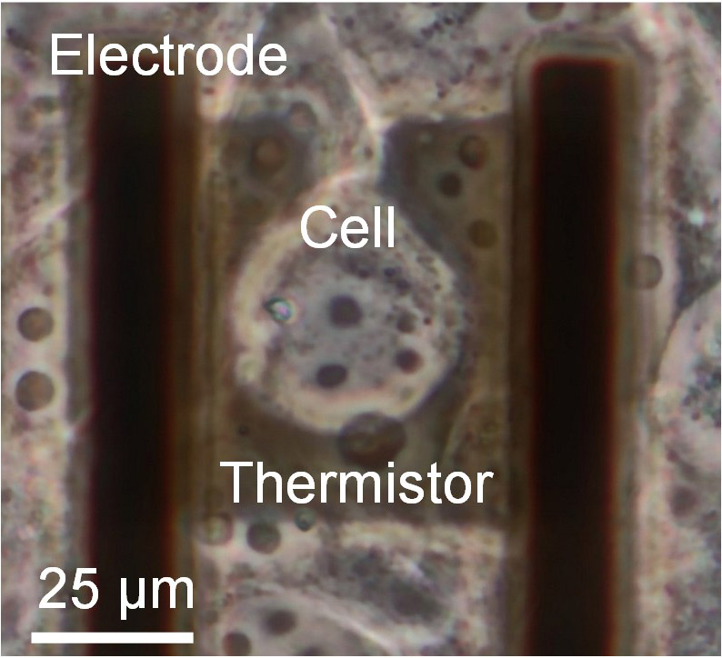
A cell positioned on the microfabricated sensor
- [1] B. D. Knapp and K. C. Huang, “The effects of temperature on cellular physiology,” Annu. Rev. Biophys., Vol.51, pp. 499-526, 2022. https://doi.org/10.1146/annurev-biophys-112221-074832
- [2] H. Matsuki, M. Goto, and N. Tamai, “Membrane states of saturated glycerophospholipids: A thermodynamic study of bilayer phase transitions,” Chem. Pharm. Bull., Vol.67, No.4, pp. 300-307, 2019. https://doi.org/10.1248/cpb.c18-00954
- [3] A. Martinière et al., “Homeostasis of plasma membrane viscosity in fluctuating temperatures,” New Phytol., Vol.192, No.2, pp. 328-337, 2011. https://doi.org/10.1111/j.1469-8137.2011.03821.x
- [4] D. L. Cano-Ramirez et al., “Plasma membrane fluidity: An environment thermal detector in plants,” Cells, Vol.10, No.10, Article No.2778, 2021. https://doi.org/10.3390/cells10102778
- [5] B. D. Sidell and J. R. Hazel, “Temperature affects the diffusion of small molecules through cytosol of fish muscle,” J. Exp. Biol., Vol.129, pp. 191-203, 1987. https://doi.org/10.1242/jeb.129.1.191
- [6] M. Guo, Y. Xu, and M. Gruebele, “Temperature dependence of protein folding kinetics in living cells,” Proc. Natl. Acad. Sci. U.S.A., Vol.109, No.44, pp. 17863-17867, 2012. https://doi.org/10.1073/pnas.1201797109
- [7] X. Fang and J. Wang, “Nonequilibrium thermodynamics in cell biology: Extending equilibrium formalism to cover living systems,” Annu. Rev. Biophys., Vol.49, pp. 227-246, 2020. https://doi.org/10.1146/annurev-biophys-121219-081656
- [8] P. P. Dzeja and A. Terzic, “Phosphotransfer networks and cellular energetics,” J. Exp. Biol., Vol.206, No.12, pp. 2039-2047, 2003. https://doi.org/10.1242/jeb.00426
- [9] C.-P. Hsieh, “Nonequilibrium thermodynamics of ion flux through membrane channels,” Entropy, Vol.19, No.1, Article No.40, 2017. https://doi.org/10.3390/e19010040
- [10] R. A. Gatenby and B. R. Frieden, “Cellular information dynamics through transmembrane flow of ions,” Sci. Rep., Vol.7, Article No.15075, 2017. https://doi.org/10.1038/s41598-017-15182-2
- [11] D. Mizuno, C. Tardin, C. F. Schmidt, and F. C. MacKintosh, “Nonequilibrium mechanics of active cytoskeletal networks,” Science, Vol.315, No.5810, pp. 370-373, 2007. https://doi.org/10.1126/science.1134404
- [12] T. Hohmann and F. Dehghani, “The cytoskeleton—A complex interacting meshwork,” Cells, Vol.8, No.4, Article No.362, 2019. https://doi.org/10.3390/cells8040362
- [13] K. Okabe and S. Uchiyama, “Intracellular thermometry uncovers spontaneous thermogenesis and associated thermal signaling,” Commun. Biol., Vol.4, Article No.1377, 2021. https://doi.org/10.1038/s42003-021-02908-2
- [14] K. Okabe et al., “Intracellular temperature mapping with a fluorescent polymeric thermometer and fluorescence lifetime imaging microscopy,” Nat. Commun., Vol.3, Article No.705, 2012. https://doi.org/10.1038/ncomms1714
- [15] A. Sedlmeier, D. E. Achatz, L. H. Fischer, H. H. Gorris, and O. S. Wolfbeis, “Photon upconverting nanoparticles for luminescent sensing of temperature,” Nanoscale, Vol.4, No.22, pp. 7090-7096, 2012. https://doi.org/10.1039/C2NR32314A
- [16] K. Oyama et al., “Walking nanothermometers: Spatiotemporal temperature measurement of transported acidic organelles in single living cells,” Lab Chip, Vol.12, No.9, pp. 1591-1593, 2012. https://doi.org/10.1039/C2LC00014H
- [17] M. J. Caterina et al., “The capsaicin receptor: A heat-activated ion channel in the pain pathway,” Nature, Vol.389, No.6653, pp. 816-824, 1997. https://doi.org/10.1038/39807
- [18] D. D. McKemy, W. M. Neuhausser, and D. Julius, “Identification of a cold receptor reveals a general role for TRP channels in thermosensation,” Nature, Vol.416, No.6876, pp. 52-58, 2002. https://doi.org/10.1038/nature719
- [19] K. Minor, A. Bjerre-Nielsen, S. S. Jonasdottir, S. Lehmann, and N. Obradovich, “Rising temperatures erode human sleep globally,” One Earth, Vol.5, No.5, pp. 534-549, 2022. https://doi.org/10.1016/j.oneear.2022.04.008
- [20] B. Janzen, “Temperature and mental health: Evidence from helpline calls,” J. Assoc. Environ. Resour. Econ., Vol.12, No.6, pp. 1431-1457, 2025. https://doi.org/10.1086/736751
- [21] B. Plitzko and S. Loesgen, “Measurement of oxygen consumption rate (OCR) and extracellular acidification rate (ECAR) in culture cells for assessment of the energy metabolism,” Bio-protocol, Vol.8, No.10, Article No.e2850, 2018. https://doi.org/10.21769/BioProtoc.2850
- [22] X. Gu, Y. Ma, Y. Liu, and Q. Wan, ‘Measurement of mitochondrial respiration in adherent cells by Seahorse XF96 Cell Mito Stress Test,’’ STAR Protoc., Vol.2, No.1, Article No.100245, 2021. https://doi.org/10.1016/j.xpro.2020.100245
- [23] H. Imamura et al., “Visualization of ATP levels inside single living cells with fluorescence resonance energy transfer-based genetically encoded indicators,” Proc. Natl. Acad. Sci. U.S.A., Vol.106, No.37, pp. 15651-15656, 2009. https://doi.org/10.1073/pnas.0904764106
- [24] S. Ley-Ngardigal and G. Bertolin, “Approaches to monitor ATP levels in living cells: Where do we stand?,” FEBS J., Vol.289, No.24, pp. 7940-7969, 2022. https://doi.org/10.1111/febs.16169
- [25] Y. Zhao et al., “In vivo monitoring of cellular energy metabolism using SoNar, a highly responsive sensor for NAD/NADH redox state,” Nat. Protoc., Vol.11, No.8, pp. 1345-1359, 2016. https://doi.org/10.1038/nprot.2016.074
- [26] T. S. Blacker et al., “Separating NADH and NADPH fluorescence in live cells and tissues using FLIM,” Nat. Commun., Vol.5, Article No.3936, 2014. https://doi.org/10.1038/ncomms4936
- [27] I. Wadsö, “Trends in isothermal microcalorimetry,” Chem. Soc. Rev., Vol.26, pp. 79-86, 1997. https://doi.org/10.1039/CS9972600079
- [28] E. Gnaiger and R. B. Kemp, “Anaerobic metabolism in aerobic mammalian cells: Information from the ratio of calorimetric heat flux and respirometric oxygen flux,” Biochim. Biophys. Acta Bioenerg., Vol.1016, No.3, pp. 328-332, 1990. https://doi.org/10.1016/0005-2728(90)90164-Y
- [29] A. Schön and I. Wadsö, “Microcalorimetric measurements on tissue cells attached to microcarriers in stirred suspension,” J. Biochem. Biophys. Methods, Vol.13, No.3, pp. 135-143, 1986. https://doi.org/10.1016/0165-022X(86)90086-2
- [30] J. S. Donner, S. A. Thompson, M. P. Kreuzer, G. Baffou, and R. Quidant, “Mapping intracellular temperature using green fluorescent protein,” Nano Lett., Vol.12, No.4, pp. 2107-2111, 2012. https://doi.org/10.1021/nl300389y
- [31] G. Kucsko et al., “Nanometre-scale thermometry in a living cell,” Nature, Vol.500, No.7460, pp. 54-58, 2013. https://doi.org/10.1038/nature12373
- [32] S. Arai, S.-C. Lee, D. Zhai, M. Suzuki, and Y. T. Chang, “A molecular fluorescent probe for targeted visualization of temperature at the endoplasmic reticulum,” Sci. Rep., Vol.4, Article No.6701, 2014. https://doi.org/10.1038/srep06701
- [33] S. Kiyonaka et al., “Genetically encoded fluorescent thermosensors visualize subcellular thermoregulation in living cells,” Nat. Methods, Vol.10, No.12, pp. 1232-1238, 2013. https://doi.org/10.1038/nmeth.2690
- [34] K. Okabe, R. Sakaguchi, B. Shi, and S. Kiyonaka, “Intracellular thermometry with fluorescent sensors for thermal biology,” Pflügers Arch., Vol.470, No.5, pp. 717-731, 2018. https://doi.org/10.1007/s00424-018-2113-4
- [35] J. Qiao, X. Mu, and L. Qi, “Construction of fluorescent polymeric nano-thermometers for intracellular temperature imaging: A review,” Biosens. Bioelectron., Vol.85, pp. 403-413, 2016. https://doi.org/10.1016/j.bios.2016.04.070
- [36] T. Sekiguchi, S. Sotoma, and Y. Harada, “Fluorescent nanodiamonds as a robust temperature sensor inside a single cell,” Biophys. Physicobiol., Vol.15, pp. 229-234, 2018. https://doi.org/10.2142/biophysico.15.0_229
- [37] G. Baffou, H. Rigneault, D. Marguet, and L. Jullien, “A critique of methods for temperature imaging in single cells,” Nat. Methods, Vol.11, No.9, pp. 899-901, 2014. https://doi.org/10.1038/nmeth.3073
- [38] S. Kiyonaka et al., “Validating subcellular thermal changes revealed by fluorescent thermosensors,” Nat. Methods, Vol.12, No.9, pp. 801-802, 2015. https://doi.org/10.1038/nmeth.3548
- [39] M. Suzuki, V. Zeeb, S. Arai, K. Oyama, and S. Ishiwata, “The gap issue between calculation and measurement in single-cell thermometry,” Nat. Methods, Vol.12, No.9, pp. 802-803, 2015. https://doi.org/10.1038/nmeth.3551
- [40] G. Baffou, H. Rigneault, D. Marguet, and L. Jullien, “Reply to: ‘Validating Subcellular Thermal Changes Revealed by Fluorescent Thermosensors’ and ‘The Gap Issue Between Calculation and Measurement in Single-Cell Thermometry’,” Nat. Methods, Vol.12, No.9, p. 803, 2015. https://doi.org/10.1038/nmeth.3552
- [41] E. A. Johannessen, J. M. R. Weaver, P. H. Cobbold, and J. M. Cooper, “Heat conduction nanocalorimeter for pl-scale single cell measurements,” Appl. Phys. Lett., Vol.80, No.11, pp. 2029-2031, 2002. https://doi.org/10.1063/1.1457532
- [42] C. Wang et al., “Determining intracellular temperature at single-cell level by a novel thermocouple method,” Cell Res., Vol.21, No.10, pp. 1517-1519, 2011. https://doi.org/10.1038/cr.2011.117
- [43] R. T. ElAfandy et al., “Nanomembrane-based, thermal-transport biosensor for living cells,” Small, Vol.13, No.7, Article No.1603080, 2017. https://doi.org/10.1002/smll.201603080
- [44] N. Inomata, M. Toda, M. Sato, A. Ishijima, and T. Ono, “Pico calorimeter for detection of heat produced in an individual brown fat cell,” Appl. Phys. Lett., Vol.100, No.15, Article No.154104, 2012. https://doi.org/10.1063/1.3701720
- [45] N. Inomata, M. Toda, and T. Ono, “Highly sensitive thermometer using a vacuum-packed Si resonator in a microfluidic chip for the thermal measurement of single cells,” Lab Chip, Vol.16, No.18, pp. 3597-3603, 2016. https://doi.org/10.1039/C6LC00949B
- [46] T. Yamada, N. Inomata, and T. Ono, “Sensitive thermal microsensor with pn junction for heat measurement of a single cell,” Jpn. J. Appl. Phys., Vol.55, No.2, Article No.027001, 2016. https://doi.org/10.7567/JJAP.55.027001
- [47] N. Inomata, R. Inaoka, K. Okabe, T. Funatsu, and T. Ono, “Short-term temperature change detections and frequency signals in single cultured cells using a microfabricated thermistor,” Sens. Bio-Sens. Res., Vol.27, Article No.100309, 2020. https://doi.org/10.1016/j.sbsr.2019.100309
- [48] N. Inomata, T. Miyamoto, K. Okabe, and T. Ono, ‘Measurement of cellular thermal properties and their temperature dependence based on frequency spectra via an on-chip-integrated microthermistor,’’ Lab Chip, Vol.23, No.10, pp. 2411-2420, 2023. https://doi.org/10.1039/D2LC01185A
- [49] N. Inomata, “Cellular temperature sensing techniques leveraging advantages of micro/nano-thermometers,” IEEJ Trans. Electr. Electron. Eng., Vol.20, No.9, pp. 1326-1332, 2025. https://doi.org/10.1002/tee.70091
This article is published under a Creative Commons Attribution-NoDerivatives 4.0 Internationa License.