
Review:
How and Why Do We Measure Cellular Heat?–Significance and History—
Naoki Inomata

Department of Robotics, Graduate School of Engineering, Tohoku University
6-6-01 aza-Aoba, Aramaki, Aoba-ku, Sendai, Miyagi 980-8579, Japan

Temperature is a fundamental physical quantity that governs chemical reaction rates, molecular diffusion, and protein stability, and is therefore intimately linked to biological function. In cells, heat is generated through a variety of nonequilibrium processes, including metabolism and mechanical activity. Cellular thermometry is increasingly recognized not only as a method for measuring temperature but also as a framework for investigating intracellular energy conversion, heat dissipation, and the spatial localization of cellular function. This review examines the significance of thermal measurement in cell-related research and traces the historical development of the field, from bulk calorimetry of cell populations to single-cell and subcellular measurements enabled by optical and microfabricated sensors. We summarize the major methodological platforms, including fluorescent polymeric thermometers, inorganic nanoparticles, fluorescent proteins, small-molecule probes, and contact-based microsensors, highlighting their respective strengths and limitations with regard to spatial and temporal resolution, targeting capability, and quantitative reliability. We also discuss the central controversy in the field, namely the “105 gap issue,” which refers to the discrepancy between experimentally reported intracellular temperature changes and theoretical predictions based on classical heat conduction. Finally, we consider future perspectives. Rather than remaining focused solely on temperature measurement, cellular thermometry is evolving toward a broader framework of cellular thermal analysis that encompasses heat production, heat transport, and cellular thermophysical properties.
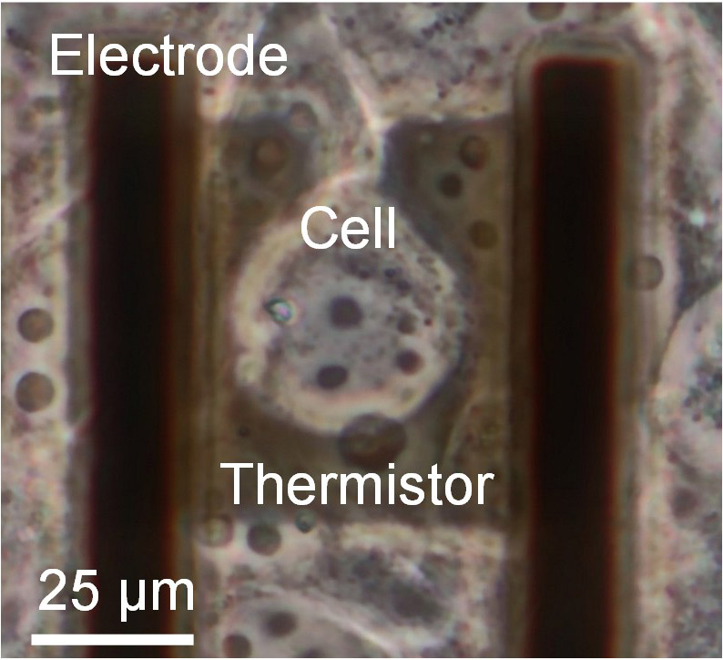
A cell positioned on the microfabricated sensor
1. Introduction
Temperature is a fundamental physical quantity that governs chemical reaction rates, molecular diffusion, phase transitions, and the structural stability of proteins 1,2,3,4,5,6. It is therefore indispensable for understanding biological phenomena. Within cells, numerous physiological processes, including ATP production, ion transport, membrane trafficking, transcription and translation, cytoskeletal dynamics, and inter-organelle interactions, involve energy conversion and heat dissipation, and their rates and functional states are influenced by temperature 1,7,8,9,10,11,12. Clarifying where heat is generated within cells, how it is transported and dissipated, and how it is linked to cellular functions is therefore essential for understanding life at the cellular level. From this perspective, Okabe and Uchiyama proposed the concept of “thermal signaling,” whereby intracellular temperature changes may function as intracellular signals 13. Traditionally, cell biology has regarded temperature primarily as an external environmental parameter. Although culture and stimulation temperatures have long been recognized as important environmental variables, the spatial and temporal distribution of temperature as a physical quantity has not been a primary focus of investigation. Since the 2010s, however, advances in fluorescent nanothermometry and highly sensitive microscale thermal sensors have made local temperature measurements in living cells increasingly feasible, and cellular thermometry has rapidly emerged as an independent research field 14,15,16. In particular, the intracellular temperature mapping reported by Okabe et al. in 2012 using a fluorescent polymeric thermometer (FPT) and fluorescence lifetime imaging microscopy (FLIM) is widely recognized as one of the earliest breakthroughs in this field 14.
Temperature is also a biological variable that broadly influences biological systems, from cells to tissues and whole organisms. At the tissue and organismal levels, temperature is not only regulated as a physiological parameter but also sensed and interpreted as biological information. For example, the heat-activated ion channel TRPV1 and the cold- and menthol-sensitive ion channel TRPM8 provide molecular bases for thermal sensation, demonstrating that thermal stimuli can be converted into neural signals 17,18. Temperature further influences tissue and organismal states through thermoregulation, immune responses, sleep, and behavioral adaptation, while ambient temperature has also been associated with psychological and mental health outcomes 19,20. Thus, temperature should be viewed not merely as an environmental condition or a passive consequence of metabolism, but as a biological information parameter that is generated, sensed, transmitted, and interpreted across cells, tissues, and organisms. From the perspective of sensor-based information transmission, cellular thermometry is particularly important because it addresses the origin of such thermal information at the smallest functional unit of life. Measuring when, where, and how heat is generated and dissipated within cells provides a foundation for understanding the links between thermal information and intracellular functions, and its integration across tissue and organismal levels.
This review first examines the need for thermal measurements in cell-related research and then outlines the historical development of the field, from bulk measurements of cell populations to measurements at the single-cell and subcellular levels. It also discusses the major current methodologies, representative controversies, unresolved issues, and future perspectives. Recent reviews have suggested that cellular thermometry is evolving beyond the simple acquisition of “temperature values” toward a broader framework of “cellular thermal analysis,” encompassing heat production, heat transport, and thermophysical properties. The present article is written from this perspective.
2. Significance of Cellular Thermal Measurement
Thermal measurement in cells is important because heat is not merely a byproduct of biological activity; rather, it serves both as a signature of intracellular energy conversion and as a parameter that helps define the local physical environment. Many intracellular processes—including metabolism, molecular transport, mechanical motion, and molecular assembly—are inherently nonequilibrium phenomena that require continuous energy input and are accompanied by energy dissipation. Heat therefore indicates not only how active a cell is, but also where energy is consumed and where it is dissipated. The significance of intracellular thermometry lies not simply in determining temperature itself, but in using heat as a probe of the spatial and temporal organization of intracellular energy conversion. In this regard, heat provides information that is complementary to conventional metabolic indicators, such as the oxygen consumption rate (OCR), ATP concentration, and the NADH/NAD\(^{+}\) ratio. Specifically, OCR primarily reflects mitochondrial respiratory activity 21,22, ATP concentration represents the cellular energetic state 23,24, and the NADH/NAD\(^{+}\) ratio indicates the cellular redox and metabolic state 25,26. All of these are useful readouts; however, they primarily reflect specific pathways or cellular states and do not directly represent the totality of energy consumption and dissipation throughout the cell. Heat, by contrast, represents the ultimate outcome of energy that is consumed and eventually dissipated within the cell. In this sense, it can be regarded as an integrated output that transcends individual pathways. Measuring heat is therefore not merely a means of tracking a single metabolic pathway, but rather a means of obtaining a broader view of the cellular mode of energy conversion.
Equally important is the concept that heat should be regarded not only as a global quantity but also as a local phenomenon. A cell is not a homogeneous reaction vessel; rather, it is a heterogeneous system that is spatially compartmentalized into structures such as the nucleus, mitochondria, endoplasmic reticulum, Golgi apparatus, lipid droplets, and the cytoskeleton. Consequently, even when heat is generated, it need not be uniformly distributed throughout the cell. Instead, heat may be concentrated near specific organelles, membrane-proximal regions, or sites enriched in molecular machinery. Measuring heat therefore entails not treating the cell as a single, spatially averaged entity, but rather asking where and through what processes energy conversion occurs. Early studies of intracellular temperature mapping attracted considerable attention precisely because they suggested the possibility of such spatial localization.
Heat may also be more than a consequence of cellular activity; in some cases, it may itself mediate cellular function. Okabe and Uchiyama proposed the concept of “thermal signaling,” suggesting that intracellular temperature changes may serve as biological signals. They further argued that high-resolution intracellular thermometry has not only enabled the visualization of thermogenic organelles but has also introduced a new perspective in which temperature fluctuations may participate in intracellular event cascades 13. Therefore, thermal measurement becomes more than a passive readout. It provides an observational axis for investigating what processes are being driven within the cell and what may be selectively regulated by temperature. If local temperature changes influence enzymatic reaction rates, phase separation, membrane permeability, molecular diffusion, or the assembly and dissociation of proteins, then measuring heat effectively amounts to probing the conditions under which intracellular reactions become possible 1,2,3,4,5,6. Cellular thermometry can therefore be viewed as an approach to interpreting biological phenomena in terms of energy flow and dissipation 7,8. Chemical analysis reveals what is present, whereas imaging reveals where it is located. Thermal measurement, in contrast, has the potential to show where energy is consumed and where it is dissipated. It may provide a means of capturing the spatial localization of cellular functions. This is because major cellular functions are not uniformly distributed throughout the cell but are instead spatially partitioned 11,12. If heat emerges as a consequence of energy conversion, then its spatial distribution may reflect this functional organization 7,8. Asking where heat is generated is therefore closely linked to asking where cellular functions occur. From this perspective, intracellular thermometry may serve as a more direct functional readout. Okabe and Uchiyama also emphasized the importance of combining intracellular thermometry with conventional molecular and cell biological approaches to establish links between temperature readouts and biological function 13. Accordingly, the primary goal of cellular thermometry is not simply to determine the average temperature of a cell. Rather, it is to decipher the spatial distribution of intracellular functions and the localization of energy conversion through the spatial distribution of heat. From this perspective, cellular thermometry should be regarded not merely as a temperature measurement technique, but as a methodology for understanding intracellular heterogeneity in terms of energy conversion and dissipation.
3. Historical Development of Cellular Thermal Measurement Technologies
3.1. Classical Calorimetry and Bulk Cellular Thermometry
Cellular thermal measurement began with bulk approaches, in which the total heat generated by cell populations—such as cell suspensions, cultured cells, or tissue fragments—was quantified as heat flow using isothermal heat-conduction microcalorimetry, typically for populations comprising on the order of \(10^{5}\)–\(10^{6}\) cells. The underlying principle is to convert the temperature difference between a sample (cells) and a reference (e.g., culture medium) under isothermal conditions into an electrical signal, thereby enabling the continuous measurement of heat flow, or heat production rate, on the microwatt (μW) scale 27. At this stage, the primary objective was to interpret cellular metabolic states through heat production. By around 1990, an analytical framework had been established in which heat flow from cultured cells was combined with oxygen consumption measurements (respirometry), and metabolic modes were characterized in terms of the heat-flow/oxygen-flow ratio (the calorimetric–respirometric ratio) 28. In 1986, cultured adherent cells attached to microcarriers—small scaffold beads employed for the large-scale suspension culture of adherent cells—could be analyzed under stirred suspension while maintaining physiological conditions that closely approximated those of conventional cell culture 29. This development made it possible to measure not only suspended cells but also cells retained under standard culture conditions. The temperature sensors used in these bulk measurements were electrical devices, such as thermocouples and thermistors. However, reports of cellular temperature measurements using comparable approaches appear to have remained relatively limited in the scientific literature until 2002.
3.2. The Shift Toward Single-Cell Measurement: Breakthroughs Enabled by Optical Methods
A major turning point in cellular thermal measurement came in 2012. In that year, Okabe et al. reported intracellular temperature mapping using an FPT in combination with fluorescence lifetime imaging 14. Sedlmeier et al. reported localized intracellular temperature measurements using upconversion nanoparticles that emit visible light under near-infrared excitation 15. Oyama et al. developed a method in which temperature-sensitive nanoparticles were introduced into organelles and used to monitor local temperature while moving within cells 16. Intracellular temperature mapping based on the fluorescence polarization anisotropy of GFP was also reported 30. Collectively, these studies suggested that even within the same cell population, individual cells may exhibit distinct thermal behaviors and that temperature may also be spatially heterogeneous within a single cell. If heat production is inherently a local phenomenon, its essential characteristics may be obscured by population-averaged measurements. These findings highlighted the need for single-cell thermometry and marked a shift in the field from an emphasis on the “average” to the “local.”
This period was followed by reports of local intracellular thermometry based on quantum sensing using nitrogen-vacancy centers in nanodiamonds by Kucsko et al. 31, endoplasmic-reticulum-selective thermal imaging employing small-molecule fluorescent probes by Arai et al. 32, and efforts to directly measure organelle-specific heat production using organelle-targeted GFP-based sensors 33. A key conceptual breakthrough of this era was that temperature was no longer measured through direct physical contact but was instead transduced into temperature-dependent optical signals—such as fluorescence intensity ratios, fluorescence lifetimes, and spectral shifts—for optical readout. This development opened the possibility of non-contact, high-spatial-resolution measurements of local temperature within living cells.
Optical approaches to intracellular thermometry can be broadly classified into thermoresponsive polymeric probes, inorganic nanoparticles, fluorescent proteins, and small-molecule fluorescent probes (organic dyes) (Fig. 1) 13,14,34. These approaches have attracted considerable interest because they enable the non-contact, high-spatial-resolution visualization of temperature changes within single cells. At the same time, they differ substantially in their temperature-sensing mechanisms, intracellular delivery strategies, targeting capabilities, and quantitative reliability.
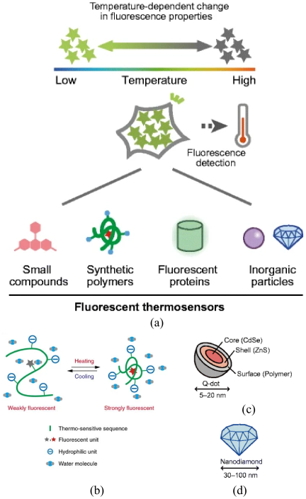
Fig. 1. Schematic illustration of intracellular thermometry using fluorescent thermosensors (a), together with schematic representations of the operating principles of FPTs in an aqueous medium (b), quantum dots (c), and fluorescent nanodiamonds (d). Panels (a), (c), and (d) are reproduced from Reference 34 under the Creative Commons Attribution 4.0 International License (CC BY 4.0; https://creativecommons.org/licenses/by/4.0/), and panel (b) is reproduced from Reference 14 under the Creative Commons Attribution-NonCommercial-ShareAlike 3.0 Unported License (CC BY-NC-SA 3.0; https://creativecommons.org/licenses/by-nc-sa/3.0/).
Thermoresponsive polymeric thermometers were among the representative tools that supported the early demonstrations of intracellular temperature mapping because they exploit temperature-dependent changes in fluorescence intensity, fluorescence lifetime, or ratiometric signals for temperature readout 14,34,35. Designs that incorporate multiple functional units into a polymer backbone provide considerable flexibility for tuning temperature sensitivity and implementing internal referencing, making them well suited for wide-field imaging and FLIM 14,35. At the same time, the influence of intracellular environmental factors other than temperature on the fluorescence response cannot always be completely excluded, and caution is therefore required when interpreting the measured signals 34,35. In addition, heterogeneity in probe distribution and localization within the cytoplasm may contribute to uncertainty in the spatial comparison of absolute temperatures.
Representative inorganic nanoparticle thermometers include upconversion nanoparticles and nanodiamonds, which generally exhibit high photostability and are well suited for long-term observation 15,31,34,36. These systems provide substantial flexibility because multiple physical quantities—such as emission wavelength ratios, luminescence lifetimes, and spin states—can serve as temperature indicators. Upconversion nanoparticles are particularly attractive because near-infrared excitation reduces background autofluorescence and light scattering 15. However, intracellular delivery pathways and localization depend on particle design and cellular uptake mechanisms, and in practice these particles may preferentially accumulate in intracellular compartments such as endosomes or lysosomes 16,34. In addition, particle–cell interactions, biocompatibility, and the influence of non-thermal factors on the temperature readout must be carefully evaluated.
Fluorescent protein-based thermometers can be genetically expressed within cells, eliminating the need for exogenous probe delivery and making them well suited for long-term observation and lineage tracing 30,33,34. Through signal peptides and fusion-protein design, they can also be targeted to specific organelles, such as mitochondria, the endoplasmic reticulum, and the nucleus, making them advantageous for organelle-selective thermal imaging. However, because fluorescence responses may also be influenced by biochemical and optical factors other than temperature, caution is required before directly interpreting fluorescence changes as absolute temperature changes. Indeed, the reliability of intracellular thermal readout using GFP-based thermometers remains a subject of ongoing debate.
Small-molecule fluorescent probes (organic dyes) offer a high degree of flexibility in molecular design, enabling the relatively facile tuning of temperature sensitivity, response kinetics, and targeting moieties 32,34. Consequently, they are well suited for local temperature measurements targeted to specific organelles, and highly sensitive endoplasmic-reticulum-selective thermal probes have indeed been reported. In addition, small-molecule probe systems are comparatively easy to introduce exogenously and can be chemically modified to suit the intended application. On the other hand, photobleaching, cellular leakage, metabolic modification or degradation, and cross-sensitivity to environmental factors other than temperature may limit their applicability, particularly for long-term observation and absolute temperature quantification. As a result, although small-molecule probes are well suited for detecting local and relative temperature changes, rigorous quantitative thermometry requires careful calibration under the specific measurement conditions employed 32,34.
3.3. A Major Point of Debate: The \(10^{5}\) Gap Issue
Any discussion of cellular thermometry must address the so-called “\(10^{5}\) gap issue.” This refers to the observation that temperature increases reported by single-cell measurements are approximately five orders of magnitude larger than those predicted by heat-conduction calculations. In other words, the experimentally measured values and theoretical estimates differ by a factor of approximately \(10^{5}\). In 2014, Baffou et al. argued that, based on simple heat-conduction calculations and scaling laws, large steady-state temperature gradients within a single cell arising solely from endogenous heat generation are difficult to justify on physical grounds 37. This argument cast doubt on the physical consistency of the large local temperature changes reported at the time using fluorescent thermometers and raised the question of whether the observed signals genuinely reflected temperature. Since then, the debate has extended to encompass measurement principles, calibration strategies, theoretical models, and the understanding of intracellular heat transport. In 2015, Kiyonaka et al. responded by emphasizing the need for careful validation when interpreting temperature changes observed within single cells, while also supporting the plausibility of subcellular thermal changes detected using fluorescent temperature sensors 38. Around the same time, Suzuki et al. explicitly framed the discrepancy between theoretical calculations and experimental measurements as the “\(10^{5}\) gap issue” and argued that it could largely be resolved by properly accounting for several factors: the cell type and the presence or type of stimulation (approximately one order of magnitude), the distance from the heat source used to evaluate the temperature rise (approximately one to two orders of magnitude), and the thermophysical properties of the cell, once these are better characterized (approximately one to two orders of magnitude) 39. Baffou et al. subsequently responded, arguing that even if local intracellular heating were inferred from experimental measurements, such observations remained inconsistent with existing theoretical estimates 40. The 2018 review by Okabe et al. further suggested that the straightforward application of macroscopic heat-diffusion models, which treat the cell interior as a homogeneous, continuous aqueous phase, may have important limitations 34. They argued that these models should be re-examined by taking intracellular heterogeneity and the spatial scale of heat sources into account, and they identified the clarification of the chemical identity of intracellular heat sources and the development of mesoscopic nonequilibrium thermodynamics as important future challenges. The issue has not yet been fully resolved.
3.4. Contact-Based Approaches Using Microfabricated Sensors
Although optical methods are now widely associated with single-cell temperature measurement, the first realization of single-cell thermometry was in fact reported by Johannessen et al. in 2002. Advances in microfabrication technology during the 2000s enabled the miniaturization of calorimetry, thereby reducing sample volumes while increasing measurement sensitivity. Johannessen et al. developed a heat-conduction nanocalorimeter based on silicon microfabrication for pL-scale samples and reported the detection of small thermal events at the level of one to a few cells 41. Another approach employed an intracellular thermocouple probe consisting of a tungsten needle and a platinum film, with a tip diameter of approximately 500 nm, to directly measure temperature changes within single cells during drug stimulation. The reported temperature resolution was below 0.1 °C, and the response time was estimated by simulation to be approximately 400 ns 42. In addition, a GaN-nanomembrane-based optical microdevice was used to evaluate the thermal transport properties of cells by detecting temperature-dependent photoluminescence shifts, and thermal conductivities of several cancer cell lines were approximately 0.5–0.6 W m\(^{-1}\) K\(^{-1}\) 43. Although several microscale temperature sensors capable of measuring small-volume biological samples, including biomolecules, have since been reported, published studies involving measurements on living cells remain extremely limited.
The authors have progressively advanced micro-temperature sensors based on microfabrication technology. By complementing the dominant optical approaches with high temperature resolution and rapid response, they have reported previously unobserved thermal phenomena in cells, as well as the quantitative evaluation of cellular thermophysical properties. In an early study published in 2012, a microsensor based on mechanical resonance was used to measure the temperature of a single suspended cell (Figs. 2(a)–(c)) 44. Conventionally, mechanical resonant sensors are operated under vacuum to minimize vibrational damping and were therefore considered incompatible with live-cell measurements in aqueous environments. This limitation was overcome by integrating the resonant sensor into a microfluidic chip such that it extended through both a fluidic region containing the cells and a physically separated vacuum region. By adopting a doubly clamped beam structure, the system ultimately achieved a temperature resolution of 79 μK and a heat resolution of 1.90 nW 45. An important biological finding of this study was that cells exhibited periodic, pulse-like heat generation even in the absence of chemical stimulation (Fig. 2(d)). While gradual temperature increases over several tens of minutes in response to chemical stimulation had already been documented during the era of bulk-calorimetry, such periodic, pulse-like heat generation had not previously been reported. Although this finding was initially met with skepticism, the same phenomenon was subsequently observed when the sensing element was replaced with a thermistor, supporting the conclusion that the measured signal originated from the cell 46.
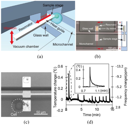
Fig. 2. Resonant thermal sensor for detecting heat generation from living cells in liquid: (a) schematic illustration of the device, (b) optical image of the fabricated device and schematic cross-sectional view, (c) a cell attached to the sample stage, and (d) representative temperature changes detected in non-stimulated cells. Panels (a)–(d) are reproduced from Figs. 1, 3, 5, and 6 of Reference 44, respectively.
The target of measurement was subsequently extended to cultured cells. In a 2020 study, a device incorporating a VO\(_{2}\) microthermistor was employed to analyze the frequency spectrum of temperature signals from a single cultured cell 47. This work was motivated by the earlier observation of periodic heat generation, and the frequency spectrum was found to vary with the ambient temperature. Specifically, when temperature signals acquired at 25 °C and 37 °C were compared, the signal intensity was greater at 37 °C, particularly in the frequency range below 2 Hz. Because 37 °C corresponds to the temperature range in which cells are highly active, these results suggested that intracellular biochemical processes may exhibit periodicity and that their dominant frequency components may lie below 2 Hz. Gradual temperature increases induced by chemical stimulation were also observed in this system, confirming its capability to detect heat generation from single cells. At that time, the system achieved a temperature resolution of 11 m°C and a response time of less than 0.1 s.
More recent studies have gone beyond the observation of temperature changes and have achieved the quantitative evaluation of thermophysical properties, such as the thermal conductivity and specific heat of single cells. In a 2023 Lab on a Chip paper, an on-chip-integrated microthermistor combined with localized heating by a focused infrared laser was employed to construct a measurement system in which thermal conductivity was derived from the steady-state temperature rise during heating, whereas specific heat was determined from the transient temperature-rise response (Figs. 3(a)–(f)) 48. Thermal conductivity at the single-cell level had been reported in several earlier studies because it can be obtained from the steady-state temperature rise. Specific heat, by contrast, requires dynamic measurements, and its determination at the single-cell level was therefore demonstrated for the first time in that study. The study further showed that these thermophysical properties depend on environmental temperature and heating frequency (Fig. 4). It also suggested that cellular temperature signals may be controlled by external heating; that is, biochemical reactions may potentially be induced or regulated by heating applied within the same frequency band as the cell’s intrinsic heat generation. In addition to the studies introduced here, single-cell thermometry using microfabricated sensors has also been reported by other groups, and readers are referred to Reference 49 for further details. Table 1 summarizes the representative cellular temperature measurement techniques discussed in this review, highlighting their differences in detection principle, temperature resolution, spatial resolution, and response time.
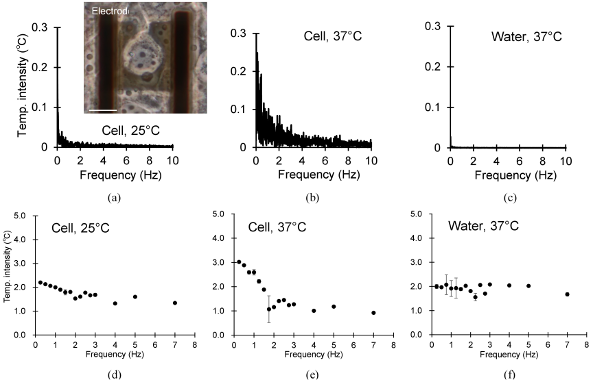
Fig. 3. Frequency spectra of spontaneous cellular thermogenesis and water under different ambient temperatures, measured without laser heating (a–c) and with periodic laser heating (d–f). The inset shows a cell positioned on the sensor. Panels (a) and (d) correspond to a cell at 25 °C, panels (b) and (e) to a cell at 37 °C, and panels (c) and (f) to water at 37 °C. These panels are reproduced from Figs. 1, 4, and 5 of Reference 48.
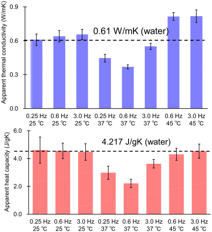
Fig. 4. Dependence of the thermophysical properties of cells on ambient temperature and laser irradiation frequency: (top) thermal conductivity as a function of temperature and laser irradiation frequency, and (bottom) specific heat as a function of temperature and local heating frequency. These panels are reproduced from Fig. 7 of Reference 48.
Another important question is why single-cell temperature measurement using microfabricated sensors did not attract widespread attention for a long time, despite having been realized earlier than thermosensitive nanoparticles. Initially, cellular temperature measurements using microfabricated devices focused on responses to known chemical stimuli and were positioned more as applications for validating device performance than as tools for discovering previously unknown phenomena or elucidating the physical principles underlying cellular behavior. As a result, the biochemical insights obtained from such measurements were relatively limited, and these studies were cited less frequently within the biophysics community. These studies subsequently helped establish microfabricated thermal sensors not only as demonstrations of device technology but also as powerful tools for investigating cellular thermal phenomena.
| Section | Materials | Detection | Resolutions | Response | Reference | |
| Temperature [°C] | Spatial | |||||
| Synthetic polymer | Polyacrylamide polymer | Fluorescence intensity ratio | 0.01–0.25 |
240 nm (diffraction limit) |
1 min | 14 |
| Fluorescent protein | Fluorescent protein | Fluorescence intensity ratio | 0.1–0.5 | NA | 50 ms | 33 |
| Inorganic materials | Nanodiamond | Normalized fluorescence | 0.044 | 50 nm | few ms | 31 |
| Nanoneedle thermocouple | Thermoelectric potential | 0.1 | appr. 500 nm |
400 ns (simulation) |
42 | |
| Thermistors | Resistivity change | 0.011 |
50 μm (sensor size) |
10 ms (sampling time) |
48 | |
4. Future Perspectives
Single-cell temperature measurement was first realized in 2002 using microfabricated sensors, and the field underwent a major breakthrough in 2012 through the development of optical approaches centered on thermosensitive nanoparticles. More than a decade has passed since the “\(10^{5}\) gap issue” was first raised. Numerous approaches based on different measurement principles have since been used to investigate thermal phenomena in cells, yet the reported magnitudes of temperature change are broadly consistent across studies employing both optical probes and microfabricated sensors 14,15,16,30,31,32,33,44,45,46,47,48. Taken together, these observations suggest that at least some cellular temperature signals have been reproducibly detected across multiple measurement platforms. At the same time, the “\(10^{5}\) gap issue” remains unresolved, suggesting that aspects of intracellular thermal behavior may not yet be fully explained by existing macroscopic physical models. Despite major advances in measurement technology, the underlying nature of this phenomenon remains unclear. Nevertheless, the field continues to make steady progress.
A recurring question is how far the fundamental performance of temperature sensors—particularly in terms of temperature resolution and temporal response—must be improved before it can be considered sufficient. At present, however, this question cannot be answered definitively because the underlying thermal phenomena occurring within cells remain incompletely understood. As sensor technology continues to advance, the required levels of temperature resolution and temporal response will likely be redefined through a recurring cycle of “observation of unknown phenomena \(\to\) quantification of unknown phenomena \(\to\) observation of new unknown phenomena.” If the immediate goal is to understand the physical phenomena occurring within cells, pursuing ever-higher temperature resolution and temporal response alone may not be the highest priority. Rather, a more pressing challenge is to elucidate the physics governing how local, and ultimately, global heat transport occurs in cells subjected to appropriate stimuli under conditions that preserve normal cellular function. Achieving this goal will require interdisciplinary research integrating measurement technology, thermal physics, cell biology, chemistry, and biophysics. Cellular thermometry is therefore expected to evolve beyond the mere observation of temperature into a methodology for integratively elucidating cellular energy conversion and the spatial localization of cellular function.
- [1] B. D. Knapp and K. C. Huang, “The effects of temperature on cellular physiology,” Annu. Rev. Biophys., Vol.51, pp. 499-526, 2022. https://doi.org/10.1146/annurev-biophys-112221-074832
- [2] H. Matsuki, M. Goto, and N. Tamai, “Membrane states of saturated glycerophospholipids: A thermodynamic study of bilayer phase transitions,” Chem. Pharm. Bull., Vol.67, No.4, pp. 300-307, 2019. https://doi.org/10.1248/cpb.c18-00954
- [3] A. Martinière et al., “Homeostasis of plasma membrane viscosity in fluctuating temperatures,” New Phytol., Vol.192, No.2, pp. 328-337, 2011. https://doi.org/10.1111/j.1469-8137.2011.03821.x
- [4] D. L. Cano-Ramirez et al., “Plasma membrane fluidity: An environment thermal detector in plants,” Cells, Vol.10, No.10, Article No.2778, 2021. https://doi.org/10.3390/cells10102778
- [5] B. D. Sidell and J. R. Hazel, “Temperature affects the diffusion of small molecules through cytosol of fish muscle,” J. Exp. Biol., Vol.129, pp. 191-203, 1987. https://doi.org/10.1242/jeb.129.1.191
- [6] M. Guo, Y. Xu, and M. Gruebele, “Temperature dependence of protein folding kinetics in living cells,” Proc. Natl. Acad. Sci. U.S.A., Vol.109, No.44, pp. 17863-17867, 2012. https://doi.org/10.1073/pnas.1201797109
- [7] X. Fang and J. Wang, “Nonequilibrium thermodynamics in cell biology: Extending equilibrium formalism to cover living systems,” Annu. Rev. Biophys., Vol.49, pp. 227-246, 2020. https://doi.org/10.1146/annurev-biophys-121219-081656
- [8] P. P. Dzeja and A. Terzic, “Phosphotransfer networks and cellular energetics,” J. Exp. Biol., Vol.206, No.12, pp. 2039-2047, 2003. https://doi.org/10.1242/jeb.00426
- [9] C.-P. Hsieh, “Nonequilibrium thermodynamics of ion flux through membrane channels,” Entropy, Vol.19, No.1, Article No.40, 2017. https://doi.org/10.3390/e19010040
- [10] R. A. Gatenby and B. R. Frieden, “Cellular information dynamics through transmembrane flow of ions,” Sci. Rep., Vol.7, Article No.15075, 2017. https://doi.org/10.1038/s41598-017-15182-2
- [11] D. Mizuno, C. Tardin, C. F. Schmidt, and F. C. MacKintosh, “Nonequilibrium mechanics of active cytoskeletal networks,” Science, Vol.315, No.5810, pp. 370-373, 2007. https://doi.org/10.1126/science.1134404
- [12] T. Hohmann and F. Dehghani, “The cytoskeleton—A complex interacting meshwork,” Cells, Vol.8, No.4, Article No.362, 2019. https://doi.org/10.3390/cells8040362
- [13] K. Okabe and S. Uchiyama, “Intracellular thermometry uncovers spontaneous thermogenesis and associated thermal signaling,” Commun. Biol., Vol.4, Article No.1377, 2021. https://doi.org/10.1038/s42003-021-02908-2
- [14] K. Okabe et al., “Intracellular temperature mapping with a fluorescent polymeric thermometer and fluorescence lifetime imaging microscopy,” Nat. Commun., Vol.3, Article No.705, 2012. https://doi.org/10.1038/ncomms1714
- [15] A. Sedlmeier, D. E. Achatz, L. H. Fischer, H. H. Gorris, and O. S. Wolfbeis, “Photon upconverting nanoparticles for luminescent sensing of temperature,” Nanoscale, Vol.4, No.22, pp. 7090-7096, 2012. https://doi.org/10.1039/C2NR32314A
- [16] K. Oyama et al., “Walking nanothermometers: Spatiotemporal temperature measurement of transported acidic organelles in single living cells,” Lab Chip, Vol.12, No.9, pp. 1591-1593, 2012. https://doi.org/10.1039/C2LC00014H
- [17] M. J. Caterina et al., “The capsaicin receptor: A heat-activated ion channel in the pain pathway,” Nature, Vol.389, No.6653, pp. 816-824, 1997. https://doi.org/10.1038/39807
- [18] D. D. McKemy, W. M. Neuhausser, and D. Julius, “Identification of a cold receptor reveals a general role for TRP channels in thermosensation,” Nature, Vol.416, No.6876, pp. 52-58, 2002. https://doi.org/10.1038/nature719
- [19] K. Minor, A. Bjerre-Nielsen, S. S. Jonasdottir, S. Lehmann, and N. Obradovich, “Rising temperatures erode human sleep globally,” One Earth, Vol.5, No.5, pp. 534-549, 2022. https://doi.org/10.1016/j.oneear.2022.04.008
- [20] B. Janzen, “Temperature and mental health: Evidence from helpline calls,” J. Assoc. Environ. Resour. Econ., Vol.12, No.6, pp. 1431-1457, 2025. https://doi.org/10.1086/736751
- [21] B. Plitzko and S. Loesgen, “Measurement of oxygen consumption rate (OCR) and extracellular acidification rate (ECAR) in culture cells for assessment of the energy metabolism,” Bio-protocol, Vol.8, No.10, Article No.e2850, 2018. https://doi.org/10.21769/BioProtoc.2850
- [22] X. Gu, Y. Ma, Y. Liu, and Q. Wan, ‘Measurement of mitochondrial respiration in adherent cells by Seahorse XF96 Cell Mito Stress Test,’’ STAR Protoc., Vol.2, No.1, Article No.100245, 2021. https://doi.org/10.1016/j.xpro.2020.100245
- [23] H. Imamura et al., “Visualization of ATP levels inside single living cells with fluorescence resonance energy transfer-based genetically encoded indicators,” Proc. Natl. Acad. Sci. U.S.A., Vol.106, No.37, pp. 15651-15656, 2009. https://doi.org/10.1073/pnas.0904764106
- [24] S. Ley-Ngardigal and G. Bertolin, “Approaches to monitor ATP levels in living cells: Where do we stand?,” FEBS J., Vol.289, No.24, pp. 7940-7969, 2022. https://doi.org/10.1111/febs.16169
- [25] Y. Zhao et al., “In vivo monitoring of cellular energy metabolism using SoNar, a highly responsive sensor for NAD/NADH redox state,” Nat. Protoc., Vol.11, No.8, pp. 1345-1359, 2016. https://doi.org/10.1038/nprot.2016.074
- [26] T. S. Blacker et al., “Separating NADH and NADPH fluorescence in live cells and tissues using FLIM,” Nat. Commun., Vol.5, Article No.3936, 2014. https://doi.org/10.1038/ncomms4936
- [27] I. Wadsö, “Trends in isothermal microcalorimetry,” Chem. Soc. Rev., Vol.26, pp. 79-86, 1997. https://doi.org/10.1039/CS9972600079
- [28] E. Gnaiger and R. B. Kemp, “Anaerobic metabolism in aerobic mammalian cells: Information from the ratio of calorimetric heat flux and respirometric oxygen flux,” Biochim. Biophys. Acta Bioenerg., Vol.1016, No.3, pp. 328-332, 1990. https://doi.org/10.1016/0005-2728(90)90164-Y
- [29] A. Schön and I. Wadsö, “Microcalorimetric measurements on tissue cells attached to microcarriers in stirred suspension,” J. Biochem. Biophys. Methods, Vol.13, No.3, pp. 135-143, 1986. https://doi.org/10.1016/0165-022X(86)90086-2
- [30] J. S. Donner, S. A. Thompson, M. P. Kreuzer, G. Baffou, and R. Quidant, “Mapping intracellular temperature using green fluorescent protein,” Nano Lett., Vol.12, No.4, pp. 2107-2111, 2012. https://doi.org/10.1021/nl300389y
- [31] G. Kucsko et al., “Nanometre-scale thermometry in a living cell,” Nature, Vol.500, No.7460, pp. 54-58, 2013. https://doi.org/10.1038/nature12373
- [32] S. Arai, S.-C. Lee, D. Zhai, M. Suzuki, and Y. T. Chang, “A molecular fluorescent probe for targeted visualization of temperature at the endoplasmic reticulum,” Sci. Rep., Vol.4, Article No.6701, 2014. https://doi.org/10.1038/srep06701
- [33] S. Kiyonaka et al., “Genetically encoded fluorescent thermosensors visualize subcellular thermoregulation in living cells,” Nat. Methods, Vol.10, No.12, pp. 1232-1238, 2013. https://doi.org/10.1038/nmeth.2690
- [34] K. Okabe, R. Sakaguchi, B. Shi, and S. Kiyonaka, “Intracellular thermometry with fluorescent sensors for thermal biology,” Pflügers Arch., Vol.470, No.5, pp. 717-731, 2018. https://doi.org/10.1007/s00424-018-2113-4
- [35] J. Qiao, X. Mu, and L. Qi, “Construction of fluorescent polymeric nano-thermometers for intracellular temperature imaging: A review,” Biosens. Bioelectron., Vol.85, pp. 403-413, 2016. https://doi.org/10.1016/j.bios.2016.04.070
- [36] T. Sekiguchi, S. Sotoma, and Y. Harada, “Fluorescent nanodiamonds as a robust temperature sensor inside a single cell,” Biophys. Physicobiol., Vol.15, pp. 229-234, 2018. https://doi.org/10.2142/biophysico.15.0_229
- [37] G. Baffou, H. Rigneault, D. Marguet, and L. Jullien, “A critique of methods for temperature imaging in single cells,” Nat. Methods, Vol.11, No.9, pp. 899-901, 2014. https://doi.org/10.1038/nmeth.3073
- [38] S. Kiyonaka et al., “Validating subcellular thermal changes revealed by fluorescent thermosensors,” Nat. Methods, Vol.12, No.9, pp. 801-802, 2015. https://doi.org/10.1038/nmeth.3548
- [39] M. Suzuki, V. Zeeb, S. Arai, K. Oyama, and S. Ishiwata, “The gap issue between calculation and measurement in single-cell thermometry,” Nat. Methods, Vol.12, No.9, pp. 802-803, 2015. https://doi.org/10.1038/nmeth.3551
- [40] G. Baffou, H. Rigneault, D. Marguet, and L. Jullien, “Reply to: ‘Validating Subcellular Thermal Changes Revealed by Fluorescent Thermosensors’ and ‘The Gap Issue Between Calculation and Measurement in Single-Cell Thermometry’,” Nat. Methods, Vol.12, No.9, p. 803, 2015. https://doi.org/10.1038/nmeth.3552
- [41] E. A. Johannessen, J. M. R. Weaver, P. H. Cobbold, and J. M. Cooper, “Heat conduction nanocalorimeter for pl-scale single cell measurements,” Appl. Phys. Lett., Vol.80, No.11, pp. 2029-2031, 2002. https://doi.org/10.1063/1.1457532
- [42] C. Wang et al., “Determining intracellular temperature at single-cell level by a novel thermocouple method,” Cell Res., Vol.21, No.10, pp. 1517-1519, 2011. https://doi.org/10.1038/cr.2011.117
- [43] R. T. ElAfandy et al., “Nanomembrane-based, thermal-transport biosensor for living cells,” Small, Vol.13, No.7, Article No.1603080, 2017. https://doi.org/10.1002/smll.201603080
- [44] N. Inomata, M. Toda, M. Sato, A. Ishijima, and T. Ono, “Pico calorimeter for detection of heat produced in an individual brown fat cell,” Appl. Phys. Lett., Vol.100, No.15, Article No.154104, 2012. https://doi.org/10.1063/1.3701720
- [45] N. Inomata, M. Toda, and T. Ono, “Highly sensitive thermometer using a vacuum-packed Si resonator in a microfluidic chip for the thermal measurement of single cells,” Lab Chip, Vol.16, No.18, pp. 3597-3603, 2016. https://doi.org/10.1039/C6LC00949B
- [46] T. Yamada, N. Inomata, and T. Ono, “Sensitive thermal microsensor with pn junction for heat measurement of a single cell,” Jpn. J. Appl. Phys., Vol.55, No.2, Article No.027001, 2016. https://doi.org/10.7567/JJAP.55.027001
- [47] N. Inomata, R. Inaoka, K. Okabe, T. Funatsu, and T. Ono, “Short-term temperature change detections and frequency signals in single cultured cells using a microfabricated thermistor,” Sens. Bio-Sens. Res., Vol.27, Article No.100309, 2020. https://doi.org/10.1016/j.sbsr.2019.100309
- [48] N. Inomata, T. Miyamoto, K. Okabe, and T. Ono, ‘Measurement of cellular thermal properties and their temperature dependence based on frequency spectra via an on-chip-integrated microthermistor,’’ Lab Chip, Vol.23, No.10, pp. 2411-2420, 2023. https://doi.org/10.1039/D2LC01185A
- [49] N. Inomata, “Cellular temperature sensing techniques leveraging advantages of micro/nano-thermometers,” IEEJ Trans. Electr. Electron. Eng., Vol.20, No.9, pp. 1326-1332, 2025. https://doi.org/10.1002/tee.70091
This article is published under a Creative Commons Attribution-NoDerivatives 4.0 Internationa License.