
Paper:
Plasmonic Coupling and Raman Enhancement in Two-Dimensional Arrays of Octahedral Au Nanoparticles
Xu Li*
 , Takeru Mizutani**, Manato Yoshikawa**, Satoshi Ogawa*
, Makoto Kuwahara*
, and Miho Tagawa*
*Institute of Materials and Systems for Sustainability, Nagoya University
Furo-cho, Chikusa-ku, Nagoya, Aichi 464-8603, Japan
**Department of Materials Science and Engineering, Nagoya University
Furo-cho, Chikusa-ku, Nagoya, Aichi 464-8603, Japan

Surface-enhanced Raman scattering (SERS) has been widely investigated as a sensitive technique for chemical and biological sensing. For applications such as intracellular sensing, nanoparticles used as SERS probes must be of an appropriate size to be introduced into cells while maintaining strong Raman enhancement. In this study, two-dimensional assemblies of octahedral gold nanoparticles (AuNPs) were investigated as a plasmonic sensing platform. The morphology and assembly structure of the octahedral AuNPs were characterized using scanning electron microscopy and scanning transmission electron microscopy. Localized plasmon modes in the nanoparticle assemblies were analyzed using electron energy loss spectroscopy. The results show that strong plasmon localization occurs in the gaps between neighboring octahedral AuNPs, indicating the formation of plasmonic hotspots. Raman measurements demonstrate that the assembled octahedral AuNPs provide clear SERS signals at analyte concentrations down to 100 nM, with a signal-to-noise ratio of 17.5 for the characteristic peak. Compared with conventional spherical nanoparticle systems, the faceted geometry of octahedral AuNPs enables smaller interparticle gaps and efficient plasmon coupling without requiring additional shell structures, or complex gap-engineering processes. These results suggest that shell-free assemblies of octahedral AuNPs provide a simple platform for correlating nanoscale LSPR localization with Raman enhancement and may serve as a basis for compact SERS probes toward future cellular sensing applications.
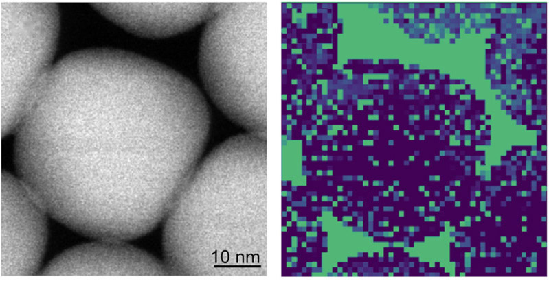
LSPR mapping of octahedral AuNPs
- [1] M. Haller et al., “Host-atom-driven transformation of a honeycomb oxide into a dodecagonal quasicrystal,” arXiv:2510.11426, 2025. https://doi.org/10.48550/arXiv.2510.11426
- [2] D. Denkova et al., “Mapping magnetic near-field distributions of plasmonic nanoantennas,” ACS Nano, Vol.7, No.4, pp. 3168-3176, 2013. https://doi.org/10.1021/nn305589t
- [3] A. J. Das, R. Shivanna, and K. S. Narayan, “Photoconductive NSOM for mapping optoelectronic phases in nanostructures,” Nanophotonics, Vol.3, Nos.1-2, pp. 19-31, 2014. https://doi.org/10.1515/nanoph-2013-0043
- [4] F. J. García-Vidal and J. B. Pendry, “Collective theory for surface enhanced Raman scattering,” Phys. Rev. Lett., Vol.77, No.6, pp. 1163-1166, 1996. https://doi.org/10.1103/PhysRevLett.77.1163
- [5] Y. Wy, H. Jung, J. W. Hong, and S. W. Han, “Exploiting plasmonic hot spots in Au-based nanostructures for sensing and photocatalysis,” Acc. Chem. Res., Vol.55, No.6, pp. 831-843, 2022. https://doi.org/10.1021/acs.accounts.1c00682
- [6] P. Wen et al., “Precise regulation and control of hotspots in nanoparticle multilayer SERS substrates,” Microchem. J., Vol.187, Article No.108371, 2023. https://doi.org/10.1016/j.microc.2022.108371
- [7] T. Ikeuchi et al., “DNA-programmed core–shell nanoparticle superlattice crystals with nanogaps for SERS sensing,” ACS Appl. Opt. Mater., Vol.4, No.7, pp. 2028-2039, 2026. https://doi.org/10.1021/acsaom.6c00173
- [8] K. Koike et al., “Quantitative drug dynamics visualized by alkyne-tagged plasmonic-enhanced Raman microscopy,” ACS Nano, Vol.14, No.11, pp. 15032-15041, 2020. https://doi.org/10.1021/acsnano.0c05010
- [9] Y. Han, S.-R. Wu, X.-D. Tian, and Y. Zhang, “Optimizing the SERS performance of 3D substrates through tunable 3D plasmonic coupling toward label-free liver cancer cell classification,” ACS Appl. Mater. Interfaces, Vol.12, No.26, pp. 28965-28974, 2020. https://doi.org/10.1021/acsami.0c04509
- [10] G. V. P. Kumar et al., “Hot spots in Ag core–Au shell nanoparticles potent for surface-enhanced Raman scattering studies of biomolecules,” J. Phys. Chem. C, Vol.111, No.11, pp. 4388-4392, 2007. https://doi.org/10.1021/jp068253n
- [11] J. Cao et al., “Fabrication of core shell Au@Ag supraparticles with 3D hotspots via evaporation self-assembly for sensitive surface enhanced Raman scattering detection,” Sens. Actuators B: Chem., Vol.382, Article No.133529, 2023. https://doi.org/10.1016/j.snb.2023.133529
- [12] T. Gao et al., “Ultrasensitive surface-enhanced Raman scattering platform for protein detection via active delivery to nanogaps as a hotspot,” ACS Nano, Vol.18, No.32, pp. 21593-21606, 2024. https://doi.org/10.1021/acsnano.4c09578
- [13] S. Park, J. Lee, and H. Ko, “Transparent and flexible surface-enhanced Raman scattering (SERS) sensors based on gold nanostar arrays embedded in silicon rubber film,” ACS Appl. Mater. Interfaces, Vol.9, No.50, pp. 44088-44095, 2017. https://doi.org/10.1021/acsami.7b14022
- [14] I. B. Becerril-Castro et al., “Gold nanostars: Synthesis, optical and SERS analytical properties,” Anal. Sens., Vol.2, No.3, Article No.e202200005, 2022. https://doi.org/10.1002/anse.202200005
- [15] L. Scarabelli, M. Coronado-Puchau, J. J. Giner-Casares, J. Langer, and L. M. Liz-Marzán, “Monodisperse gold nanotriangles: Size control, large-scale self-assembly, and performance in surface-enhanced Raman scattering,” ACS Nano, Vol.8, No.6, pp. 5833-5842, 2014. https://doi.org/10.1021/nn500727w
- [16] H.-L. Wu et al., “A comparative study of gold nanocubes, octahedra, and rhombic dodecahedra as highly sensitive SERS substrates,” Inorg. Chem., Vol.50, No.17, pp. 8106-8111, 2011. https://doi.org/10.1021/ic200504n
- [17] M. Mulvihill, A. Tao, K. Benjauthrit, J. Arnold, and P. Yang, “Surface-enhanced Raman spectroscopy for trace arsenic detection in contaminated water,” Angew. Chem. Int. Ed., Vol.47, No.34, pp. 6456-6460, 2008. https://doi.org/10.1002/anie.200800776
- [18] J. Kim et al., “Ensemble hot-spots in 3D supercrystals of plasmonic octahedral nanoparticles in tip-to-tip configured superlattices,” Nat. Commun., Vol.16, Article No.2762, 2025. https://doi.org/10.1038/s41467-025-58029-5
- [19] Y. Hu, J. Liao, D. Wang, and G. Li, “Fabrication of gold nanoparticle-embedded metal–organic framework for highly sensitive surface-enhanced Raman scattering detection,” Anal. Chem., Vol.86, No.8, pp. 3955-3963, 2014. https://doi.org/10.1021/ac5002355
- [20] C.-C. Chang, H.-L. Wu, C.-H. Kuo, and M. H. Huang, “Hydrothermal synthesis of monodispersed octahedral gold nanocrystals with five different size ranges and their self-assembled structures,” Chem. Mater., Vol.20, No.24, pp. 7570-7574, 2008. https://doi.org/10.1021/cm8021984
- [21] J. Czochralski, “Ein neues Verfahren zur Messung der Kristallisationsgeschwindigkeit der Metalle,” Z. Phys. Chem., Vol.92U, No.1, pp. 219-221, 1918 (in German). https://doi.org/10.1515/zpch-1918-9212
- [22] N. R. Jana, L. Gearheart, and C. J. Murphy, “Evidence for seed-mediated nucleation in the chemical reduction of gold salts to gold nanoparticles,” Chem. Mater., Vol.13, No.7, pp. 2313-2322, 2001. https://doi.org/10.1021/cm000662n
- [23] Y. Pu, Y. Zhao, P. Zheng, and M. Li, “Elucidating the growth mechanism of plasmonic gold nanostars with tunable optical and photothermal properties,” Inorg. Chem., Vol.57, No.14, pp. 8599-8607, 2018. https://doi.org/10.1021/acs.inorgchem.8b01354
- [24] D. J. Wales and J. P. K. Doye, “Global optimization by basin-hopping and the lowest energy structures of Lennard-Jones clusters containing up to 110 atoms,” J. Phys. Chem. A, Vol.101, No.28, pp. 5111-5116, 1997. https://doi.org/10.1021/jp970984n
- [25] J. R. Lombardi and R. L. Birke, “Theory of surface-enhanced Raman scattering in semiconductors,” J. Phys. Chem. C, Vol.118, No.20, pp. 11120-11130, 2014. https://doi.org/10.1021/jp5020675
- [26] E. C. Le Ru and B. Auguié, “Enhancement factors: A central concept during 50 years of surface-enhanced Raman spectroscopy,” ACS Nano, Vol.18, No.14, pp. 9773-9783, 2024. https://doi.org/10.1021/acsnano.4c01474
This article is published under a Creative Commons Attribution-NoDerivatives 4.0 Internationa License.