
Paper:
Salt-Dependent Crystallization of DNA-Functionalized Nanoparticle Superlattices in Microdroplets Toward Cellular Applications
Saki Kozawa*
 , Hiroya Arai*, Xu Li**
, Naotomo Tottori***
, Yoko Yamanishi***
, and Miho Tagawa**
*Department of Materials Science and Engineering, Nagoya University
Furo-cho, Chikusa-ku, Nagoya, Aichi 464-8603, Japan
**Institute of Materials and Systems for Sustainability, Nagoya University
Furo-cho, Chikusa-ku, Nagoya, Aichi 464-8603, Japan
***Department of Mechanical Engineering, Kyushu University
744 Motooka, Nishi-ku, Fukuoka, Fukuoka 819-0395, Japan

DNA-functionalized nanoparticle (DNA–NP) superlattices have attracted attention as programmable nanomaterials for sensing and bio-related applications, because DNA hybridization enables programmable interactions between nanoparticles and thus control of interparticle spacing. Such applications require controlling the size, shape, and crystallinity to maintain structural stability and achieve consistent physical properties. In this study, we investigated the crystallization behavior of DNA–NP assemblies formed in microdroplets under different NaCl concentrations. Crystallization occurred in water-in-oil droplets, which provided spatially confined reaction environments for DNA–NP assembly and enabled crystallization to proceed in numerous spatially isolated microreactors under well-defined conditions. Optical microscopy revealed that crystals were typically formed individually within droplets and exhibited narrow size distributions under each condition. Within the same NaCl concentration, the average crystal size generally varied with droplet diameter. Small-angle X-ray scattering analysis showed that increasing NaCl concentration resulted in broader diffraction peaks and fewer observable higher-order reflections, indicating a reduction in crystalline order. Scanning electron microscopy further revealed differences in crystal morphology. Well-defined faceted crystals were formed at lower NaCl concentrations, whereas rounded crystals were frequently formed at higher concentrations. Overall, salt concentration is an important factor governing the crystalline order and morphology of DNA–NP superlattices formed in microdroplets.
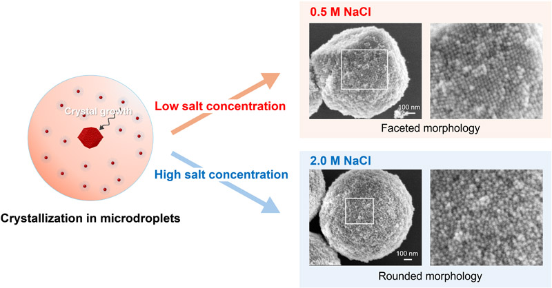
DNA-NP crystallization in microdroplets
- [1] A. Taleb, C. Petit, and M. P. Pileni, “Optical Properties of Self-Assembled 2D and 3D Superlattices of Silver Nanoparticles,” The J. of Physical Chemistry B, Vol.102, No.12, pp. 2214-2220, 1998. https://doi.org/10.1021/jp972807s
- [2] G. Rainò, M. A. Becker, M. I. Bodnarchuk, R. F. Mahrt, M. V. Kovalenko, and T. Stöferle, “Superfluorescence from lead halide perovskite quantum dot superlattices,” Nature, Vol.563, No.7733, pp. 671-675, 2018. https://doi.org/10.1038/s41586-018-0683-0
- [3] N. S. Mueller, E. Pfitzner, Y. Okamura, G. Gordeev, P. Kusch, H. Lange, J. Heberle, F. Schulz, and S. Reich, “Surface-Enhanced Raman Scattering and Surface-Enhanced Infrared Absorption by Plasmon Polaritons in Three-Dimensional Nanoparticle Supercrystals,” ACS Nano, Vol.15, No.3, pp. 5523-5533, 2021. https://doi.org/10.1021/acsnano.1c00352
- [4] D. Nykypanchuk, M. M. Maye, D. Van der Lelie, and O. Gang, “DNA-guided crystallization of colloidal nanoparticles,” Nature, Vol.451, pp. 549-552, 2008. https://doi.org/10.1038/nature06560
- [5] R. J. Macfarlane, B. Lee, M. R. Jones, N. Harris, G. C. Schatz, and C. A. Mirkin, “Nanoparticle Superlattice Engineering with DNA,” Science, Vol.334, No.6053, pp. 204-208, 2011. https://doi.org/10.1126/science.1210493
- [6] V. Kumari, S. Vishwas, R. Kumar, V. Kakoty, R. Khursheed, M. R. Babu, V. Harish, N. Mittal, P. K. Singh, N. S. Alharthi, M. A. Hakami, F. F. Aba Alkhayl, G. Gupta, G. D. Rubis, K. R. Paudel, M. Singh, M. Zandi, B. G. Oliver, K. Dua et al., “An overview of biomedical applications for gold nanoparticles against lung cancer,” J. of Drug Delivery Science and Technology, Vol.86, Article No.104729, 2023. https://doi.org/10.1016/j.jddst.2023.104729
- [7] N. Ramakrishnan, M. Lee, Y. Choi, S. Kim, J. Kim, and Y. Kwon, “Deciphering Gold Nanoparticle-Induced Cytotoxicity for Safer Nanomedicine Applications: Exploring the Impact of Conjugation Chemistry, Functional Groups, and Metal Core Properties,” ACS Applied Nano Materials, Vol.8, No.51, pp. 24301-24313, 2025. https://doi.org/10.1021/acsanm.5c03906
- [8] B. Yan, Y.-y. Zhu, Y. Wei, and H. Pei, “Study on surface enhanced Raman scattering of Au and Au@AlO spherical dimers based on 3D finite element method,” Scientific Reports, Vol.11, No.1, Article No.8391, 2021. https://doi.org/10.1038/s41598-021-87997-z
- [9] H. Liu, Z. Yang, L. Meng, Y. Sun, J. Wang, L. Yang, J. Liu, and Z. Tian, “Three-Dimensional and Time-Ordered Surface-Enhanced Raman Scattering Hotspot Matrix,” J. of the American Chemical Society, Vol.136, No.14, pp. 5332-5341, 2014. https://doi.org/10.1021/ja501951v
- [10] T. Ikeuchi, S. Kozawa, H. Niinomi, X. Li, S. Ogawa, M. Kuwahara, T. Oshikiri, K. Hiramatsu, and M. Tagawa, “DNA-Programmed Core–Shell Nanoparticle Superlattice Crystals with Nanogaps for SERS Sensing,” ACS Applied Optical Materials, Vol.4, No.7, pp. 2028-2039, 2026. https://doi.org/10.1021/acsaom.6c00173
- [11] M. B. Ross, J. C. Ku, V. M. Vaccarezza, G. C. Schatz, and C. A. Mirkin, “Nanoscale form dictates mesoscale function in plasmonic DNA-nanoparticle superlattices,” Nature Nanotechnology, Vol.10, No.5, pp. 453-458, 2015. https://doi.org/10.1038/nnano.2015.68
- [12] M. B. Ross, J. C. Ku, M. G. Blaber, C. A. Mirkin, and G. C. Schatz, “Defect tolerance and the effect of structural inhomogeneity in plasmonic DNA-nanoparticle superlattices,” Proc. of the National Academy of Sciences, Vol.112, No.33, pp. 10292-10297, 2015. https://doi.org/10.1073/pnas.1513058112
- [13] S. Nangia and R. Sureshkumar, “Effects of Nanoparticle Charge and Shape Anisotropy on Translocation through Cell Membranes,” Langmuir, Vol.28, No.51, pp. 17666-17671, 2012. https://doi.org/10.1021/la303449d
- [14] X. W. Gu, X. Ye, D. M. Koshy, S. Vachhani, P. Hosemann, and A. P. Alivisatos, “Tolerance to structural disorder and tunable mechanical behavior in self-assembled superlattices of polymer-grafted nanocrystals,” Proc. of the National Academy of Sciences, Vol.114, No.11, pp. 2836-2841, 2017. https://doi.org/10.1073/pnas.1618508114
- [15] C. Schildkraut and S. Lifson, “Dependence of the melting temperature of DNA on salt concentration,” Biopolymers, Vol.3, No.2, pp. 195-208, 1965. https://doi.org/10.1002/bip.360030207
- [16] X. Zhang, J.-S. Zhang, Y.-Z. Shi, X.-L. Zhu, and Z.-J. Tan, “Potential of mean force between like-charged nanoparticles: Many-body effect,” Scientific Reports, Vol.6, No.1, Article No.23434, 2016. https://doi.org/10.1038/srep23434
- [17] Q. Yu, J. Chen, D. Shi, and M. Chen, “Ordered self-assembly of DNA-modified nanoparticles in salt solutions,” Colloids and Surfaces A: Physicochemical and Engineering Aspects, Vol.671, Article No.131669, 2023. https://doi.org/10.1016/j.colsurfa.2023.131669
- [18] Y. Li, M. Girard, M. Shen, J. A. Millan, and M. O. de la Cruz, “Strong attractions and repulsions mediated by monovalent salts,” Proc. of the National Academy of Sciences, Vol.114, No.45, pp. 11838-11843, 2017. https://doi.org/10.1073/pnas.1713168114
- [19] S. E. Seo, B. Lee, T. Oh, M. Girard, and C. A. Mirkin, “Salt-Induced Polymorphs Observed in Colloidal Single Crystals,” J. of the American Chemical Society, Vol.147, No.38, pp. 34831-34839, 2025. https://doi.org/10.1021/jacs.5c10844
- [20] S. E. Seo, M. Girard, M. O. de la Cruz, and C. A. Mirkin, “The Importance of Salt-Enhanced Electrostatic Repulsion in Colloidal Crystal Engineering with DNA,” ACS Central Science, Vol.5, No.1, pp. 186-191, 2019. https://doi.org/10.1021/acscentsci.8b00826
- [21] C. Brandel and J. H. ter Horst, “Measuring induction times and crystal nucleation rates,” Faraday Discussions, Vol.179, pp. 199-214, 2015. https://doi.org/10.1039/C4FD00230J
- [22] T. Karmakar, P. M. Piaggi, and M. Parrinello, “Molecular Dynamics Simulations of Crystal Nucleation from Solution at Constant Chemical Potential,” J. of Chemical Theory and Computation, Vol.15, No.12, pp. 6923-6930, 2019. https://doi.org/10.1021/acs.jctc.9b00795
- [23] F. Bourgeois, S. Teychené, and B. Biscans, “Applicability of Probabilistic Nucleation Modelling for the Analysis of Microfluidics Data,” KONA Powder and Particle J., Vol.35, No.0, pp. 258-272, 2018. https://doi.org/10.14356/kona.2018017
- [24] A. Hensley, W. M. Jacobs, and W. B. Rogers, “Self-assembly of photonic crystals by controlling the nucleation and growth of DNA-coated colloids,” Proc. of the National Academy of Sciences, Vol.119, No.1, Article No.e2114050118, 2022. https://doi.org/10.1073/pnas.2114050118
- [25] A. Hensley, T. E. Videbæk, H. Seyforth, W. M. Jacobs, and W. B. Rogers, “Macroscopic photonic single crystals via seeded growth of DNA-coated colloids,” Nature Communications, Vol.14, No.1, Article No.4237, 2023. https://doi.org/10.1038/s41467-023-39992-3
- [26] M. Maeki, Y. Teshima, S. Yoshizuka, H. Yamaguchi, K. Yamashita, and M. Miyazaki, “Controlling Protein Crystal Nucleation by Droplet-Based Microfluidics,” Chemistry – A European J., Vol.20, No.4, pp. 1049-1056, 2014. https://doi.org/10.1002/chem.201303270
- [27] S. V. Koniakhin, I. E. Eliseev, I. N. Terterov, A. V. Shvidchenko, E. D. Eidelman, and M. V. Dubina, “Molecular dynamics-based refinement of nanodiamond size measurements obtained with dynamic light scattering,” Microfluidics and Nanofluidics, Vol.18, Nos.5-6, pp. 1189-1194, 2015. https://doi.org/10.1007/s10404-014-1512-x
- [28] S. Dhakal, K. L. Kohlstedt, G. C. Schatz, C. A. Mirkin, and M. O. de la Cruz, “Growth Dynamics for DNA-Guided Nanoparticle Crystallization,” ACS Nano, Vol.7, No.12, pp. 10948-10959, 2013. https://doi.org/10.1021/nn404476f
- [29] H. Sumi, N. Ohta, H. Sekiguchi, S. Harada, T. Ujihara, K. Tsukamoto, and M. Tagawa, “Two-Step Nanoparticle Crystallization via DNA-Guided Self-Assembly and the Nonequilibrium Dehydration Process,” Crystal Growth & Design, Vol.21, No.8, pp. 4506-4515, 2021. https://doi.org/10.1021/acs.cgd.1c00398
- [30] H. Sumi, N. Ohta, H. Sekiguchi, S. Harada, T. Ujihara, K. Tsukamoto, and M. Tagawa, “Designing a High-Crystallinity Nano-Gapped Particle Superlattice via DNA-Guided Colloidal Crystallization and Dehydration,” Crystal Growth & Design, Vol.22, No.6, pp. 3708–3718, 2022. https://doi.org/10.1021/acs.cgd.2c00075
- [31] N. Tottori, M. Tagawa, A. Takao, L. Zhang, M. Yokomori, S. Sakuma, and Y. Yamanishi, “Thermally Regulated Droplet Microfluidics Enables Uniform, Size Controllable DNA Nanoparticle Superlattices,” Small, Vol.22, No.54, Article No.e74787, 2026. https://doi.org/10.1002/smll.74787
- [32] T. Oh, S. S. Park, and C. A. Mirkin, “Stabilization of Colloidal Crystals Engineered with DNA,” Advanced Materials, Vol.31, No.1, Article No.1805480, 2019. https://doi.org/10.1002/adma.201805480
- [33] R. Hosemann and A. M. Hindeleh, “Structure of crystalline and paracrystalline condensed matter,” J. of Macromolecular Science, Part B, Vol.34, No.4, pp. 327-356, 1995. https://doi.org/10.1080/00222349508219497
- [34] Y. Xia, X. Xia, and H.-C. Peng, “Shape-Controlled Synthesis of Colloidal Metal Nanocrystals: Thermodynamic versus Kinetic Products,” J. of the American Chemical Society, Vol.137, No.25, pp. 7947-7966, 2015. https://doi.org/10.1021/jacs.5b04641
- [35] J. Kim, S. Kang, F. Cheng, Y. Wang, X. Ye, and J. Park, “Recent advances in liquid phase transmission electron microscopy of nanoparticle growth and self-assembly,” MRS Bulletin, Vol.49, No.4, pp. 365-376, 2024. https://doi.org/10.1557/s43577-024-00702-z
- [36] C. A. Orme, A. Noy, A. Wierzbicki, M. T. McBride, M. Grantham, H. H. Teng, P. M. Dove, and J. J. DeYoreo, “Formation of chiral morphologies through selective binding of amino acids to calcite surface steps,” Nature, Vol.411, No.6839, pp. 775-779, 2001. https://doi.org/10.1038/35081034
- [37] L. D. Marks and L. Peng, “Nanoparticle shape, thermodynamics and kinetics,” J. of Physics: Condensed Matter, Vol.28, No.5, Article No.053001, 2016. https://doi.org/10.1088/0953-8984/28/5/053001
- [38] I. Canton and G. Battaglia, “Endocytosis at the nanoscale,” Chemical Society Reviews, Vol.41, No.7, pp. 2718-2739, 2012. https://doi.org/10.1039/c2cs15309b
- [39] M. V. Baranov, M. Kumar, S. Sacanna, S. Thutupalli, and G. Van Den Bogaart, “Modulation of Immune Responses by Particle Size and Shape,” Frontiers in Immunology, Vol.11, Article No.607945, 2021. https://doi.org/10.3389/fimmu.2020.607945
- [40] Y.-C. Wu, T.-H. Wu, D. L. Clemens, B.-Y. Lee, X. Wen, M. A. Horwitz, M. A. Teitell, and P.-Y. Chiou, “Massively parallel delivery of large cargo into mammalian cells with light pulses,” Nature Methods, Vol.12, No.5, pp. 439-444, 2015. https://doi.org/10.1038/nmeth.3357
- [41] E. Auyeung, R. J. Macfarlane, C. H. J. Choi, J. I. Cutler, and C. A. Mirkin, “Transitioning DNA-Engineered Nanoparticle Superlattices from Solution to the Solid State,” Advanced Materials, Vol.24, No.38, pp. 5181-5186, 2012. https://doi.org/10.1002/adma.201202069
This article is published under a Creative Commons Attribution-NoDerivatives 4.0 Internationa License.