
Paper:
Three-Dimensional Characterization of AC Electrothermal Flow in Microfluidic Devices
Daisuke Ichimura*, Akihiko Ishida*, and Masahiro Motosuke**,***

*Department of Mechanical Engineering, Graduate School of Engineering, Tokyo University of Science
6-3-1 Niijuku, Katsushika-ku, Tokyo 125-8585, Japan
**Department of Mechanical Engineering, Faculty of Engineering, Tokyo University of Science
6-3-1 Niijuku, Katsushika-ku, Tokyo 125-8585, Japan
***Water Frontier Research Center, Research Institute for Science and Technology, Tokyo University of Science
1-3 Kagurazaka, Shinjuku-ku, Tokyo 162-8601, Japan

In microfluidic systems, liquid flow structure near the substrate is crucial for regulating the mechanical microenvironment of cells cultured on the surface and mass transport toward the substrate. Therefore, externally induced flow is expected not only to enhance surface reactions but also to modulate cellular responses through controlled shear stress. However, the direct evaluation of the velocity component normal to the substrate remains challenging in monolithic microfluidic devices. In this study, a three-dimensional (3D) flow velocity measurement method based on orthogonal-plane micro-particle image velocimetry (micro-PIV) was developed to evaluate alternating-current electrothermal flow modulation. The 3D velocity field was reconstructed from two orthogonal velocity fields obtained by scanning with different observation planes: one parallel to the bottom and the other perpendicular to the bottom through a sidewall of a polydimethylsiloxane microchannel. The validity of the orthogonal-plane micro-PIV was confirmed through measurements of three-component velocities in a tilted rectangular channel. Using this method, alternating-current electrothermal (ACET) flow generated by temperature-dependent gradients in fluid properties under an electric field was investigated. The results revealed a 3D circular stirring flow that transports fluid from the bulk region toward the surface. This flow structure enhances the transport of suspended species toward the interface and is expected to modulate the local shear environment, demonstrating that ACET-driven 3D flow can simultaneously enhance surface reactions and regulate the cellular mechanical environment in microfluidic systems.
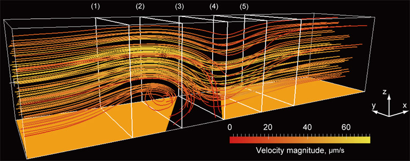
3D ACET flow in a microchannel
- [1] S. Scarano, M. Mascini, A. P. F. Turner, and M. Minunni, “Surface plasmon resonance imaging for affinity-based biosensors,” Biosensors and Bioelectronics, Vol.25, No.5, pp. 957-966, 2010. https://doi.org/10.1016/j.bios.2009.08.039
- [2] G. Chen, Y. Lv, P. Guo, C. Lin, X. Zhang, L. Yang, and Z. Xu, “Matrix mechanics and fluid shear stress control stem cells fate in three dimensional microenvironment,” Current Stem Cell Research & Therapy, Vol.8, No.4, pp. 313-323, 2013. https://doi.org/10.2174/1574888X11308040007
- [3] C. P. Gayer and M. D. Basson, “The effects of mechanical forces on intestinal physiology and pathology,” Cell Signalling, Vol.21, No.8, pp. 1237-1244, 2009. https://doi.org/10.1016/j.cellsig.2009.02.011
- [4] W. J. Polacheck, R. Li, S. G. M. Uzel, and R. D. Kamm, “Microfluidic platforms for mechanobiology,” Lab on a Chip, Vol.13, No.12, pp. 2252-2267, 2013. https://doi.org/10.1039/C3LC41393D
- [5] H. Lu, L. Y. Koo, W. M. Wang, D. A. Lauffenburger, L. G. Griffith, and K. F. Jensen, “Microfluidic shear devices for quantitative analysis of cell adhesion,” Analytical Chemistry, Vol.76, No.18, pp. 5257-5264, 2004. https://doi.org/10.1021/ac049837t
- [6] X. Gao and Y. Li, “Biofluid pumping and mixing by an AC electrothermal micropump embedded with a spiral microelectrode pair in a cylindrical microchannel,” Electrophoresis, Vol.39, Issue 24, pp. 3156-3170, 2018. https://doi.org/10.1002/elps.201800162
- [7] A. Abdelghany, K. Yamasaki, Y. Ichikawa, and M. Motosuke, “Efficient nanoparticle focusing utilizing cascade AC electroosmotic flow,” Electrophoresis, Vol.43, Issues 16-17, pp. 1755-1764, 2022. https://doi.org/10.1002/elps.202200054
- [8] A. Ramos, H. Morgan, N. G. Green, and A. Castellanos, “AC electrokinetics: a review of forces in microelectrode structures,” J. of Physics D: Applied Physics, Vol.31, pp. 2338-2353, 1998. https://doi.org/10.1088/0022-3727/31/18/021
- [9] I. Kawata, S. Kobayashi, Y. Ichikawa, and M. Motosuke, “On-chip AC electrothermal pump for pulsatile perfusion,” Micromachines, Vol.17, No.4, Article No.492, 2026. https://doi.org/10.3390/mi17040492
- [10] J. G. Santiago, S. T. Wereley, C. D. Meinhart, D. J. Beebe, and R. J. Adrian, “A particle image velocimetry system for microfluidics,” Experiments in Fluids, Vol.25, pp. 316-319, 1998. https://doi.org/10.1007/s003480050235
- [11] C. D. Meinhart, S. T. Wereley, and J. G. Santiago, “A PIV algorithm for estimating time-averaged velocity fields,” J. of Fluids Engineering, Vol.122, No.2, pp. 285-289, 2000. https://doi.org/10.1115/1.483256
- [12] R. Lindken, J. Westerweel, and B. Wieneke, “Stereoscopic micro particle image velocimetry,” Experiments in Fluids, Vol.41, pp. 161-171, 2006. https://doi.org/10.1007/s00348-006-0154-5
- [13] M. R. Bown, J. M. MacInnes, R. W. K. Allen, and W. B. J. Zimmerman, “Three-dimensional, three-component velocity measurements using stereoscopic micro-PIV and PTV,” Measurement Science and Technology, Vol.17, pp. 2175-2185, 2006. https://doi.org/10.1088/0957-0233/17/8/017
- [14] C.-T. Yang and H.-S. Chuang, “Measurement of a microchamber flow by using a hybrid multiplexing holographic velocimetry,” Experiments in Fluids, Vol.39, pp. 385-396, 2005. https://doi.org/10.1007/s00348-005-1022-4
- [15] S. Kim and S. J. Lee, “Measurement of 3D laminar flow inside a micro tube using micro digital holographic particle tracking velocimetry,” J. of Micromechanics and Microengineering, Vol.17, No.10, pp. 2157-2162, 2007. https://doi.org/10.1088/0960-1317/17/10/030
- [16] T. A. Ooms, R. Lindken, and J. Westerweel, “Digital holographic microscopy applied to measurement of a flow in a T-shaped micromixer,” Experiments in Fluids, Vol.47, pp. 941-955, 2009. https://doi.org/10.1007/s00348-009-0683-9
- [17] R. Barnkob and M. Rossi, “General defocusing particle tracking: fundamentals and uncertainty assessment,” Experiments in Fluids, Vol.61, Article No.110, 2020. https://doi.org/10.1007/s00348-020-2937-5
- [18] Y. Ichikawa, R. Kikuchi, K. Yamamoto, and M. Motosuke, “Determining particle depth positions and evaluating dispersion using astigmatism PTV with a neural network,” Applied Optics, Vol.60, Issue 22, pp. 6538-6546, 2021. https://doi.org/10.1364/AO.427571
- [19] C. D. Meinhart, S. T. Wereley, and J. G. Santiago, “PIV measurements of a microchannel flow,” Experiments in Fluids, Vol.27, pp. 414-419, 1999. https://doi.org/10.1007/s003480050366
- [20] C. D. Meinhart, S. T. Wereley, and M. H. B. Gray, “Volume illumination for two-dimensional particle image velocimetry,” Measurement Science and Technology, Vol.11, No.6, pp. 809-814, 2000. https://doi.org/10.1088/0957-0233/11/6/326
- [21] F. M. White, “Viscous Fluid Flow,” p. 123, McGraw-Hill, 1974.
- [22] M. Motosuke, K. Yamasaki, A. Ishida, H. Toki, and S. Honami, “Improved particle concentration by cascade AC electroosmotic flow,” Microfluidics and Nanofluidics, Vol.14, No.6, pp. 1021-1030, 2013. https://doi.org/10.1007/s10404-012-1109-1
This article is published under a Creative Commons Attribution-NoDerivatives 4.0 Internationa License.