
Paper:
Development of a Microchannel with Tapered Funnels for Evaluating Liposome Membrane Tension
Kaoru Uesugi*1,*2,†
 , Haruto Obuchi*3, Kazuaki Nagayama*2, and Taro Toyota*3,*4
*1Department of Robotics, Graduate School of Science and Engineering, Ritsumeikan University
1-1-1 Nojihigashi, Kusatsu, Shiga 525-8577, Japan
*2Department of Mechanical Systems Engineering, Graduate School of Science and Engineering, Ibaraki University
Nakanarusawa-cho, Hitachi, Ibaraki 316-8511, Japan
*3Department of Basic Science, Graduate School of Arts and Sciences, The University of Tokyo
3-8-1 Komaba, Meguro-ku, Tokyo 153-8902, Japan
*4Universal Biology Institute, The University of Tokyo
7-3-1 Hongo, Bunkyo-ku, Tokyo 113-0033, Japan
†Corresponding author

Amoeba-type molecular robots, which are deformed by internal actuator molecules, use micrometer-sized liposomes as their flexibly deformable housings. To design amoeba-type molecular robots, it is crucial to evaluate the liposome membrane tension because the deformation of these robots is influenced by the membrane tension of the housing liposomes. In this study, we proposed a microchannel that enabled the evaluation of liposome membrane tension by observing the radii of liposomes trapped in tapered funnels integrated into the microchannel. Because the funnels were connected in series, the liposome membrane tension could be evaluated in a single experiment without requiring advanced experimental techniques or considerable experimental time. In addition, the evaluated liposomes were trapped in the funnels while adhering to the funnel walls, which may prevent damage due to stress concentrations and reduce the influence of dispersion leakage and fabrication inaccuracy of the microchannel. The membrane tension was derived by substituting the radii of the trapped liposomes and the pressure difference between the inlet and outlet of the funnel into the Young–Laplace equation. We evaluated the membrane tension under two conditions: (1) single-liposome trapping condition (0.57 and 0.34 mN/m) and (2) multiple-liposome trapping condition (0.41 mN/m). The difference in membrane tension between these conditions was not significant. However, we observed variability in the contact angle between the liposome and funnel wall, as well as in the membrane curvature, under the multiple-trapping condition. Therefore, we deduced that the single-liposome trapping condition was more suitable for membrane tension evaluation using the proposed microchannel.
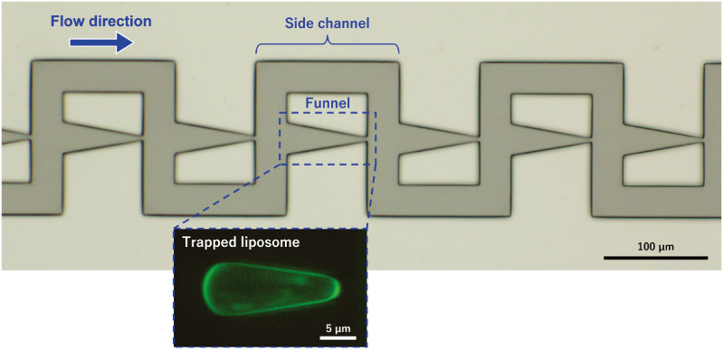
Microchannel with tapered funnels for evaluating liposome membrane tension
- [1] M. Hagiya, A. Konagaya, S. Kobayashi, H. Saito, and S. Murata, “Molecular robots with sensors and intelligence,” Acc. Chem. Res., Vol.47, Issue 6, pp. 1681-1690, 2014. https://doi.org/10.1021/ar400318d
- [2] M. Hayashi, M. Nishiyama, Y. Kazayama, T. Toyota, Y. Harada, and K. Takiguchi, “Reversible morphological control of tubulin-encapsulating giant liposomes by hydrostatic pressure,” Langmuir, Vol.32, Issue 15, pp. 3794-3802, 2016. https://doi.org/10.1021/acs.langmuir.6b00799
- [3] S. Tanaka, K. Takiguchi, and M. Hayashi, “Repetitive stretching of giant liposomes utilizing the nematic alignment of confined actin,” Commun. Phys., Vol.1, Article No.18, 2018. https://doi.org/10.1038/s42005-018-0019-2
- [4] Y. Sato, Y. Hiratsuka, I. Kawamata, S. Murata, and S. M. Nomura, “Micrometer-sized molecular robot changes its shape in response to signal molecules,” Sci. Robot., Vol.2, No.4, Article No.aal3735, 2017. https://doi.org/10.1126/scirobotics.aal3735
- [5] K. Uesugi, “Evaluating mechanical properties of liposomes,” S. Murata, T. Nakakuki, S. Nomura, and A. Kuzuya (Eds.), “Molecular robotics II: Toward chemical AI,” pp. 135-150, Springer Nature Singapore, 2026.
- [6] R. Kwok and E. Evans, “Thermoelasticity of large lecithin bilayer vesicles,” Biophys. J., Vol.35, Issue 3, pp. 637-652, 1981. https://doi.org/10.1016/S0006-3495(81)84817-5
- [7] E. Evans and D. Needham, “Physical properties of surfactant bilayer membranes: Thermal transitions, elasticity, rigidity, cohesion, and colloidal interactions,” J. Phys. Chem., Vol.91, Issue 16, pp. 4219-4228, 1987. https://doi.org/10.1021/j100300a003
- [8] D. Needham and E. Evans, “Structure and mechanical properties of giant lipid (DMPC) vesicle bilayers from 20°C below to 10°C above the liquid crystal-crystalline phase transition at 24°C,” Biochemistry, Vol.27, Issue 21, pp. 8261-8269, 1988. https://doi.org/10.1021/bi00421a041
- [9] E. Evans and W. Rawicz, “Entropy-driven tension and bending elasticity in condensed-fluid membranes,” Phys. Rev. Lett., Vol.64, No.17, pp. 2094-2097, 1990. https://doi.org/10.1103/PhysRevLett.64.2094
- [10] D. Needham and R. S. Nunn, “Elastic deformation and failure of lipid bilayer membranes containing cholesterol,” Biophys. J., Vol.58, No.4, pp. 997-1009, 1990. https://doi.org/10.1016/S0006-3495(90)82444-9
- [11] K. Olbrich, W. Rawicz, D. Needham, and E. Evans, “Water permeability and mechanical strength of polyunsaturated lipid bilayers,” Biophys. J., Vol.79, Issue 1, pp. 321-327, 2000. https://doi.org/10.1016/S0006-3495(00)76294-1
- [12] W. Rawicz, K. C. Olbrich, T. Mcintosh, D. Needham, and E. Evans, “Effect of chain length and unsaturation on elasticity of lipid bilayers,” Biophys. J., Vol.79, Issue 1, pp. 328-339, 2000. https://doi.org/10.1016/S0006-3495(00)76295-3
- [13] M. Elias, A. Dutoya, A. Laborde, A. Lecestre, C. Montis, L. Caselli, D. Berti, B. Lonetti, C. Roux, and P. Joseph, “Microfluidic characterization of biomimetic membrane mechanics with an on-chip micropipette,” Micro Nano Eng., Vol.8, Article No.100064, 2020. https://doi.org/10.1016/j.mne.2020.100064
- [14] S. Landiech et al., “Parallel on-chip micropipettes enabling quantitative multiplexed characterization of vesicle mechanics and cell aggregates rheology,” APL Bioeng., Vol.8, Issue 2, Article No.026122, 2024. https://doi.org/10.1063/5.0193333
- [15] Q. Guo, S. M. McFaul, and H. Ma, “Deterministic microfluidic ratchet based on the deformation of individual cells,” Phys. Rev. E, Vol.83, No.5, Article No.051910, 2011. https://doi.org/10.1103/PhysRevE.83.051910
- [16] Q. Guo, S. Park, and H. Ma, “Microfluidic micropipette aspiration for measuring the deformability of single cells,” Lab Chip, Vol.12, Issue 15, pp. 2687-2695, 2012. https://doi.org/10.1039/c2lc40205j
- [17] Q. Guo, S. J. Reiling, P. Rohrbach, and H. Ma, “Microfluidic biomechanical assay for red blood cells parasitized by Plasmodium falciparum,” Lab Chip, Vol.12, Issue 6, pp. 1143-1150, 2012. https://doi.org/10.1039/c2lc20857a
- [18] W. B. Haines, “Studies in the physical properties of soil. V. The hysteresis effect in capillary properties, and the modes of moisture distribution associated therewith,” J. Agric. Sci., Vol.20, Issue 1, pp. 97-116, 1930. https://doi.org/10.1017/S002185960008864X
- [19] L. M. Lee and A. P. Liu, “A microfluidic pipette array for mechanophenotyping of cancer cells and mechanical gating of mechanosensitive channels,” Lab Chip, Vol.15, Issue 1, pp. 264-273, 2015. https://doi.org/10.1039/c4lc01218f
- [20] Y.-J. Li et al., “A microfluidic micropipette aspiration device to study single-cell mechanics inspired by the principle of Wheatstone bridge,” Micromachines, Vol.10, Issue 2, Article No.131, 2019. https://doi.org/10.3390/mi10020131
- [21] W.-H. Tan and S. Takeuchi, “A trap-and-release integrated microfluidic system for dynamic microarray applications,” Proc. Natl. Acad. Sci. U.S.A., Vol.104, No.4, pp. 1146-1151, 2007. https://doi.org/10.1073/pnas.0606625104
- [22] F. M. White, “Viscous Flow in Ducts,” F. M. White and H. Xue (Eds.), “Fluid Mechanics, 7th ed.,” pp. 347-454, McGraw-Hill, 2011.
- [23] K. Tsumoto, H. Matsuo, M. Tomita, and T. Yoshimura, “Efficient formation of giant liposomes through the gentle hydration of phosphatidylcholine films doped with sugar,” Colloids Surfaces B: Biointerfaces, Vol.68, Issue 1, pp. 98-105, 2009. https://doi.org/10.1016/j.colsurfb.2008.09.023
- [24] F. G. Woodhouse and R. E. Goldstein, “Shear-driven circulation patterns in lipid membrane vesicles,” J. Fluid Mech., Vol.705, pp. 165-175, 2012. https://doi.org/10.1017/jfm.2012.118
- [25] A. R. Honerkamp-Smith, F. G. Woodhouse, V. Kantsler, and R. E. Goldstein, “Membrane viscosity determined from shear-driven flow in giant vesicles,” Phys. Rev. Lett., Vol.111, Article No.038103, 2013. https://doi.org/10.1103/PhysRevLett.111.038103
- [26] B. Sebastian, T. Favero, and P. S. Dittrich, “The effects of shear force transmission across vesicle membranes,” J. Phys. Chem. Lett., Vol.8, Issue 24, pp. 6128-6134, 2017. https://doi.org/10.1021/acs.jpclett.7b02676
- [27] H. Sugiyama, T. Osaki, S. Takeuchi, and T. Toyota, “Perfusion chamber for observing a liposome-based cell model prepared by a water-in-oil emulsion transfer method,” ACS Omega, Vol.5, Issue 31, pp. 19429-19436, 2020. https://doi.org/10.1021/acsomega.0c01371
- [28] I. Biswas, A. Kumar, and M. Sadrzadeh, “Microfluidic membrane filtration systems to study biofouling,” M. S. Kandelousi (Ed.), “Microfluidics and Nanofluidics,” pp. 293-308, IntechOpen, 2018. https://doi.org/10.5772/intechopen.75006
- [29] H.-J. Butt et al., “Contact angle hysteresis,” Curr. Opin. Colloid Interface Sci., Vol.59, Article No.101574, 2022. https://doi.org/10.1016/j.cocis.2022.101574
- [30] S. D. Shoemaker and T. K. Vanderlick, “Intramembrane electrostatic interactions destabilize lipid vesicles,” Biophys. J., Vol.83, Issue 4, pp. 2007-2014, 2002. https://doi.org/10.1016/S0006-3495(02)73962-3
- [31] Video 1, “Fluorescence microscopy movie of the microchannel into which a fluorescent liposome dispersion was introduced.” https://youtu.be/X_pEGb2fLvY [Accessed April 15, 2026]
- [32] Video 2, “Fluorescence microscopy movie showing a liposome dispersion flowing through the device, with all 30 funnels successfully trapping liposomes.” https://youtu.be/ucs1pth_EFU [Accessed April 15, 2026]
This article is published under a Creative Commons Attribution-NoDerivatives 4.0 Internationa License.