
Paper:
A Novel Supercooling-Based Nonfreezing Preservation Method for Early Embryos Using Plant-Derived Substances to Maintain Cellular Integrity
Yumi Hoshino
 and Masashi Ikeuchi
Department of Precision Biomedical Engineering, Institute of Science Tokyo
2-3-10 Kanda Surugadai, Chiyoda-ku, Tokyo 101-0062, Japan

Maintaining cellular integrity during storage is essential for the stable handling and quality control of living cells in future sensor-integrated biological systems. In this study, we investigated whether plant-derived substances improve the effectiveness of a supercooling-based nonfreezing preservation method for early embryos while maintaining cellular integrity. Mouse 2-cell embryos were used as a model system, and the University of Wisconsin (UW) solution was used as the basal preservation medium. Coffee lees extract was examined as a plant-derived preservation agent, and its representative components, caffeine and chlorogenic acid, were evaluated. Embryos preserved in UW solution alone at -6°C for 72 h showed residual membrane deformation, whereas supplementation with coffee lees extract, particularly at 10%, improved post-preservation morphology. Caffeine showed concentration-dependent protective and adverse effects, with the intermediate concentration providing the most favorable preservation outcome. Additional supporting experiments demonstrated that coffee lees extract and caffeine maintained stable supercooling without freezing under prolonged storage conditions and improved cytoskeletal integrity and post-preservation developmental competence, as assessed by blastocyst development after culture. These findings demonstrate that natural plant-derived substances enhance the nonfreezing preservation of early embryos under supercooling conditions while maintaining cellular integrity. The proposed method may provide a practical basis for the storage, transport, and quality control of living cells and support future applications involving sensor-integrated biological systems.
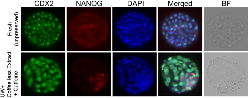
Blastocysts after 7-day supercooling
1. Introduction
Recent advances in biohybrid engineering have increased interest in the use of living cells and tissues as functional materials for sensing, monitoring, and information transmission systems 1. In such applications, the condition of the biological material before use is critical because structural instability and functional deterioration directly affect the reproducibility, quantitative reliability, and stable performance. Therefore, technologies that facilitate controlled storage and handling of living cells and tissues are gaining importance as supporting elements for future sensor-integrated biological systems 2, particularly for cells and tissues that incorporate intracellular nanoscale sensors or microdevices. In such device-integrated biological materials, conventional cryopreservation may pose additional challenges because intracellular device materials may promote ice nucleation, whereas freezing and thawing may impair both the cellular integrity and structural or functional stability of the embedded devices. Considering this viewpoint, implementing strategies to reduce ice-related damage could be beneficial in preserving both the biological integrity and the sensing capability during the phases of storage and transportation.
Although cryopreservation is widely used for the long-term storage of cells and tissues, several limitations remain, such as membrane injury caused by ice crystal formation, osmotic stress during cooling and warming, and cytotoxicity of cryoprotective agents 3,4. These issues are particularly important when the preserved cells are to be used in a structurally and functionally controlled manner. In this context, nonfreezing hypothermic preservation under supercooled conditions is an attractive alternative for reducing storage-related damage, maintaining living cells in a usable state 5 and facilitating the preservation, quality control, transport, and pre-use stabilization of sensor-bearing cells and tissues.
Plant-derived materials with anti-ice-nucleation or supercooling-promoting properties have recently attracted attention as low-damage preservation agents 6. Coffee lees extract contains compounds that may stabilize the supercooled state and protect cells during low-temperature storage 7. Among its components, caffeine and chlorogenic acid are of particular interest because of their potential protective effects 8,9. Although plant-derived substances and their components exhibit protective potential in several preservation settings, their usefulness in nonfreezing preservation of early embryos under supercooling conditions remains unclear. This unresolved issue provides the rationale for the present study.
In this study, we evaluated a novel supercooling-based nonfreezing preservation method for early embryos using coffee lees extract and its major components. We examined the post-preservation structural integrity and developmental competence to determine the potential utility of this approach for the low-damage preservation of biological materials.
2. Materials and Methods
2.1. Study Design
Mouse 2-cell embryos were used as the primary experimental models. University of Wisconsin (UW; ViaSpan; Astellas Pharma Inc., Tokyo, Japan) solution was used as the basal preservation medium, and coffee lees extract was evaluated as a natural plant-derived preservative. The representative components of the extract, including caffeine and chlorogenic acid, were evaluated to identify the candidate factors associated with preservation efficacy. This study included preservation experiments for the short-term assessment of post-preservation morphology and embryo experiments to evaluate cytoskeletal integrity and post-preservation developmental competence.
2.2. Preparation and Characterization of Plant-Derived Substances
Plant-derived supercooling-facilitating substances were prepared using coffee lees extract. The isolation, purification, and compositional analyses of the extract were performed in collaboration with Professor Hidehisa Kawahara (Kansai University). The major components identified in the extract were caffeine, caffeic acid, chlorogenic acid, and coumaric acid 7. Among these components, caffeine exhibited the highest anti-ice-nucleation activity and was selected for subsequent preservation experiments. Coffee lees extract was used as a crude plant-derived preservative, and caffeine and chlorogenic acid were individually tested as candidate functional components.
2.3. Collection and Preparation of Embryos
Mouse embryos were obtained via in vitro fertilization (IVF) according to a conventional protocol 10. ICR mice purchased from Japan SLC, Inc. (Shizuoka, Japan) were used in this study. All animal procedures were conducted in accordance with the institutional guidelines for animal experimentation of Japan Women’s University and were approved by the Animal Experiment Committee of Japan Women’s University (approval no. II20-2). Mature oocytes were collected from superovulated female mice and IVF was performed using human tubal fluid medium (ARK Resource, Kumamoto, Japan). After IVF, the fertilized oocytes were transferred to potassium simplex optimized medium (KSOM; ARK Resource, Kumamoto, Japan) and cultured in vitro. Normally cleaved 2-cell embryos were selected the following day and used for preservation experiments. In the main experiments, embryos were used for short-term preservation and post-preservation morphological evaluation. In the supporting embryo experiments, preserved embryos were cultured to evaluate their developmental progression to the blastocyst stage.
2.4. Preservation Media and Experimental Groups
UW solution was used as the basal preservation medium throughout the study 5. In the main embryo experiments, coffee lees extract was added to the UW solution at a final concentration of 5% or 10%. Caffeine (Fujifilm Wako, Osaka, Japan) and chlorogenic acid (Tokyo Chemical Industry, Tokyo, Japan) were individually tested to evaluate the contributions of the representative components of the extract.
2.5. Main Nonfreezing Preservation Experiments
For the main embryo preservation experiments, 2-cell embryos were immersed in 100 μL of each preservation solution. Five to 10 embryos were placed in each tube during the experimental run. The samples were placed in a temperature-controlled aluminum block (Cryo Porter, Scinics Corp., Tokyo, Japan), and the temperature was reduced stepwise from 20°C to \(-\)6°C at a rate of 2°C/min and then maintained at \(-\)6°C for 72 h. After preservation, samples were rewarmed stepwise from \(-\)6°C to 20°C at 2°C/min, rapidly transferred to 37°C, and allowed to recover at 37°C for 1 h before morphological and cytological analyses (Fig. 1).
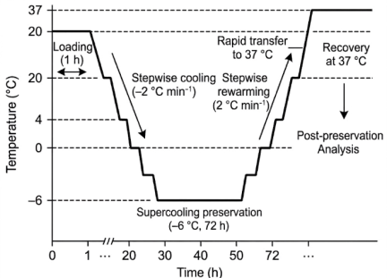
Fig. 1. Schematic temperature profile for supercooling-based nonfreezing preservation and recovery in the main embryo experiments. Embryos were cooled from 20°C to \(-\)6°C at 2°C/min, maintained at \(-\)6°C for 72 h, rewarmed stepwise to 20°C at the same rate, and then rapidly transferred to 37°C for recovery before morphological and cytological analyses.
2.6. Supporting Prolonged Preservation Experiments
Preliminary experiments demonstrated relatively high variability in morphological outcomes after 24 h of preservation, whereas a 72-h preservation period produced more reproducible differences among the preservation solutions tested. Therefore, a fixed storage period of 72 h was selected for the main experiments.
Additional embryo experiments were performed to further evaluate the preservation performance under prolonged storage conditions. The prolonged preservation experiments included four groups: UW solution alone, UW solution supplemented with 10% coffee lees extract, UW solution supplemented with caffeine at 173 μg/mL, and UW solution containing both 10% coffee lees extract and caffeine at 173 μg/mL. The 2-cell embryos were immersed in 100 μL of each preservation solution and cooled from 20°C to \(-\)6°C at a rate of 2°C/min using a temperature-controlled system. The samples were stored at \(-\)6°C for 1, 3, 7, or 14 days and then rewarmed to 20°C at the same rate. The temperature was controlled using the method described by Berendsen et al. 5. After rewarming, embryos were fixed for cytoskeletal assessment or cultured in KSOM for developmental evaluation.
Prolonged preservation experiments were designed as a stepwise extension of the main 72-h protocol. The 1-day condition was included as the earliest reference point, followed by progressive extension to 3, 7, and 14 days. This staged approach was adopted because the validation of preservation for 1 month or longer requires substantially more time to obtain sufficient embryos and complete repeated experimental runs.
2.7. Morphological and Cytological Assessment
After preservation, embryos from the main experiments were fixed in 2% paraformaldehyde (Fujifilm Wako Pure Chemical Corporation, Osaka, Japan), permeabilized, and stained with phalloidin (Cytoskeleton, Inc., Denver, CO, USA) to visualize F-actin and propidium iodide (PI; Dojindo Laboratories, Kumamoto, Japan) and assess nuclear morphology. The samples were examined using a confocal laser-scanning microscope (FV1200; Olympus, Tokyo, Japan) 11.
Embryonic integrity was evaluated based on the cortical F-actin distribution and membrane contour, nuclear morphology, blastomere shape, and zona pellucida morphology. Membrane deformation was defined as an irregular or discontinuous cortical F-actin pattern and/or the loss of a smooth blastomere contour. Blastomere abnormalities were defined as a distortion of the normal rounded shape, marked asymmetry, cytoplasmic separation or leakage, or loss of the original blastomere outline. Zona pellucida damage was defined as visible distortion, discontinuity, or rupture of the zona structure. Nuclear abnormalities were identified by abnormally condensed or intensely stained PI-positive nuclei.
For quantitative morphological assessment, embryos were classified as morphologically normal when all four parameters were comparable to those of fresh control embryos. Embryos with abnormalities in one or more parameters were classified as morphologically abnormal. Only embryos in which all four parameters could be reliably evaluated were included in the overall classification.
2.8. Developmental Assessment After Preservation
In the main embryo experiments, the preserved embryos were evaluated based primarily on their post-preservation morphology. In the supporting embryo experiments, preserved 2-cell embryos were transferred to KSOM and cultured for 4 days at 37°C in a CO\(_{2}\) incubator (PHC, Tokyo, Japan). Developmental competence was assessed based on the rate of development to the blastocyst stage. Blastocyst formation was evaluated under a stereomicroscope, based on the presence of a blastocoel cavity and the characteristic morphology of the blastocysts. Representative blastocysts obtained after prolonged preservation were examined by immunofluorescence staining for CDX2 and NANOG, representative markers of the trophectoderm (TE) and inner cell mass (ICM), respectively, according to a conventional procedure as previously described 12. For this analysis, anti-CDX2 (\(1:50\); BioGenex, MU392AUC) and anti-NANOG (\(1:100\); Cell Signaling Technology, #8822) were used as primary antibodies, followed by Alexa Fluor 488-conjugated anti-mouse immunoglobulin G (\(1:200\)) and Cy3-conjugated anti-rabbit secondary antibodies. Nuclei were counterstained with 4\('\),6-diamidino-2-phenylindole (DAPI). This analysis was used to visualize blastocyst organization and illustrate the structural features observed under each preservation condition.
2.9. Assessment of Supercooling Stability
To examine whether plant-derived substances stabilize the supercooled state, coffee lees extract and caffeine were added to the UW solution, which was then maintained under subzero conditions. Aliquots of 100, 500, and 1,000 μL were placed in 1.5-mL tubes under temperature-controlled conditions using the Cryo Porter system, as described above. The ability of the solution to remain unfrozen during prolonged storage was visually evaluated over a temperature range of approximately \(-\)6°C to \(-\)12°C. In this assessment, storage was continued for at least 24 h, and in the supporting setting, the maintenance of the supercooled state without visible freezing for up to 14 days was used as an indicator of supercooling-promoting activity.
2.10. Statistical Analysis
Blastocyst development, which was analyzed as a categorical outcome, was defined as the number of embryos that reached the blastocyst stage relative to the total number of cultured embryos. The group differences at each storage period were evaluated using the chi-square test. When a significant overall group difference was detected, pairwise comparisons were performed using Fisher’s exact test, and \(p\)-values were adjusted using the Holm method. Statistical significance was set at \(p < 0.05\). Data were obtained from three independent experiments.
Morphological outcomes were summarized descriptively as the number of morphologically normal or abnormal embryos relative to the total number of evaluable embryos under each condition (\(n/N\) [%]). No inferential statistical testing was performed for the morphological assessment because of the limited and unequal sample sizes.
3. Results
3.1. Coffee Lees Extract and Caffeine Supported Stable Supercooling Without Freezing
To determine whether plant-derived substances could stabilize the supercooled state, coffee lees extract or caffeine was added to the UW solution, and their supercooling-promoting effects were examined. Both substances maintained a stable supercooled state under subzero conditions, and the preservation solution remained unfrozen over a temperature range of approximately \(-\)6°C to \(-\)12°C. In the prolonged preservation setting, the preservation solution did not freeze even after 14 days of storage, indicating that both coffee lees extract and caffeine were effective supercooling-facilitating substances.
3.2. UW Solution Alone Enabled Nonfreezing Preservation of 2-Cell Embryos, Despite Residual Morphological Damage
In the main embryo preservation experiments, 2-cell embryos were stored for 72 h in the UW solution alone at \(-\)6°C under supercooling conditions. After rewarming, the embryos recovered without evident nuclear condensation, suggesting that severe cell death did not occur during preservation. However, morphological abnormalities, particularly membrane deformation and blastomere shape distortion, were still observed in the UW alone group (Fig. 2). Thus, UW solution alone provided a certain level of protection during nonfreezing storage but was insufficient to fully preserve normal embryo morphology.
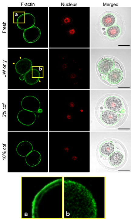
Fig. 2. Representative fluorescence images of 2-cell embryos after nonfreezing preservation in UW solution with or without coffee lees extract. Embryos were preserved at \(-\)6°C for 72 h under supercooling conditions in UW solution alone (UW only) or UW solution supplemented with 5% (5% cof) or 10% (10% cof) coffee lees extract. A fresh 2-cell embryo is shown as the normal control (top row). F-actin (green) and nuclei (red) were visualized by phalloidin and PI staining, respectively. Membrane integrity was evaluated based on the F-actin staining pattern. Magnified views of the cell membrane are shown in panels (a) and (b). In the fresh control, the F-actin-stained membrane appeared smooth and uniform (panel (a)), whereas in the UW-only group, the irregular distribution of F-actin along the cell surface (panel (b)) indicates membrane distortion. The yellow arrows indicate representative regions of membrane distortion. Scale bars: 50 μm.
3.3. Coffee Lees Extract Improved the Morphological Preservation of 2-Cell Embryos in a Concentration-Dependent Manner
The effect of coffee lees extract on embryo preservation was examined by supplementing the UW solution with the extract at final concentrations of 5% and 10%. The embryos preserved in the presence of coffee lees extract showed improved post-preservation morphology compared with those preserved in the UW solution alone. The effect was concentration-dependent, with the 10% group exhibiting greater reduction in membrane deformation than the 5% group (Fig. 2). The embryos preserved in UW solution supplemented with 10% coffee lees extract included a higher proportion of morphologically normal embryos and fewer abnormalities than those in the UW-alone and 5% coffee lees extract groups. Therefore, one or more components of coffee lees extract may protect embryos against low-temperature injury during nonfreezing preservation.
3.4. Caffeine Exerted Protective Effects, Whereas Chlorogenic Acid Alone Was Less Suitable
Caffeine and chlorogenic acid were individually tested to identify the active components responsible for the preservation effect of coffee lees extract. Because the caffeine concentration in the coffee lees extract was 173 μg/mL, its effect was evaluated at three concentrations based on this value: high (1730 μg/mL), intermediate (173 μg/mL), and low (17.3 μg/mL, corresponding to the concentration present in the 10% coffee lees extract group). The intermediate and low caffeine concentrations increased the proportion of morphologically normal embryos compared to the UW solution alone, whereas the high concentration provided only limited protection and was associated with blastomere deformation and zona pellucida fragility (Fig. 3). These findings indicate that caffeine may be beneficial within an appropriate concentration range, whereas excessive concentrations may exert adverse effects.
Next, the effectiveness of chlorogenic acid was evaluated. Because the concentration of chlorogenic acid in the coffee lees extract was 62.3 μg/mL, its effect was evaluated at three concentrations based on this value: high (623 μg/mL), intermediate (62.3 μg/mL), and low (6.23 μg/mL, corresponding to the concentration present in the 10% coffee lees extract group). Unlike caffeine, chlorogenic acid alone exhibited limited suitability as a preservative. Despite a slight protective effect under certain conditions, membrane damage was detectable, and the overall effect was weaker than that of the coffee lees extract or caffeine. Therefore, chlorogenic acid alone was insufficient for the stable preservation of 2-cell embryos under the present conditions.
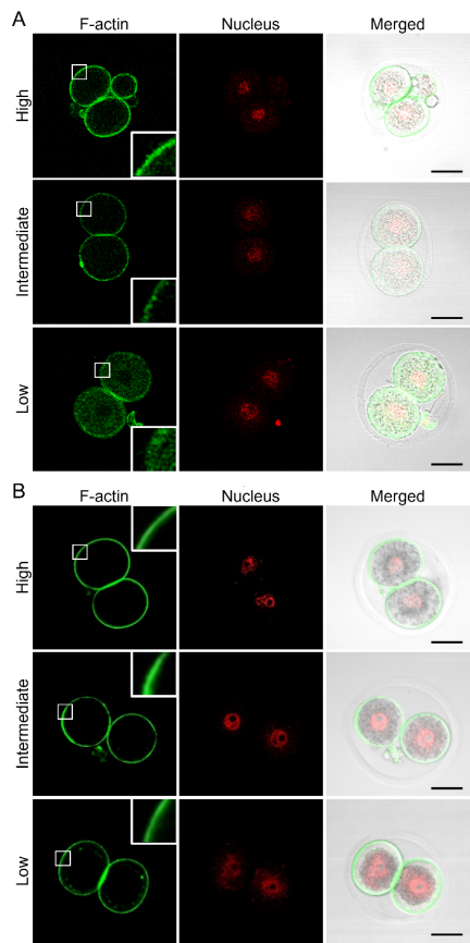
Fig. 3. Representative fluorescence images of 2-cell embryos after nonfreezing preservation in UW solution containing caffeine or chlorogenic acid at different concentrations. Embryos were preserved at \(-\)6°C for 72 h under supercooling conditions in UW solution containing caffeine (A) or chlorogenic acid (B). Representative images of the high, intermediate, and low concentration groups are shown from top to bottom in each panel. F-actin (green) and nuclei (red) were visualized by phalloidin and PI staining, respectively. Insets show magnified views of the boxed membrane regions, highlighting representative differences in the F-actin staining pattern after preservation. Scale bars: 50 μm.
A simple quantitative assessment supported these qualitative morphological observations (Table 1). The percentage of embryos classified as morphologically normal increased from 22.2% (4/18) in the UW-alone group to 41.2% (7/17) and 71.4% (10/14) in the 5% and 10% coffee lees extract groups, respectively. Among the caffeine conditions, the intermediate concentration showed the highest percentage of morphologically normal embryos at 68.2% (15/22), compared to 25.0% (6/24) and 46.7% (7/15) in the high- and low-concentration groups, respectively. In contrast, the percentage in the chlorogenic acid groups remained between 25.0% and 37.5%.
| Preservation condition |
No. of embryos, \(n\) |
No. of morphologically normal, \(n\) [%] |
| UW alone | 18 | 4 (22.2) |
| Coffee lees extract, 5% | 17 | 7 (41.2) |
| Coffee lees extract, 10% | 14 | 10 (71.4) |
| Caffeine, high | 24 | 6 (25.0) |
| Caffeine, intermediate | 22 | 15 (68.2) |
| Caffeine, low | 15 | 7 (46.7) |
| Chlorogenic acid, high | 24 | 9 (37.5) |
| Chlorogenic acid, intermediate | 32 | 9 (28.1) |
| Chlorogenic acid, low | 32 | 8 (25.0) |
3.5. Plant-Derived Substance-Based Preservation Improved Developmental Competence After Storage
When the preserved embryos were subsequently cultured in KSOM, blastocyst development rates differed significantly among the four groups at each storage period (Fig. 4; Day 1, \(p = 0.00086\); Day 3, \(p = 6.02 \times 10^{-8}\); Day 7, \(p = 3.93 \times 10^{-22}\); Day 14, \(p = 5.49 \times 10^{-17}\); chi-square test using pooled data from three independent experiments). Both coffee lees extract and caffeine improved developmental competence after rewarming compared with UW solution alone, and this effect became more pronounced after storage for 3 days or longer. On Days 7 and 14, the combined treatment group (a) exhibited significantly higher blastocyst development rates than either coffee lees extract alone or caffeine alone (b), whereas the UW-alone group (c) consistently showed the lowest developmental outcome. These findings indicated that the combination of coffee lees extract and caffeine provided the strongest overall protective effect.
Representative post-culture images obtained after prolonged preservation are shown in Fig. 4. Fluorescence staining for CDX2 and NANOG was used to visualize blastocyst organization under each condition. In the representative image from the UW-alone group, fewer DAPI-positive nuclei were visually apparent, while CDX2 staining showed an uneven distribution. In contrast, more uniform CDX2 and NANOG staining patterns were observed in the groups containing coffee lees extract and/or caffeine.
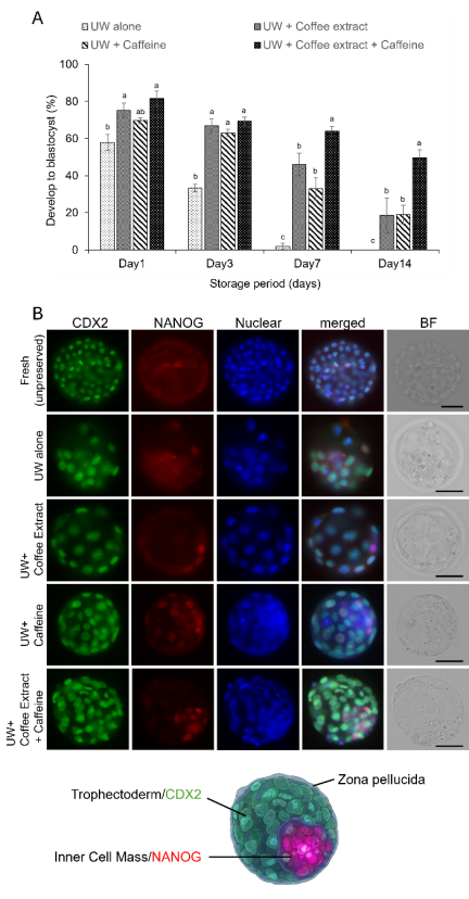
Fig. 4. Rate of development to the blastocyst stage after 1, 3, 7, and 14 days of supercooling preservation. (A) Two-cell embryos were preserved at \(-\)6°C for 1, 3, 7, or 14 days in UW solution alone, UW solution supplemented with 10% coffee lees extract, UW solution supplemented with 173 μg/mL caffeine, or UW solution containing both substances. After rewarming, the embryos were cultured in KSOM for 4 days, and the percentage developing to the blastocyst stage was determined. Data were obtained from three independent experiments and are presented as mean \(\pm\) standard deviation (SD). The different letters indicate significant differences among groups within each storage period (\(p < 0.05\)). (B) Representative fluorescence and bright-field images of blastocysts obtained after 7-day preservation under each condition. A fresh, non-preserved blastocyst is shown for comparison. CDX2, NANOG, and nuclei are shown in green, red, and blue, respectively. The bottom schematic illustrates the major structural features of a mouse blastocyst, including the CDX2-positive TE, NANOG-positive ICM, and blastocoel cavity. Scale bars: 50 μm.
3.6. Overall Preservation Performance Supported the Feasibility of the Novel Nonfreezing Preservation Method
Collectively, the present findings demonstrate that plant-derived substances, particularly coffee lees extract and caffeine, improve supercooling-based nonfreezing preservation of early embryos. Coffee lees extract stabilized embryo morphology after storage; caffeine contributed substantially to the protective effect; and plant-derived substance-based preservation maintained cytoskeletal organization and developmental potential after prolonged storage. These results support the feasibility of a novel nonfreezing preservation method for early embryos that uses plant-derived substances to maintain cellular integrity.
4. Discussion
In the present study, we examined whether coffee lees extract and its representative components could improve supercooling-based nonfreezing preservation of mouse 2-cell embryos in UW solution. The broader objective was to establish a low-damage preservation strategy that could support the stable handling and quality control of living cells intended for future sensor-integrated biological systems.
Early embryos were selected as experimental models because they provided a sensitive and informative system for evaluating preservation-induced damage. At the 2-cell stage, individual blastomeres are clearly distinguishable, which allows the direct assessment of membrane integrity, blastomere morphology, and cytoskeletal organization after preservation. In addition, embryos at this stage can be further cultured, which enables the evaluation of both structural preservation and post-preservation developmental competence.
The present results demonstrate that supplementation with plant-derived substances improves the preservation of 2-cell embryos under subzero nonfreezing conditions. In the main experiments, the addition of coffee lees extract to the UW solution reduced morphological damage after storage at \(-\)6°C for 72 h. In addition, the substance-treated groups exhibited better maintenance of cellular organization and higher developmental competence after rewarming during the supporting prolonged preservation experiments. These findings suggest that the proposed preservation method may reduce storage-associated damage to highly sensitive living cells.
An important implication of the present findings is that effective nonfreezing preservation depends on both the basal preservation medium and optimization of plant-derived substances that stabilize the supercooled state and protect cellular structures 5,7. The UW solution alone permitted embryo recovery after preservation, indicating a degree of baseline protection. However, residual membrane and blastomere deformation remained evident, suggesting that UW solution alone was insufficient to maintain embryonic integrity. The improvement observed after adding coffee lees extract indicates that its plant-derived components provide complementary protection during subzero storage. This interpretation is consistent with the maintenance of preservation solutions in a visibly unfrozen state during storage.
Among the major components of coffee lees extract, caffeine appeared to play an important role. Caffeine exhibits strong anti-ice-nucleation activity 7,13, and in this study, caffeine alone improved the post-preservation embryo morphology, particularly at the intermediate concentration. These findings support the possibility that suppression of ice nucleation contributes to the protective effect of caffeine. In addition, caffeine may mitigate oxidative and low-temperature stress or help maintain membrane and cytoskeletal stability. However, these potential mechanisms were not directly examined in the present study. Notably, the highest caffeine concentration produced more frequent morphological abnormalities, indicating that preservation efficacy depends on the concentration and that excessive caffeine exposure may be detrimental.
In contrast, chlorogenic acid alone exhibited limited suitability as a preservative under the experimental conditions. Despite its antioxidant properties, its protective effect on embryo morphology was relatively weak, and membrane abnormalities remained evident. This suggests that oxidative stress is unlikely to be the sole cause of preservation injury in this model. Damage during prolonged supercooling may involve multiple processes, including membrane destabilization, cytoskeletal disruption, and broader low-temperature stress responses 4,13. Therefore, the superior effect of coffee lees extract may reflect cooperative or complementary actions among several constituents rather than the effect of a single compound. Consistent with this interpretation, the combined treatment yielded the highest developmental competence in the prolonged preservation experiments.
The descriptive quantitative assessment was consistent with the representative images, particularly the improvement observed with 10% coffee lees extract and the intermediate caffeine concentration. However, because the morphological analysis included a limited number of evaluable embryos, these results should be interpreted as supportive descriptive evidence.
Embryos preserved with plant-derived substances retained greater developmental competence after rewarming, as demonstrated by improved blastocyst formation after storage for 3 days or longer. In addition, representative CDX2 and NANOG staining also suggested that the general organization of the TE and ICM was better maintained in the coffee lees extract- and caffeine-containing groups than in the UW-alone group. These results indicate that the benefits of the preservation method were not limited to immediate post-preservation morphology but extended to preimplantation developmental potential.
Nevertheless, the possible effects of caffeine on developmental kinetics and blastocyst quality remain unclear. In the present study, caffeine exposure was limited to the preservation period, and the embryos were subsequently cultured in caffeine-free KSOM. However, cleavage timing, total blastocyst cell number, lineage-specific cell allocation, and implantation competence were not quantitatively assessed. Future studies should examine these endpoints to determine the developmental safety of caffeine exposure during supercooling preservation.
This study has several limitations. First, although a descriptive quantitative morphological assessment was included, the number of evaluable embryos differed among the conditions and was relatively limited. Studies using larger and more balanced sample sizes are required to confirm the reproducibility and statistical robustness of the morphological findings. Second, the molecular mechanisms underlying the protective effects of coffee lees extract and caffeine remain unclear. Further studies should directly examine the oxidative stress, membrane integrity, cytoskeletal organization, metabolic status, and other cellular responses during low-temperature preservation. Third, developmental competence was only evaluated up to the blastocyst stage. Although blastocyst formation and representative CDX2 and NANOG expression patterns provide information on pre-implantation development and lineage organization, they do not establish complete embryonic normality. Morphologically normal blastocysts may exhibit subtle chromosomal, epigenetic, metabolic, or cellular abnormalities that become apparent only after implantation. Future in vivo validation should, therefore, include embryo transfer into recipient females and the assessment of implantation, embryo resorption, fetal and placental development, live-birth rate, and postnatal health of the offspring.
Fourth, the main and prolonged preservation experiments differed in storage duration and experimental endpoint, although the same cooling and rewarming rates were used. The 72-h period in the main experiments was selected because preliminary 24-h experiments produced variable morphological outcomes, whereas 72-h preservation yielded more reproducible differences among the tested solutions. The prolonged experiments included a 1-day reference point, followed by progressive extensions to 3, 7, and 14 days. This staged approach enabled the evaluation of longer preservation intervals while ensuring that sufficient embryos were collected for repeated experiments. Nevertheless, future studies should adopt a unified experimental design and further extend the storage period to assess the practical applicability of this method.
These findings may also be relevant for future applications in which living cells and tissues are used as sensing elements, signal carriers, or functional components in biohybrid systems. In such applications, the preservation quality before use may influence the structural uniformity, functional reproducibility, quantitative reliability, and performance after device integration 2. Cells or tissues containing intracellular sensors or microdevices may be particularly vulnerable to conventional freezing and thawing. Embedded materials can potentially promote ice nucleation, whereas ice formation, thermal expansion and contraction, and mechanical stress could cause device displacement, deformation, disruption of the device–cell interface, or changes in sensor sensitivity and calibration. By avoiding ice formation and phase transition-associated stress, supercooling-based nonfreezing preservation may facilitate the maintenance of both cellular integrity and device functionality during storage, transport, handling, and pre-use stabilization. However, the structural and functional integrity of integrated sensing devices after preservation must be directly evaluated in future studies.
In conclusion, coffee lees extract and caffeine improved the supercooling-based nonfreezing preservation of mouse 2-cell embryos. The protective effect of coffee lees extract reflects the cooperative action of multiple constituents, rather than a single mechanism. Although further validation using larger sample sizes, unified preservation conditions, and in vivo developmental assessment is required, the present findings support the potential of plant-derived substance-based nonfreezing preservation as a low-damage strategy for sensitive living biological materials and provide a conceptual basis for future preservation systems intended for sensor-integrated biological applications.
Conflict of Interest The authors declare no conflicts of interest.
Acknowledgments
This study was supported by a research grant from Ito Foundation of Japan. The authors are grateful to Professor Hidehisa Kawahara of Kansai University for providing the coffee lees extract used in this study.
- [1] N. Gupta, V. Renugopalakrishnan, D. Liepmann, R. Paulmurugan, and B. D. Malhotra, “Cell-based Biosensors: Recent Trends, Challenges and Future Perspectives,” Biosens. Bioelectron., Vol.141, Article No.111435, 2019. https://doi.org/10.1016/j.bios.2019.111435
- [2] S. Wang and G. D. Elliott, “Synergistic Development of Biochips and Cell Preservation Methodologies: A Tale of Converging Technologies,” Curr. Stem Cell Rep., Vol.3, No.1, pp. 45-53, 2017. https://doi.org/10.1007/s40778-017-0074-8
- [3] J. Liebermann, J. Dietl, P. Vanderzwalmen, and M. J. Tucker, “Recent Developments in Human Oocyte, Embryo and Blastocyst Vitrification: Where Are We Now?,” Reprod. Biomed. Online, Vol.7, No.6, pp. 623-633, 2003. https://doi.org/10.1016/s1472-6483(10)62084-6
- [4] B. P. Best, “Cryoprotectant Toxicity: Facts, Issues, and Questions,” Rejuvenation Res., Vol.18, No.5, pp. 422-436, 2015. https://doi.org/10.1089/rej.2014.1656
- [5] T. A. Berendsen, B. G. Bruinsma, C. F. Puts, N. Saeidi, O. B. Usta, B. E. Uygun, M. L. Izamis, M. Toner, M. L. Yarmush, and K. Uygun, “Supercooling Enables Long-Term Transplantation Survival Following 4 Days of Liver Preservation,” Nat. Med., Vol.20, No.7, pp. 790-793, 2014. https://doi.org/10.1038/nm.3588
- [6] S. Fujikawa, C. Kuwabara, J. Kasuga, and K. Arakawa, “Supercooling-Promoting (Anti-ice Nucleation) Substances,” M. Iwaya-Inoue, M. Sakurai, and M. Uemura (Eds.), “Survival Strategies in Extreme Cold and Desiccation,” pp. 289-320, 2018. https://doi.org/10.1007/978-981-13-1244-1_16
- [7] H. Kawahara, E. Tagawa, C. Watanabe, J. Hamada, and S. Hamada, “Characterization of Anti-Ice Nucleation Activity of the Extract from Coffee Refuse,” Biocontrol. Sci., Vol.22, No.4, pp. 205-211, 2017. https://doi.org/10.4265/bio.22.205
- [8] F. Ariu, L. Bogliolo, G. Leoni, L. Falchi, D. Bebbere, S. M. Nieddu, M. T. Zedda, S. Pau, and S. Ledda, “Effect of Caffeine Treatment Before Vitrification on MPF and MAPK Activity and Spontaneous Parthenogenetic Activation of In Vitro Matured Ovine Oocytes,” Cryo Letters, Vol.35, No.6, pp. 530-536, 2014.
- [9] B. A. Pereira, B. R. Chaves, M. C. Teles, T. P. Pontelo, C. R. Oliveira, R. V. de Souza, J. E. Rodriguez-Gil, and M. G. Zangeronimo, “Chlorogenic Acid Improves the Quality of Boar Semen Subjected to Cooled Storage at 15°C,” Andrologia, Vol.50, No.5, Article No.e12978, 2018. https://doi.org/10.1111/and.12978
- [10] Y. Hoshino and E. Sato, “Protein Kinase B (PKB/Akt) Is Required for the Completion of Meiosis in Mouse Oocytes,” Dev. Biol., Vol.314, No.1, pp. 215-223, 2008. https://doi.org/10.1016/j.ydbio.2007.12.005
- [11] Y. Hoshino and T. Uchida, “Prolyl Isomerase, Pin1, Controls Meiotic Progression in Mouse Oocytes,” Cells, Vol.11, No.23, Article No.3772, 2022. https://doi.org/10.3390/cells11233772
- [12] S. Bessonnard, S. Vandormael-Pournin, S. Coqueran, M. Cohen-Tannoudji, and J. Artus, “PDGF Signaling in Primitive Endoderm Cell Survival Is Mediated by PI3K-mTOR Through p53-Independent Mechanism,” Stem Cells, Vol.37, No.7, pp. 888-898, 2019. https://doi.org/10.1002/stem.3008
- [13] G. D. Elliott, S. Wang, and B. J. Fuller, “Cryoprotectants: A Review of the Actions and Applications of Cryoprotective Solutes that Modulate Cell Recovery from Ultra-Low Temperatures,” Cryobiology, Vol.76, pp. 74-91, 2017. https://doi.org/10.1016/j.cryobiol.2017.04.004
This article is published under a Creative Commons Attribution-NoDerivatives 4.0 Internationa License.