
Paper:
Engineering of LNA-Modified DNA-AuNP Nanomachines for Intracellular miRNA Imaging
Maasa Yokomori*,**
 , Keitaro Yoshimoto**
, and Yoko Yamanishi***
*Department of Precision Mechanical Engineering, School of Science and Engineering, Chuo University
1-13-27 Kasuga, Bunkyo-ku, Tokyo 112-8551, Japan
**Department of Life Sciences, Graduate School of Arts and Sciences, The University of Tokyo
3-8-1 Komaba, Meguro-ku, Tokyo 153-8902, Japan
***Department of Mechanical Engineering, Faculty of Engineering, Kyushu University
744 Motooka, Nishi-ku, Fukuoka, Fukuoka 819-0395, Japan

DNA nanomachines based on gold nanoparticles (AuNPs) are promising platforms for intracellular sensing of microRNAs (miRNAs), which are biomarkers involved in a wide range of diseases. However, achieving robust performance under intracellular conditions remains challenging. In this study, we investigated the effect of locked nucleic acid (LNA) modification on an entropy-driven DNA-AuNP nanomachine designed for miRNA detection. By incorporating LNA into probe sequences, we evaluated signal generation, sensitivity, and selectivity under cytosol-mimicking conditions. Consequently, appropriately designed LNA modifications substantially restored nanomachine functionality under intracellularly relevant conditions. In contrast, excessive or unfavorably positioned LNA incorporation led to increased nonspecific background signals. Under optimized conditions, the LNA-modified nanomachine achieved a detection limit of approximately 10 pM for miRNA-21 with high sequence specificity. Furthermore, when delivered into MCF-7 cancer cells using a transfection reagent, the LNA-modified DNA-AuNP nanomachine showed a higher fraction of fluorescence-positive cells compared with the unmodified system. This study clarifies the critical role of probe modification in the design of DNA-AuNP nanomachines operating within living cells and presents an effective strategy for realizing live-cell miRNA imaging based on molecular robotics concepts.
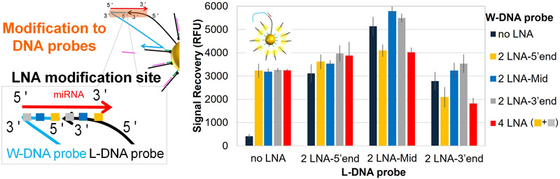
LNA positions affect signal recovery
1. Introduction
Analytical technologies for biomolecular information are critically important for understanding biological systems and diagnosing diseases and have attracted considerable research attention 1,2,3,4. MicroRNAs (miRNAs) are short, non-coding RNAs that play pivotal roles in the post-transcriptional regulation of gene expression 5,6. The aberrant expression of specific miRNAs has been implicated in a wide range of pathological conditions, including cancer, cardiovascular diseases, and neurodegenerative disorders. miRNAs have emerged as promising biomarkers for early diagnosis and therapeutic monitoring owing to their disease-specific expression profiles and intracellular localization 6,7,8. Consequently, the development of sensitive and selective tools for detecting intracellular miRNAs is of great biomedical interest.
DNA nanomachines constructed from rationally designed oligonucleotides offer a versatile platform for molecular sensing 9. From the perspective of molecular robotics 10,11, such systems can be regarded as molecular-scale robotic systems that integrate sensing, information processing, and actuation using biomolecular components. The inherent programmability of DNA, together with its high sequence specificity and ability to undergo conformational changes in response to molecular inputs, makes DNA nanomachines ideal candidates for intracellular biosensing 9,12. Gold nanoparticles (AuNPs) have been widely used as scaffolds to enhance the functionality and stability of DNA nanomachines in biological environments 12,13. AuNPs provide numerous advantages such as efficient fluorescence quenching 14, nuclease resistance by sterically protecting DNA strands 15, and high-density DNA loading for amplified signal output and improved sensitivity 16.
Notably, entropy-driven DNA-AuNP nanomachines, such as those reported by Liang et al.16, are well-suited for intracellular sensing owing to their ability to undergo structural changes in response to target molecules and generate real-time signal outputs. AuNPs decorated with DNA probes and fluorophore-modified DNA strands are used in this nanomachine. The DNA probe complementary to the target miRNA is split into two parts: one anchored to the AuNP surface, and the other designed as a free single-stranded probe. Upon hybridization with the target miRNA, the free probe is brought into proximity to the AuNP surface and initiates a strand displacement reaction, releasing the fluorophore-modified strand and restoring fluorescence. This sensing mechanism exploits the excellent distance-dependent quenching properties of AuNPs, whereby the fluorescence from dyes near the AuNP surface is efficiently quenched and can be restored by increasing the separation distance 14. Subsequently, the free probe catalyzes successive reactions on adjacent substrates, generating an amplified fluorescence signal. Liang et al. demonstrated that, compared to conventional nanoflare sensors and entropy-driven designs lacking a walking mechanism, this nanomachine exhibits accelerated reaction kinetics and superior sensitivity owing to the high local effective concentration of DNA components on the AuNP surface, achieving a detection limit of approximately 8 pM.
However, DNA-AuNP nanomachines often exhibit robust activity under conditions of higher ionic strength than that of the cytosol, raising concerns that their reactivity may be reduced in intracellular environments. This is because DNA duplex formation and the associated trigger reactions are sensitive to factors, such as ionic strength and DNA sequence design 17,18. In DNA-AuNP nanomachines, in particular, DNA strands are densely arranged on the surface of AuNPs, resulting in a high local negative charge density 19,20,21. Under low ionic strength conditions, the electrostatic repulsion between DNA strands is enhanced, making hybridization more prone to destabilization 19,20,21.
A promising strategy for enhancing duplex formation under such conditions involves the use of chemically modified nucleotides 22,23. For instance, locked nucleic acid (LNA) is a nucleotide analog in which a methylene bridge between the 2\('\)-O and 4\('\)-C positions of the ribose locks the sugar into a C3\('\)-endo conformation, thereby increasing rigidity and thermal stability of nucleic acid duplex 24,25. LNA exhibits exceptionally high hybridization affinity with DNA and RNA; molecular dynamics simulations and in situ hybridization experiments have demonstrated that it enables stable duplex formation, even under low ionic strength conditions. Indeed, LNA-modified DNA probes have been reported to significantly improve intracellular miRNA detection efficiency compared to conventional DNA probes 26.
However, the effect of LNA modification on DNA immobilized on AuNPs has not been systematically investigated, and knowledge regarding its influence and behavior remains limited. According to Xu and Craig 19, the thermodynamics of duplex formation for DNA immobilized on AuNP surfaces differ markedly from those of freely diffusing DNA, particularly at high surface coverage where both enthalpy and entropy are reduced, leading to a substantial decrease in free energy. These findings suggest that the hybridization behavior between immobilized DNA strands is fundamentally different from that between free DNA strands, and that the stabilizing effect of LNA modification may also behave differently in such contexts. Therefore, further investigation is required to determine whether LNA modification can effectively enhance the hybridization performance of DNA-AuNP systems under intracellular conditions.
In this study, we investigated the effect of incorporating LNA modifications into DNA-AuNP nanomachines under conditions that mimic the intracellular environment, specifically, sodium and potassium ionic strengths combined with molecular crowding induced by high-molecular-weight polyethylene glycol 27. Using a previously reported DNA-AuNP nanomachine design 16, we systematically evaluated the effects of LNA modifications on signal generation, reaction kinetics, sensitivity, and specificity in the presence of target miRNAs. This approach is expected to preserve and enhance the functionality of DNA-AuNP nanomachines under cytosol-like conditions, thereby enabling sensitive detection of miRNAs.
2. Materials and Methods
2.1. Reagents
Thiol-modified DNA was purchased from the Japan Gene Research Laboratories (Sendai, Japan). LNA-modified DNA and cyanine 5 (Cy5)-modified DNA were purchased from Integrated DNA Technologies, Inc. (Coralville, IA, USA). miRNAs were obtained from FASMAC Co., Ltd. (Kanagawa, Japan). Unmodified DNA and DNA containing phosphorothioate linkages were purchased from Eurofins Genomics (Tokyo, Japan). The sequences of all oligonucleotides used in this study are listed in Table 1 (in Appendix A).
AuNP solution (20 nm in diameter), borate buffer, and sodium chloride (NaCl) solution were purchased from Nanopartz Inc. (Loveland, CO, USA), Thermo Fisher Scientific (Waltham, MA, USA), and Promega Corp. (Madison, WI, USA), respectively. Phosphate buffer powder (pH 7.2) (FUJIFILM Wako Pure Chemical Corporation, Tokyo, Japan) and polyethylene glycol 20000 powder (Tokyo Chemical Industry Co., Ltd., Tokyo, Japan) were dissolved in ultrapure water (Thermo Fisher Scientific Inc., USA).
The human breast cancer cell line MCF-7 was obtained from the RIKEN BRC Cell Bank (Tsukuba, Japan). Dulbecco’s modified Eagle’s medium (DMEM) and fetal bovine serum (FBS) were purchased from FUJIFILM Wako Pure Chemical Corporation (Osaka, Japan). Opti-MEM, trypsin/EDTA (0.25%), and Lipofectamine-2000 were purchased from Gibco (Gaithersburg, USA), Nacalai Tesque, Inc. (Kyoto, Japan), and Invitrogen (Carlsbad, CA, USA), respectively.
2.2. Preparation of DNA-AuNP Nanomachines
DNA-AuNPs were prepared according to previously reported methods 16,28. First, the deprotected thiol-modified oligonucleotides were added to the AuNP solution at a molar ratio of \(1000:500:1\) (\(\textrm{A}:\textrm{L}:\textrm{AuNP}\)). Subsequently, borate buffer was added and the mixture was incubated for 15 min with gentle shaking. The NaCl concentration was then gradually increased from 0.05 M to 0.5 M using 5 M NaCl. After each NaCl addition, the nanoparticle solution was sonicated for approximately 10 s and stirred for 15 min. Finally, the solution was incubated overnight with shaking.
Excess oligonucleotides were removed by repeated centrifugation at \(5000 \times {g}\) for 90 min four times. After removing the supernatant, the pellet was resuspended in phosphate buffer. The final concentration of DNA-AuNPs was determined by UV-visible spectroscopy (UV-2600i; Shimadzu Corporation, Tokyo, Japan).
PBS (1\(\times\)) containing B (final concentration, 1 μM) and C (final concentration, 0.6 μM) (final MgCl\(_2\) concentration, 2.5 mM; final NaCl concentration, 50 mM) was added to the DNA-AuNP solution (final concentration, 0.8 nM). The mixture was heated at 65°C for 15 min and then incubated overnight at room temperature in the dark. After incubation, the resulting solution was washed three times by centrifugation at \(5000 \times {g}\) for 90 min at 4°C using 1\(\times\) PBS.
2.3. In Vitro Evaluation of DNA-AuNP Nanomachine Reactions
Reaction solutions were prepared such that the final concentrations of DNA-AuNPs (A/B/C-Cy5), W-DNA, and DNA strand F were 100 pM, 5 nM, and 320 nM, respectively. While incubating at 37°C, the fluorescence intensity for blank samples and target miRNA was monitored for approximately 20 h using a microplate reader (Spark, Tecan Group Ltd., Zurich, Switzerland).
2.4. Intracellular Imaging
The transfection reactions were performed as previously described 16. An aliquot of Lipofectamine-2000 was diluted in Opti-MEM (solution I). Cy5-labeled DNA-AuNPs assembled with A/B1/C1 (solution II) and a mixture of nucleic acid strand F and W-DNA probes (solution III) were diluted using Opti-MEM. After incubation at room temperature for 10 min, half of solution I was mixed with solution II, and the remaining half of solution I was mixed with solution III. After incubation at 37°C for 20 min, the two mixtures were combined, diluted in DMEM containing 10% FBS, and added to the cells. In the final mixture, the concentrations of Cy5-labeled DNA-AuNPs, DNA strand F, and W-DNA probes were 0.1 nM, 20 nM, and 10 nM, respectively. The cells were incubated at 37°C in a humidified incubator. After washing them twice with 1\(\times\) PBS, fluorescent images of the cells were acquired using a fluorescence microscope (Eclipse Ti, Nikon Corporation, Tokyo, Japan) or a confocal microscope (TCS-SP8, Leica Microsystems GmbH, Wetzlar, Germany). Fluorescence images were analyzed using ImageJ software (NIH). Fluorescence-positive cells were identified using ImageJ software (NIH). The fluorescence channel was thresholded using a fixed intensity range (60–115 nm) to exclude background signals. Regions of interest corresponding to fluorescent signals were extracted using the “Analyze Particles” function with a minimum size threshold of 2000 pixels\(^2\). Cells exhibiting intracellular fluorescence signals above this threshold were classified as fluorescence-positive. The total number of cells was manually counted from the corresponding bright-field images, and the percentage of fluorescence-positive cells was calculated.
3. Results and Discussion
3.1. Design of LNA-Modified DNA-AuNP Nanomachines
Figure 1 illustrates the design and operating principles of the entropy-driven DNA-AuNP nanomachines used in this study. The nanomachine consisted of AuNPs functionalized with DNA probes (L-DNA probes) and fluorescence-modified DNA strands (C-Cy5) together with W-DNA probes (walking legs) and a fuel strand (F) in solution. The DNA substrate on the AuNP surface is composed of a three-stranded complex (A/B/C-Cy5), where strand A is immobilized on the AuNP surface and strand B acts as an intermediate strand bridging A and C-Cy5. In this system, the fluorescence of C-Cy5 was quenched because of its proximity to the AuNP surface, and the fluorescence is restored by increasing the distance between the fluorophore and the nanoparticle. When the target miRNA is present, the nanomachine is activated through a strand displacement process mediated by short single-stranded regions (toeholds) within the DNA structure, resulting in the continuous release of C-Cy5 and recovery of the fluorescence signal (Fig. 1(A)).
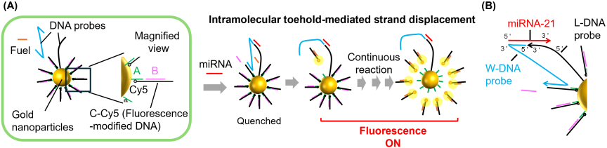
Fig. 1. Schematic representation of an entropy-driven DNA-AuNP nanomachine for miRNA analysis. (A) Gold nanoparticles are functionalized with L-DNA probes and fluorescence-modified DNA strands (C-Cy5). In the presence of a miRNA template, an intramolecular toehold-mediated strand displacement process is triggered, leading to continuous disassembly of C-Cy5 and recovery of the fluorescence signal. (B) Detailed structural diagram showing the interaction between miRNA, W-DNA probe, and L-DNA probe on the surface of gold nanoparticles.
Figures 1(B) and 7 (in Appendix A) show how the miRNA, W-DNA probe, and L-DNA probe interact near the nanoparticle surface. The L-DNA probe contains a sequence that is partially complementary to the target miRNA, anchoring the miRNA close to the nanoparticle. The W-DNA probe also binds to the same miRNA, forming a ternary complex that brings the W-DNA into proximity to the AuNP surface. This proximity enables W-DNA to initiate strand displacement on the nanoparticle surface, where W-DNA first binds to an exposed toehold region (toehold 1), displacing strand B and exposing a second toehold (toehold 2). Subsequently, the fuel strand binds and hybridizes to the exposed region, triggering branch migration and complete disassembly of the DNA structure. During this process, the W-DNA strand regenerates and participates in the subsequent reactions to form a catalytic cycle that enables repeated signal amplification. Consequently, the fluorescence previously quenched by the AuNP surface is restored. This catalytic cycle repeats as W-DNA “walks” along the AuNP surface, resulting in continuous signal amplification. This intramolecular reaction on the AuNP surface proceeds more efficiently than conventional intermolecular reactions because of the high local concentration of DNA components.
In this study, LNA modifications were introduced into DNA probes that formed complementary duplexes with miRNAs to enhance the double-strand stability under cytosol-mimicking conditions. Specifically, four types of W-DNA probes containing LNA modifications in the miRNA-complementary region were designed and synthesized: probes modified with two LNA bases at the 5\('\) end, two LNA bases in the middle region, two LNA bases at the 3\('\) end, and a probe modified with two LNA bases at both termini (Table 1 in Appendix A). To examine the effect of the number of LNA modifications, two types of W-DNA probes containing three LNA modifications in the miRNA complementary region and two types of L-DNA probes containing three or four LNA modifications were designed and synthesized (Table 1).
3.2. Evaluation of the Effect of LNA Modification on DNA-AuNP Nanomachine Performance under Intracellular-Like Conditions
First, the fluorescence response of the non-modified DNA-AuNP nanomachine was evaluated in the presence and absence of target miRNA-21 (miR-21) under cytosol-mimicking environments (100 mM K\(^{+}\), 10 mM Na\(^{+}\), 0.5 mM Mg\(^{2+}\), and 10% PEG20000) (Fig. 2(A)). Fluorescence recovery was negligible even in the presence of the target miRNA, suggesting a significant loss of function under physiologically relevant conditions. To overcome this issue, experiments were conducted under cytoplasm-mimicking conditions using four types of W-DNA probes with LNA modifications introduced into the miRNA-complementary region: three probes containing two LNA nucleotides at different positions and one probe containing four LNA nucleotides. As illustrated in Fig. 2(B), the LNA-modified DNA-AuNP nanomachines demonstrated a marked improvement in signal generation, reaching a plateau at approximately 400 min, and showing a clear fluorescence signal difference compared to the blank. Furthermore, the reaction kinetics were slightly affected by the position of LNA incorporation (Fig. 8 in Appendix A). However, an increase in the number of LNA modifications did not significantly accelerate this response.
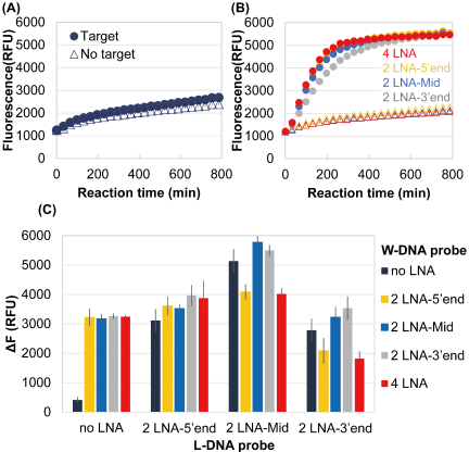
Fig. 2. Time-course fluorescence measurements of the DNA nanomachine in the presence (filled circles) and absence (open triangle) of the target miR-21 (1 nM). (A) DNA-AuNP nanomachines without LNA modification. (B) DNA-AuNP nanomachines with LNA modification in the W-DNA probe. Yellow, blue, and gray markers represent two LNA nucleotides introduced at the 5\('\) end (2 LNA-5\('\) end), middle (2 LNA-Mid), and 3\('\) end (2 LNA-3\('\) end) of the sequence complementary to the target miRNA, respectively. Red markers represent four LNA nucleotides incorporated at both the 5\('\) and 3\('\) ends (4 LNA). (C) Effects of both the position and number of LNA modifications on the performance of DNA nanomachines targeting miR-21. \(\Delta\)F represents the fluorescence change, defined as the fluorescence intensity measured in the presence of miRNA minus that measured in its absence, recorded at the final measurement point. Each data point (or bar) represents the average of three independent measurements. Error bars indicate the standard deviation (SD, \(n = 3\)).
Next, we systematically analyzed the effects of introducing LNA modifications into both W-DNA and L-DNA probes. Fig. 2(C) summarizes the experimental results of the effects of the position and number of LNA modifications on the performance of the DNA-AuNP nanomachines targeting miR-21. \(\Delta\)F represents the fluorescence change, defined as the fluorescence intensity measured in the presence of miRNA minus that measured in its absence, evaluated at the final measurement point. The use of LNA-modified DNA probes resulted in a substantial improvement in performance under cytosol-mimicking conditions. Notably, the extent of this improvement strongly depended on the specific combination of modifications of the two DNA probes. Interestingly, when LNA modifications were introduced at the 3\('\) terminus or the central region of L-DNA, their combination with an LNA modification at the 5\('\) terminus of W-DNA led to a reduced \(\Delta\)F compared with combinations involving modifications at other positions. The decrease in \(\Delta\)F was attributed to an increase in nonspecific fluorescence intensity. Collectively, these results demonstrate that LNA modification is an effective strategy for restoring the function of DNA-AuNP nanomachines under cytosol-mimicking conditions; however, its effect is highly position-dependent. Excessive nonspecific stabilization caused by dual modifications may compromise the specificity.
Furthermore, we examined the effect of the number of LNA modifications introduced into DNA probes (Fig. 3). As shown in Fig. 3(A), pronounced differences in the final \(\Delta\)F were observed depending on the modification position. In the case of W-DNA-modified with LNA at the 5\('\) terminus, a significant increase in fluorescence was observed even in the absence of the target (Fig. 3(B)). As a result, \(\Delta\)F was found to approach zero after approximately 20 h. Therefore, to achieve a robust performance in miRNA detection, optimization of both the number and positional arrangement of LNA modifications is essential. Notably, the requirements for LNA modification are not universal and should be evaluated based on the target miRNA. We also evaluated the effects of introducing LNA on DNA probes targeting miRNA-155 (miR-155). Consequently, even when unmodified DNA probes were used, a target miRNA dependent increase in fluorescence intensity was observed under cytosol-mimicking conditions and the introduction of LNA did not lead to a significant increase in \(\Delta\)F (Fig. 9 in Appendix A). Depending on the target molecule, this variation may be attributed to differences in miRNA sequences. The L-DNA probe for miR-155 has an approximately 1.5-fold higher GC content than that for miR-21, indicating greater duplex stability. Therefore, DNA-AuNP nanomachine targeting miR-155 can likely function under cytosol-mimicking conditions, even without additional stabilization of the duplex provided by LNA modifications.
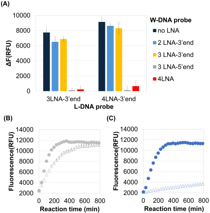
Fig. 3. Effects of increasing the number of LNA modifications on the performance of DNA-AuNP nanomachines targeting miR-21. (A) Bar plots summarizing the fluorescence change, \(\Delta\)F, measured at the final measurement point. (B, C) Time-course fluorescence were measured for the DNA-AuNP nanomachines with 3LNA-3\('\) ends L-DNA probes in the presence (filled circles) and absence (open triangles) of the target miR-21 (1 nM). 3LNA-5\('\) ends (B) and 3LNA-3\('\) ends (C) DNA probes were employed as W-DNA probes, respectively. Each data point (or bar) represents the average of three independent measurements. Error bars indicate the standard deviation (SD, \(n = 3\)).
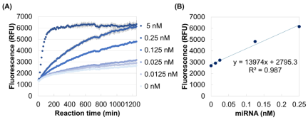
Fig. 4. Evaluation of the sensitivity of LNA-modified DNA nanomachines in a simulated cytoplasmic ionic environment. (A) Monitoring the fluorescence increase over time in response to various concentrations of miR-21 using the proposed LNA-modified DNA nanomachine. (B) Fluorescence intensity plotted against miR-21 concentration at the final measurement point, demonstrating the nanomachine’s sensitivity and ability to discriminate miRNA levels quantitatively. Each data point represents the mean of three independent measurements. Error bars indicate the standard deviation (SD, \(n = 3\)).
3.3. Evaluation of Sensitivity
The sensitivity of the proposed LNA-modified DNA-AuNP nanomachine was also evaluated. The fluorescence intensity was monitored over time for miR-21 concentrations ranging from 0 to 5 nM, revealing a clear concentration-dependent increase, which demonstrates the ability of the nanomachine to respond proportionally to target levels (Fig. 4(A)). The fluorescence intensity at the final measurement point was plotted against miR-21 concentration, yielding a strong linear correlation (\(R^2 = 0.987\)) (Fig. 4(B)). Based on this standard curve, the limit of detection was calculated to be approximately 10 pM (3\(\sigma\)b/slope, where \(\sigma\)b is the standard deviation of blank samples) using the standard curve, which corresponds to approximately six copies of miRNA per 10 μm cell.
3.4. Evaluation of Selectivity
The selectivity of the proposed DNA-AuNP nanomachine toward different miRNA sequences was evaluated using miR-21 (target), miR-10b, miR-195, miR-M1 (single-base mutation), miR-M2 (two-base mutation), and a control sample without miRNA (Fig. 5). The nanomachine clearly discriminated miR-21 from miR-10b and miR-195, demonstrating high sequence specificity for these targets. In contrast, miR-M1 and miR-M2, which contain one or two base mutations relative to miR-21, exceeded the threshold and exhibited considerable variability, indicating limited discrimination for closely related sequences and highlighting the need for further optimization of probe design.
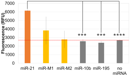
Fig. 5. Evaluation of the specificity of LNA-modified DNA nanomachines in a simulated cytoplasmic ionic environment. Fluorescence intensity (RFU) was measured for miR-21 (target miRNA), miR-10b, miR-195, miR-M1, miR-M2, and a control sample without miRNA (no miRNA). Each bar represents the standard deviation of three independent measurements. The statistical significance was tested using the Welch’s \(t\)-test (\(^{****}\): \(p <0.001\), \(^{***}\): \(p <0.01\)). The red dashed line represents the fluorescence threshold determined from the no miRNA control, used to assess the specificity of the nanomachine. Each bar represents the mean of three independent measurements. Error bars indicate the standard deviation (SD, \(n = 3\)).
3.5. Intracellular Performance of DNA-AuNP Nanomachines for miRNA Imaging
Finally, to evaluate the effectiveness of the DNA-AuNP nanomachines for miRNA imaging in living cells, DNA-AuNP nanomachines complexed with lipofectamine, a cationic liposome-based transfection reagent, were co-incubated with human breast cancer cells (MCF-7), which are known to highly express miR-21. Based on confocal z-stack imaging, the fluorescence was not restricted to the cell surface but was distributed within MCF-7 cells, exhibiting behavior consistent with cellular internalization (Fig. 10 in Appendix A). Furthermore, compared with unmodified DNA-AuNP nanomachines (Fig. 6(A)), the use of LNA-modified DNA-AuNP nanomachines resulted in an increased number of cells exhibiting detectable fluorescence signals, suggesting that LNA modification is effective in enhancing intracellular performance (Fig. 6(B)). The total percentages of fluorescence-positive cells were (A) 4/298 cells \(= 1.3\%\) and (B) 12/317 cells \(= 3.8\%\) (Fig. 11 in Appendix A).
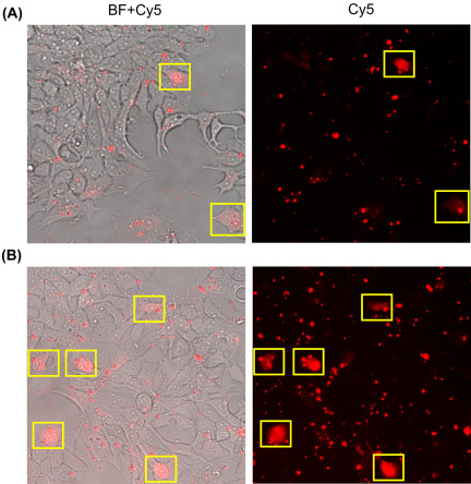
Fig. 6. Intracellular imaging of miR-21 using DNA-AuNP nanomachines. Representative microscopic images of human breast cancer (MCF-7) cells incubated for 12 h with (A) the non-modified nanomachines and (B) the LNA-modified nanomachines. Merged bright-field (BF) and Cy5 images, together with fluorescence-only (Cy5) images, are presented. The yellow squares indicate cells in which fluorescence was detected.
4. Conclusion
In this study, we systematically investigated the effects of LNA modification on the performance of DNA-AuNP nanomachines under intracellularly relevant conditions. Although conventional DNA-AuNP nanomachines may fail to exhibit target miRNA-dependent signal responses under cytosol-mimicking environments, the introduction of LNA into miRNA-complementary regions effectively restored signal generation. The effect of the LNA modification was found to be strongly dependent on its position and number. Appropriate incorporation enhanced the sensitivity and intracellular robustness, whereas unfavorable combinations led to increased nonspecific background signals. Under optimized conditions, the LNA-modified nanomachine enabled sensitive and selective detection of miR-21 at picomolar levels. Furthermore, fluorescence imaging of living MCF-7 cells suggested that the LNA-modified nanomachines showed improved intracellular performance compared to unmodified nanomachines. Collectively, these results suggest that the probe modification conditions represent an important design parameter for DNA-AuNP nanomachines and provide useful insights for their application in intracellular biosensing and live-cell imaging.
Acknowledgments
This work was supported by the JST Moonshot Research and Development Program (Grant Numbers JPMJMS2217-2-1 and JPMJMS2217-3-1).
- [1] A. Van Gool et al., “Analytical techniques for multiplex analysis of protein biomarkers,” Expert Rev. Proteomics, Vol.17, No.4, pp. 257-273, 2020. https://doi.org/10.1080/14789450.2020.1763174
- [2] M. Yokomori, O. Gotoh, Y. Murakami, K. Fujimoto, and A. Suyama, “A multiplex RNA quantification method to determine the absolute amounts of mRNA without reverse transcription,” Anal. Biochem., Vol.539, pp. 96-103, 2017. https://doi.org/10.1016/j.ab.2017.10.006
- [3] Y. Zhao et al., “Nucleic Acids Analysis,” Sci. China Chem., Vol.64, No.2, pp. 171-203, 2021. https://doi.org/10.1007/s11426-020-9864-7
- [4] S. Zhou et al., “Optimization of aptamer-triggered hybridization chain reaction for rapid visual ATP detection using gold nanoparticles,” Analytical Methods, Vol.18, No.7, pp. 1541-1547, 2026. https://doi.org/10.1039/D5AY01738F
- [5] D. P. Bartel, “MicroRNAs Genomics, Biogenesis, Mechanism, and Function,” Cell, Vol.116, pp. 281-297, 2004. https://doi.org/10.1016/S0092-8674(04)00045-5
- [6] L. F. R. Gebert and I. J. MacRae, “Regulation of microRNA function in animals,” Nat. Rev. Mol. Cell Biol., Vol.20, No.1, pp. 21-37, 2019. https://doi.org/10.1038/s41580-018-0045-7
- [7] Y. Wu, Q. Li, R. Zhang, X. Dai, W. Chen, and D. Xing, “Circulating microRNAs: Biomarkers of disease,” Clinica Chimica Acta, Vol.516, pp. 46-54, 2021. https://doi.org/10.1016/j.cca.2021.01.008
- [8] H. Dong, J. Lei, L. Ding, Y. Wen, H. Ju, and X. Zhang, “MicroRNA: Function, detection, and bioanalysis,” Chem. Rev., Vol.113, No.8, pp. 6207-6233, 2013. https://doi.org/10.1021/cr300362f
- [9] H. Peng et al., “Signal amplification in living cells: A review of microRNA detection and imaging,” Anal. Chem., Vol.92, No.1, pp. 292-308, 2020. https://doi.org/10.1021/acs.analchem.9b04752
- [10] H. Shibuya, S. Okada, and K. Shoji, “Control of Osmotic-Engine-Driven Liposomes Using Biological Nanopores,” J. Robot. Mechatron., Vol.35, No.5, pp. 1213-1218, 2023. https://doi.org/10.20965/jrm.2023.p1213
- [11] M. Akter and A. Kakugo, “Biomolecular Motor-Based Swarm Robot: An Innovation in Molecular Delivery,” J. Robot. Mechatron., Vol.35, No.4, pp. 1047-1051, 2023. https://doi.org/10.20965/jrm.2023.p1047
- [12] S. Yang, X. Zhan, L. Yuan, M. L. de la Chapelle, W. Fu, and X. Yang, “Entropy driven-based catalytic biosensors for bioanalysis: From construction to application—A review,” Anal. Chim. Acta, Vol.1338, Article No.343549, 2025. https://doi.org/10.1016/j.aca.2024.343549
- [13] Q. Zhang, S. Xu, J. Zheng, J.-R. Zhang, and J.-J. Zhu, “Advances in self-assembled Au-DNA nanomachines,” iScience, Vol.26, No.4, Article No.106327, 2023. https://doi.org/10.1016/j.isci.2023.106327
- [14] E. Dulkeith et al., “Fluorescence Quenching of Dye Molecules near Gold Nanoparticles: Radiative and Nonradiative Effects,” Phys. Rev. Lett., Vol.89, No.20, Article No.203002, 2002. https://doi.org/10.1103/PhysRevLett.89.203002
- [15] D. S. Seferos, A. E. Prigodich, D. A. Giljohann, P. C. Patel, and C. A. Mirkin, “Polyvalent DNA Nanoparticle Conjugates Stabilize Nucleic Acids,” Nano Lett., Vol.9, No.1, pp. 308-311, 2009. https://doi.org/10.1021/nl802958f
- [16] C. Liang, P. Ma, H. Liu, X. Guo, B. Yin, and B. Ye, “Rational Engineering of a Dynamic, Entropy-Driven DNA Nanomachine for Intracellular MicroRNA Imaging,” Angewandte Chemie, Vol.129, No.31, pp. 9205-9209, 2017. https://doi.org/10.1002/ange.201704147
- [17] J. Marmur and P. Doty, “Determination of the base composition of deoxyribonucleic acid from its thermal denaturation temperature,” J. Mol. Biol., Vol.5, No.1, pp. 109-118, 1962. https://doi.org/10.1016/S0022-2836(62)80066-7
- [18] M. Yokomori, M. Murasawa, M. Matsuo, M. Nagano, and K. Yoshimoto, “Design and Evaluation of Sticky End-Type Bivalent DNA Aptamers Containing M08s-1 as Anticoagulant Agents,” ChemMedChem, Vol.20, No.23, Article No.e202500688, 2025. https://doi.org/10.1002/cmdc.202500688
- [19] J. Xu and S. L. Craig, “Thermodynamics of DNA hybridization on gold nanoparticles,” J. Am. Chem. Soc., Vol.127, No.38, pp. 13227-13231, 2005. https://doi.org/10.1021/ja052352h
- [20] S. J. Hurst, H. D. Hill, and C. A. Mirkin, “‘Three-dimensional hybridization’ with polyvalent DNA–gold nanoparticle conjugates,” J. Am. Chem. Soc., Vol.130, No.36, pp. 12192-12200, 2008. https://doi.org/10.1021/ja804266j
- [21] P. S. Randeria et al., “What Controls the Hybridization Thermodynamics of Spherical Nucleic Acids?,” J. Am. Chem. Soc., Vol.137, No.10, pp. 3486-3489, 2015. https://doi.org/10.1021/jacs.5b00670
- [22] T. Mana, B. Bhattacharya, H. Lahiri, and R. Mukhopadhyay, “XNAs: A Troubleshooter for Nucleic Acid Sensing,” ACS Omega, Vol.7, No.18, pp. 15296-15307, 2022. https://doi.org/10.1021/acsomega.2c00581
- [23] M. Petersen and J. Wengel, “LNA: A versatile tool for therapeutics and genomics,” Trends Biotechnol., Vol.21, No.2, pp. 74-81, 2003. https://doi.org/10.1016/S0167-7799(02)00038-0
- [24] B. Vester and J. Wengel, “LNA (Locked Nucleic Acid): High-affinity targeting of complementary RNA and DNA,” Biochemistry, Vol.43, No.42, pp. 13233-13241, 2004. https://doi.org/10.1021/bi0485732
- [25] S. K. Singh, A. A. Koshkin, J. Wengel, and P. Nielsen, “LNA (locked nucleic Acids): Synthesis and high-affinity nucleic acid recognition,” Chemical Communications, Vol.34, No.4, pp. 455-456, 1998. https://doi.org/10.1039/a708608c
- [26] R. Thomsen, P. S. Nielsen, and T. H. Jensen, “Dramatically improved RNA in situ hybridization signals using LNA-modified probes,” RNA, Vol.11, No.11, pp. 1745-1748, 2005. https://doi.org/10.1261/rna.2139705
- [27] S. B. Zimmerman and A. P. Minton, “Macromolecular Crowding: Biochemical, Biophysical, and Physiological Consequences,” Annu. Rev. Biophys. Biomol. Struct., Vol.22, No.1, pp. 27-65, 1993. https://doi.org/10.1146/annurev.bb.22.060193.000331
- [28] M. Yokomori, H. Suzuki, A. Nakamura, S. S. Sugano, and M. Tagawa, “DNA-functionalized colloidal crystals for macromolecular encapsulation,” Soft Matter, Vol.18, No.36, pp. 6954-6964, 2022. https://doi.org/10.1039/D2SM00949H
Appendix
Appendix A. Additional Information
This appendix provides supplementary details, including oligonucleotide sequences (Table 1), a comprehensive reaction schematic (Fig. 7), and additional experimental results (Figs. 8–11) to support the findings discussed in the main text.
| Name | \(^{a}\)Sequence (\(5 ' \to 3 ' )\) |
| A | SH-TTTTTTTTTTTCTCACTAACTTACGG |
| L | SH-TTTTTTTTTTTTTTTTTTTTTTTTTTCAACATCAGT |
| L (2 LNA-5\('\) end) | SH-TTTTTTTTTTTTTTTTTTTTTTTTTTCAACATCAGT |
| L (2 LNA-Mid) | SH-TTTTTTTTTTTTTTTTTTTTTTTTTTCAACATCAGT |
| L (2 LNA-3\('\) end) | SH-TTTTTTTTTTTTTTTTTTTTTTTTTTCAACATCAGT |
| L (3 LNA-3\('\) end) | SH-TTTTTTTTTTTTTTTTTTTTTTTTTTTTTTTTTTTTTTTTTCAACATCAGT |
| L (4 LNA-3\('\) end) | SH-TTTTTTTTTTTTTTTTTTTTTTTTTTTTTTTTTTTTTTTTTCAACATCAGT |
| L-miR155 | SH-TTTTTTTTTTTTTTTTTTTTTTTTTACCCCTATCAC |
| W | CTGATAAGCTATTTTTTTTTTTTTTTTTTTTTTTTTTTTTTGTCCTATATGCGACATGTC |
| W (2 LNA-5\('\) end) | CTGATAAGCTATTTTTTTTTTTTTTTTTTTTTTTTTTTTTTGTCCTATATGCGACATGTC |
| W (2 LNA-Mid) | CTGATAAGCTATTTTTTTTTTTTTTTTTTTTTTTTTTTTTTGTCCTATATGCGACATGTC |
| W (2 LNA-3\('\) end) | CTGATAAGCTATTTTTTTTTTTTTTTTTTTTTTTTTTTTTTGTCCTATATGCGACATGTC |
| W (3 LNA-5\('\) end) | CTGATAAGCTATTTTTTTTTTTTTTTTTTTTTTTTTTTTTTGTCCTATATGCGACATGTC |
| W (3 LNA-3\('\) end) | CTGATAAGCTATTTTTTTTTTTTTTTTTTTTTTTTTTTTTTGTCCTATATGCGACATGTC |
| W (4 LNA) | CTGATAAGCTATTTTTTTTTTTTTTTTTTTTTTTTTTTTTTGTCCTATATGCGACATGTC |
| W-miR155 | GATTAGCATTAATTTTTTTTTTTTTTTTTTTTTTTTTTTTTTGTCCTATATGCGACATGTC |
| W (2 LNA-5\('\) end)-miR155 | GATTAGCATTAATTTTTTTTTTTTTTTTTTTTTTTTTTTTTTGTCCTATATGCGACATGTC |
| W (2 LNA-Mid)-miR155 | GATTAGCATTAATTTTTTTTTTTTTTTTTTTTTTTTTTTTTTGTCCTATATGCGACATGTC |
| W (2 LNA-3\('\) end)-miR155 | GATTAGCATTAATTTTTTTTTTTTTTTTTTTTTTTTTTTTTTGTCCTATATGCGACATGTC |
| B | CCCTAGTCCTATATGCGAC |
| C-Cy5 | GACATGTCGCATATAGGACTAGGGCCGTAAGTTAGTGAGA-Cyanine 5\('\)(Cy5) |
| F | T*A*A*CTTACGGCCCTAGTCCTATATGC*G*A*C |
| miR-21 | UAGCUUAUCAGACUGAUGUUGA |
| miR-M1 | UAGCUGAUCAGACUGAUGUUGA |
| miR-M2 | UAACUGAUCAGACUGAUGUUGA |
| miR-10b | UACCCUGUAGAACCGAAUUUGUG |
| miR-195 | UAGCAGCACAGAAAUAUUGGC |
| miR-155 | UUAAUGCUAAUCGUGAUAGGGGU |
\(^{a}\) SH represents thiolate modification. Red characters represent the LNA modification. Asterisks represent the phosphorothioate bonds.
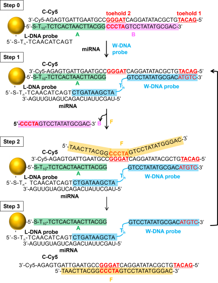
Fig. 7. Schematic illustration of the catalytic intramolecular strand displacement reaction on the AuNP surface, delineating the respective roles of strands A, B, and C-Cy5, the spatial positions of toehold 1 and toehold 2, and the involvement of W-DNA and fuel strand (F) in the reaction cycle.
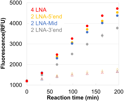
Fig. 8. Detailed comparison of reaction kinetics for different LNA incorporation positions. This figure shows an expanded view of Fig. 2 over the time range up to 200 min. Fluorescence intensity (RFU) is plotted as a function of reaction time for each condition. Filled circles represent reactions in the presence of target miRNA, while triangle markers indicate control conditions without target. Each data point represents the average of three independent measurements. Error bars indicate the standard deviation (SD, \(n = 3\)).
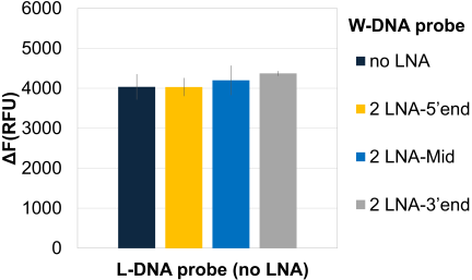
Fig. 9. Effects of LNA modifications on the performance of DNA-AuNP nanomachines targeting miR-155. Time-course fluorescence was measured for the DNA-AuNP nanomachines in the presence and absence of the target miR-155 (1 nM). Bar plots summarize the \(\Delta\)F measured at the final measurement point. Each bar represents the average of three independent measurements. Error bars indicate the standard deviation (SD, \(n = 3\)).
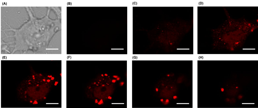
Fig. 10. Confocal imaging of MCF-7 cells incubated with DNA-AuNP nanomachines. (A) Bright-field image of an MCF-7 cell. (B–H) Confocal fluorescence images of Cy5 acquired at different focal planes with a z-step size of 2 μm. Scale bar \(=10\) μm.
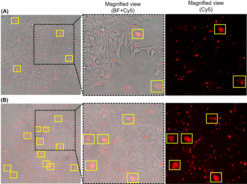
Fig. 11. Microscopic images of MCF-7 cells incubated for 12 h with (A) the non-modified nanomachines and (B) the LNA-modified nanomachines. The left panels show full BF and Cy5 merged images, with regions of interest indicated by dashed boxes. The middle panels show corresponding magnified views, and the right panels show Cy5 images. Cells classified as fluorescence-positive are highlighted by yellow squares based on the presence of distinct intracellular fluorescence signals above background levels.
This article is published under a Creative Commons Attribution-NoDerivatives 4.0 Internationa License.