
Paper:
Impact of Micropillar-Induced Mechanical Confinement of the Cell Nucleus on DNA Damage Response to UV Irradiation
Kazuaki Nagayama and Koki Miura
Micro-Nano Biomechanics Laboratory, Department of Mechanical Systems Engineering Ibaraki University
4-12-1 Nakanarusawa-cho, Hitachi, Ibaraki 316-8511, Japan

DNA damage induced by ultraviolet (UV) radiation adversely affects genome stability and contributes to various diseases. Therefore, biochemical or biomechanical methods for protecting DNA from such damage are highly important. In this study, we investigated the effects of mechanical factors, specifically the deformation of the cell nucleus using polydimethylsiloxane (PDMS)-based microfabricated arrays of micropillars with circular and elliptical cross-sections, on the UV resistance of DNA in cultured mammalian vascular smooth muscle cells. The cells spread normally within the spaces between the micropillars, where their nuclei exhibited significant deformation and appeared mechanically “trapped” between the pillars. This entrapment was accompanied by a decrease in nuclear volume and enhanced Hoechst fluorescence intensity, indicating increased DNA density within the nucleus. Evaluation of UV-induced DNA damage via the fluorescent intensity of phospho-histone γ-H2AX revealed that damage was significantly inhibited in the nuclei deformed between the pillars. These inhibitory effects were particularly pronounced on substrates with circular cross-section micropillars compared to elliptical cross-sections. Our results suggest that the suppression of UV-induced DNA damage may be associated with reduced cell motility and increased intranuclear DNA density induced by mechanical confinement of the cell nucleus by the micropillars.
Mechanical cell trapping enhances DNA damage resistance
1. Introduction
The cytotoxicity of optical radiation on cells and tissues has been well-documented through both in vivo and in vitro studies 1,2,3. Exposure to ultraviolet (UV) radiation invariably damages living cells within tissues, inducing DNA damage through photochemical stress. In particular, UVC radiation (100–280 nm) can be directly absorbed by DNA, adversely affecting genomic stability 4. Furthermore, UV radiation is known to generate free radicals that induce DNA strand breaks 5,6. Considering that DNA damage disrupts normal biological functions, accelerates aging, and causes various diseases, effective strategies for DNA repair and protection are essential.
From a biomechanical perspective, recent studies have indicated that biophysical and biomechanical cues are critical factors in regulating cellular events such as proliferation, differentiation, apoptosis, and gene expression 7,8,9,10. The nucleus, which is the largest and stiffest organelle in eukaryotic cells, is subjected to mechanical forces transmitted from the extracellular environment through the cytoskeleton 11,12. It has been suggested that the nucleus itself acts as a cellular mechanosensor. For instance, changes in nuclear shape induced by controlled cell adhesion areas may influence the regulation of cell proliferation 13,14. Our previous work demonstrated that nuclear deformation significantly inhibits cell migration, proliferation, and protein synthesis 15,16. Consequently, we hypothesized that large-scale deformation of the nucleus would alter the intranuclear mechanical environment in association with chromatin DNA condensation, potentially enhancing the radiation resistance of DNA. In our earlier study using microfabricated substrates with micropillar arrays (several micrometers in diameter and height), we observed that amphibian-derived cells (Xenopus laevis cell line derived from a tadpole) cultured on these micropillar substrates exhibited significant nuclear deformation as they were compressed between the pillars, which subsequently suppressed UV-induced DNA strand breaks 17.
Beyond their biological significance, it is important to understand how artificial microstructured materials mechanically regulate nuclear morphology and intracellular responses for the rational design of implantable sensors, biointerfaces, and other artificial devices introduced into cells or tissues. Knowledge of the influence of microstructured surfaces on nuclear mechanics and genomic stability may contribute to improving long-term biocompatibility while enabling stable intracellular signal readouts in future biomedical devices.
In the present study, we extended this investigation to mammalian cells (embryonic rat aortic smooth muscle cells) to examine the effects of nuclear deformation on cellular UV radiation resistance. Specifically, we fabricated micropillar substrates with both circular and elliptical cross sections to compare the differences in mechanical transitions imposed by the pillar geometry. The cell nuclei exhibited dramatic deformation, which matched the surface topography of the micropillar arrays. We characterized the nuclear morphological changes and confirmed the localization of intranuclear DNA and deformation of the nuclear envelope using confocal microscopy. We also assessed the inhibitory effects of mechanical confinement on cell migration using live cell imaging. Finally, the UV-induced DNA damage was quantified using fluorescence imaging, and we discussed the influence of nuclear deformation by micropillar arrays on the radiation resistance of cells.
2. Materials and Methods
2.1. Micropillar Array Substrates
Micropillar array substrates were fabricated using a replica-molding technique 18,19,20. Briefly, an array of photoresist posts was patterned onto a silicon wafer using standard photolithography to serve as a male mold. To create a female mold, poly(dimethylsiloxane) (PDMS) (Sylgard 184; Dow Corning, Midland, MI, USA) was poured over the male mold, cured at 70 °C for 6 h, and subsequently peeled off. The resulting female mold was silanized to facilitate the subsequent release of PDMS. Finally, the PDMS was cast into a female mold, cured at 100 °C for 1 h, and peeled off to obtain the final micropillar array substrate.
To investigate the effects of mechanical confinement on the cellular behavior, PDMS micropillar arrays with two distinct geometries were fabricated in a hexagonal arrangement. The circular pillars, based on our previous work 17, had a diameter, height, and center-to-center spacing of 3, 9, and 9 μm, respectively (Fig. 1(A)). The elliptical pillars featured a major axis of 7 μm and a minor axis of 3 μm (Fig. 1(B)). The height for both pillar types was maintained at 9 μm.
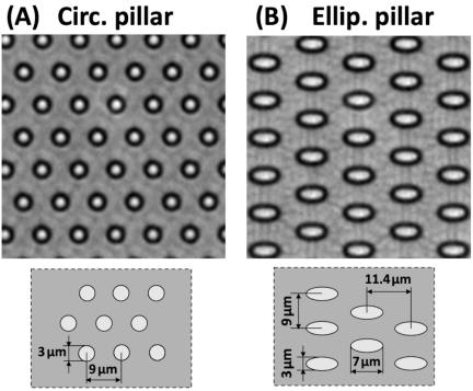
Fig. 1. Representative bright-field images of PDMS micropillar substrates with hexagonal arrangements of pillars having a circular cross-section (A) and an elliptical cross-section (B). Lower schematics indicate the pillar dimensions.
2.2. Cell Culture
An embryonic rat aortic smooth muscle cell line with a fibroblast-like phenotype (A7r5, CRL-1444; ATCC), which is frequently used in studies on cellular responses to mechanical forces, was employed as an experimental model in this study. The cells were cultured in Dulbecco’s modified Eagle’s medium (DMEM; Wako, Japan) containing 10% fetal bovine serum (FBS; JRH Bioscience, USA), 100 U/mL penicillin, and 100 μg/mL streptomycin (Sigma) at 37 °C in an atmosphere of 5% CO\(_2\) and 95% air. The cells were passaged at a \({1:4}\) split ratio upon reaching approximately 80% confluence. The flat substrates and two types of micropillar array substrates (featuring circular and elliptical cross-sections), fabricated from the same lot of PDMS, were placed in 35 mm glass-bottom dishes (No.0; Matsunami, Osaka, Japan). All the substrate surfaces were treated with oxygen plasma (5 mA, 10 Pa) for 2 min using a plasma generator (SEDE-P; Meiwafosis, Tokyo, Japan). To facilitate cell adhesion, the entire surface of each substrate was coated with 50 μg/mL fibronectin (Sigma). A7r5 cells were seeded onto the three types of substrates at an initial density of approximately 50 cells/mm\(^2\) and cultured for three days under the conditions described above.
In previous reports, including our studies, fibronectin was selectively coated only on the top surfaces of the micropillars to ensure that cells adhered strictly to the tops for the measurement of cellular traction forces 18,19,20. However, in the present study, the entire surface of the micropillars was coated with fibronectin. Consequently, the cells seeded on the micropillars were entirely inserted into the spaces between the pillars during the cell spreading process. The nuclei in the cells were also inserted between the micropillars, which was likely due to the nuclei being pressed against the substrate base by the tension generated by the actomyosin cytoskeleton during cell spreading.
2.3. Measurement of the Cells and Nucleus Morphology
The cells cultured on the PDMS pillars and flat substrates for three days were fixed with phosphate-buffered saline (PBS(\(-\)); Nissui, Tokyo, Japan) containing 3.7% formaldehyde for 5 min, permeabilized with PBS(\(-\)) containing 0.5% Triton X-100 (ICN Biomedicals, Irvine, CA, USA) for 5 min, and then rinsed with PBS(\(-\)) supplemented with 1% bovine serum albumin (BSA) to block the non-specific binding of proteins. Actin filaments were stained by incubating the cells with approximately 200 nM Alexa Fluor 546-conjugated phalloidin (Molecular Probes, Eugene, OR, USA) for 30 min. Intranuclear DNA was stained with Hoechst 33342 (\({1:1000}\) dilution, Invitrogen, Carlsbad, CA, USA) for 30 min. Immunofluorescent staining was performed to visualize Lamin A/C, a key protein in the nuclear lamina that maintains nuclear mechanical integrity. The cells were incubated with a primary anti-lamin A/C antibody (\({1:200}\) dilution, stock concentration: 0.285 mg/mL, ab133256; Abcam) for 60 min, followed by incubation with a secondary goat anti-rabbit IgG H&L Alexa Fluor 488 antibody (\({1:200}\) dilution, stock concentration: 2 mg/mL, ab150077; Abcam) for 60 min. All antibodies were diluted in PBS containing 1% BSA.
To examine the overall morphology of actin filaments and the nuclei, fluorescence images were acquired using an imaging system consisting of an inverted fluorescence microscope (IX71; Olympus), an electron-multiplying CCD camera (C9100-12; Hamamatsu Photonics, Japan), and a \(40{\times}\) objective lens (UPLXAPO40X, \(\textrm{NA}=0.95\); Olympus, Japan).
For detailed three-dimensional analysis of the nucleus and nuclear lamina, confocal fluorescence image slices were obtained at 0.5 μm intervals over the thickness of the nucleus (approximately 10 μm). This was performed using a \(60{\times}\) oil-immersion objective lens (UPLSAPO60XO, \(\textrm{NA}=1.35\); Olympus, Japan), a confocal unit (CSU-X1; Yokogawa Electric, Tokyo, Japan), a multicolor fluorescence light source system (Light Engine Spectra-X; Opto-line, Osaka, Japan), and a digital CMOS camera (ORCA-Flash4.0 V2; Hamamatsu Photonics, Japan). Nuclear outlines were automatically traced, and the projected area, shape index, and volume of the nuclei were analyzed using ImageJ/FIJI software (National Institutes of Health). The shape index expressed by Eq. (1) is defined as 1.0 for a circle and approaches zero for highly elongated and irregular shapes.
2.4. Measurement of Cell Migration on the Micropillar Substrates
The cells were precultured on PDMS pillars and flat substrates for three days. Then, the time course phase-contrast images of these cells were obtained for 24 h at 10 min intervals using a microscope imaging system with a compact CO\(_2\) incubator (Stage top incubator, Tokai Hit, Japan) to maintain the culture conditions (37 °C, 5% CO\(_2\) and 95% air). To analyze cell migration on the substrates, individual cells in the image stacks were tracked manually using the ImageJ/FIJI software.
2.5. Estimation of UV Radiation-Induced DNA Damage
The cells cultured on the PDMS pillars and flat substrates for three days were subjected to UV-C irradiation. Immediately after removing the culture medium using an aspirator, the cells were exposed to UV-C light for 20 s at an intensity of approximately 0.4 mW/cm\(^2\) using a conventional germicidal lamp (peak emission at 254 nm), resulting in a total fluence of 8 mJ/cm\(^2\). The incident fluence rate was measured using a radiometer (NOVA II, Ophir Optronics). UV-induced DNA damage was evaluated by immunofluorescence staining of phosphorylated histone 2AX (\(\gamma\)-H2AX), a primary marker of DNA double-strand breaks (DSBs). As DSB-induced \(\gamma\)-H2AX expression is known to occur in a time-dependent manner 21, fresh medium was added after irradiation, and the cells were returned to the incubator. The cells were then fixed and permeabilized at 0.5, 1, 2, and 6 h post-irradiation, followed by the blocking of non-specific binding sites with BSA, as described in the previous section. The samples were incubated with mouse anti-\(\gamma\)-H2AX primary antibodies (\({1:200}\) dilution, 05-636-I, clone JBW301, Merck Millipore) for 1 h at room temperature (25 °C) and subsequently treated with a secondary antibody (goat anti-mouse Alexa Fluor 546, \({1:200}\) dilution, Invitrogen) for 1 h at room temperature. All antibodies were diluted in PBS containing 1% BSA. The intranuclear DNA was stained with Hoechst 33342. Fluorescence images were captured using an inverted fluorescence microscope with a CMOS camera and \(40{\times}\) objective lens (UPLXAPO40X, \(\textrm{NA}=0.95\), Olympus, Japan). For quantitative analysis, background noise was subtracted from the images using ImageJ/FIJI software, and the fluorescence intensity of \(\gamma\)-H2AX within each nucleus was measured as an index of UV-induced DNA damage.
2.6. Statistical Analysis
Data are expressed as mean \(\pm\) standard deviation (SD). Statistical significance was assessed using ANOVA with correction for multiple comparisons, followed by Student’s paired and unpaired \(t\)-tests, using the statistical analysis program MEPHAS (https://alain003.phs.osaka-u.ac.jp/mephas/index_jp.html). Statistical significance was set at \({p<0.05}\) in all analyses.
3. Results
Typical fluorescence images of A7r5 cells cultured on flat PDMS and PDMS micropillar substrates are shown in Fig. 2. On both the fibronectin-coated flat and pillar substrates, the cells spread completely and were characterized by well-developed linear actin filaments (Figs. 2(A)–(C)). Cell nuclei on the flat substrate had an elliptical shape and a smooth surface (Fig. 2(D)). In contrast, on both circular and elliptical micropillar substrates, the nuclei were fully inserted and squeezed between the pillars. These nuclei were significantly deformed according to the pillar geometry, appearing to be mechanically “trapped” within the pillar arrays (Figs. 2(E), (F), (H), and (I)).
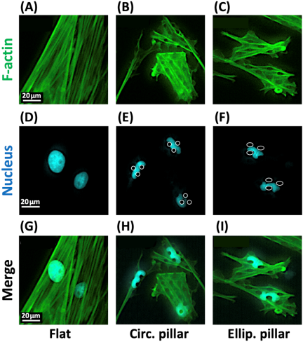
Fig. 2. Representative fluorescence images of A7r5 cells stained for actin filaments (green) and intranuclear DNA (cyan), including their corresponding merged images. Cells were cultured for three days on flat substrates (A, D, G), circular micropillar substrates (B, E, H), and elliptical micropillar substrates (C, F, I).
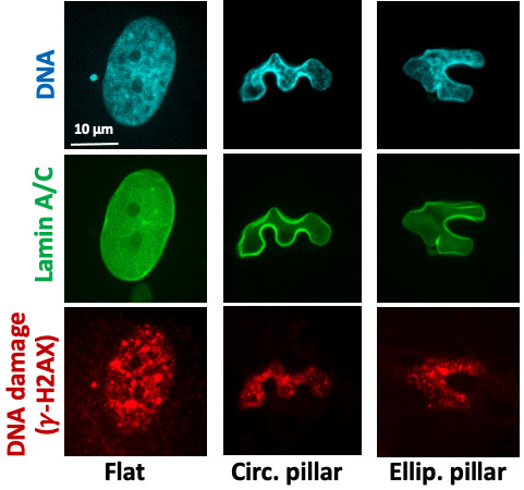
Figure 3 shows representative confocal fluorescence microscopy images of intranuclear DNA, nuclear lamina morphology, and DNA damage sites in the cells on each substrate after UV irradiation. In the nuclei on the flat substrate, DNA appeared to be distributed throughout the nucleoplasm (Fig. 3(A)), while the nuclear lamina (Lamin A/C) formed a thin, relatively uniform envelope along the nuclear periphery (Fig. 3(D)). In contrast, in the nuclei trapped between the micropillars, a high DNA density was observed along the regions significantly indented by the lateral surfaces of the pillars (Figs. 3(B) and (C)). At these sites, the fluorescence intensity of the nuclear lamina increased (Figs. 3(E) and (F)). Regarding the DNA damage sites visualized by \(\gamma\)-H2AX, discrete foci were scattered within the nuclei in all groups, and no specific trend was identified. However, the distribution of these damage sites appeared to be relatively heterogeneous in the nuclei trapped between micropillars (Figs. 3(H) and (I)).
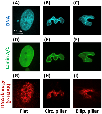
Fig. 3. Representative confocal fluorescence image slices of A7r5 cells stained for DNA (Hoechst 33342), nuclear lamina (Lamin A/C), and \(\gamma\)-H2AX. Cells were cultured for three days on flat substrates (A, D, G), circular micropillar substrates (B, E, H), and elliptical micropillar substrates (C, F, I), and then fixed and stained 1 h after UV exposure.
Quantitative morphological analysis of the cell nuclei, based on confocal fluorescence microscopy data, showed that the projected area, major axis length, and minor axis length of the nuclei on the flat PDMS substrates were \(337{\pm}81\) μm\(^2\), \(24.1{\pm}3.8\) μm, and \(16.8{\pm}2.4\) μm, respectively. In comparison, the nuclear projected area on both the circular and elliptical micropillar substrates was significantly reduced to approximately 60% of that on the flat PDMS substrates (Fig. 4(A)). Furthermore, the nuclear shape index decreased to below 0.5 on these pillar substrates, indicating a significant shift toward an elongated and irregular morphology (Fig. 4(B)). Regarding nuclear volume, the nuclei on the circular micropillar substrates showed a significant decrease to approximately 60% compared with those on the flat substrates. In contrast, the nuclear volume on the elliptical micropillar substrates remained comparable to that on the flat substrates (Fig. 4(C)). The fluorescence intensity of intranuclear DNA was significantly increased on both the circular and the elliptical micropillar substrates compared to those on the flat substrates (Fig. 4(D)).
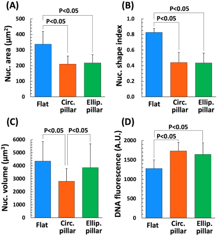
Fig. 4. Nucleus morphology ((A) area, (B) shape index, and (C) volume) and intranuclear DNA fluorescence intensity (D) of A7r5 cells obtained with confocal image slice data. Graphs are represented by mean \(+\) SD. More than 90 nuclei were analyzed for each experimental group (flat, \({n=101}\); circ. pillar, \({n=104}\); and ellip. pillar, \({n=98}\)).
Figure 5 shows the cell migration analysis of A7r5 cells on the PDMS micropillar substrates, where significant nuclear deformation occurred, in comparison with cells on the flat PDMS substrates. On the flat PDMS substrates, the cells moved in random directions (Fig. 5(A)) with a migration speed of approximately 10 μm/h (Fig. 5(D), light blue bar). In contrast, cell migration was markedly restricted on the circular micropillar substrates (Fig. 5(B)), and the migration speed was reduced by less than half (Fig. 5(D), orange bar). On elliptical micropillar substrates, the cell migration speed was also significantly reduced (Fig. 5(D), green bar) by approximately 60% compared with that on the flat substrates. However, in some cases, the cells exhibited linear movement, as if they had passed through the gaps between the pillars (Fig. 5(C)), suggesting that the inhibitory effect on cell migration was lower than that of the circular micropillars.
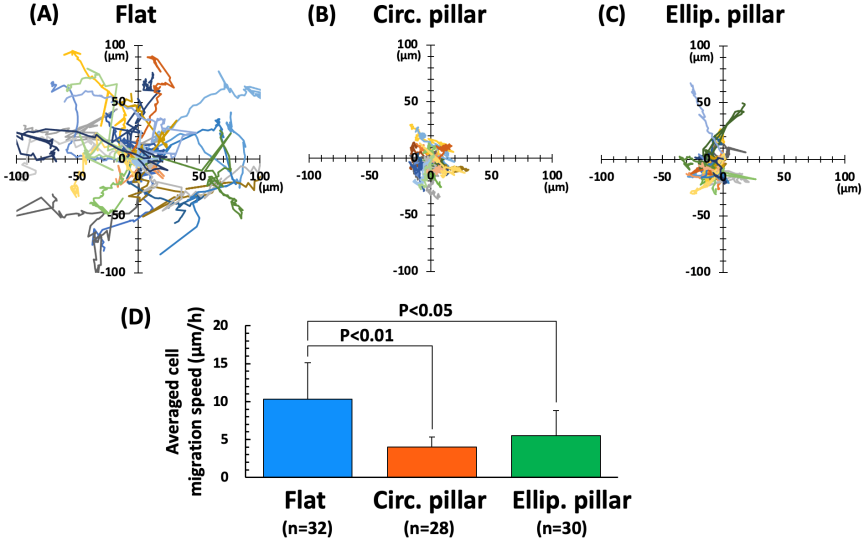
Fig. 5. (A–C) Migration trajectories of the three experimental groups and (D) the corresponding average cell migration speeds quantified from these trajectories.
Finally, A7r5 cells on each substrate were cultured for 0.5 to 6 h following UV irradiation, and fluorescence imaging of intranuclear DNA and phosphorylated histone \(\gamma\)-H2AX was performed (Figs. 6(A)–(C)). In cells on the flat substrates, the fluorescence intensity of \(\gamma\)-H2AX gradually increased after UV irradiation, peaking at 2 h, and then showed a decreasing trend while maintaining relatively high intensity levels even after 6 h (Fig. 6(D), light blue circles). In contrast, in the nuclei deformed by the micropillars, on both the circular and elliptical substrates, the expression of \(\gamma\)-H2AX peaked at 0.5 h post-irradiation. Subsequently, the intensity gradually decreased, reaching levels comparable to those of the non-irradiated control group after 6 h (Fig. 6(D), red squares and green triangles). Notably, the inhibitory effect on DNA damage was most pronounced in the cell nuclei trapped between the circular micropillars (Fig. 6(D), red squares).
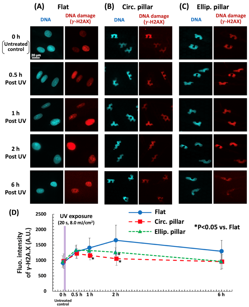
Fig. 6. (A–C) Representative fluorescence images of intranuclear DNA and DNA damage (\(\gamma\)-H2AX) in A7r5 cells cultured for specific periods following UV irradiation. (D) Time course of DNA damage development after UV irradiation.
4. Discussion
In this study, the effects of nuclear deformation on the UV resistance of A7r5 cells were investigated using PDMS micropillar substrates. These results suggested that large-scale nuclear deformation mediated by mechanical trapping between micropillars significantly suppressed UV-induced DNA damage. These findings are consistent with our previous results using amphibian cells 17 and are the first to demonstrate that the inhibitory effect of nuclear deformation on UV-induced DNA damage also occurs in mammalian cells. Furthermore, micropillar substrates with circular and elliptical cross sections were fabricated to compare the differences in mechanical cell confinement and their respective inhibitory effects. The suppression of UV-induced DNA damage was more pronounced in cells in which the nuclei were trapped between the circular micropillars.
Mechanical trapping of the cell nucleus not only caused significant deformation of the nuclear lamina and heterogeneity in intranuclear DNA (Fig. 3) but also induced a reduction in nuclear size (Figs. 4(A) and (C)). This mechanical stimulation led to an increase in the fluorescence intensity of Hoechst-stained DNA, which was evident from the increased intranuclear DNA density (Fig. 4(D)). The effects of nuclear reduction and DNA condensation were particularly prominent in cells trapped between the circular micropillars. Versaevel et al. 14 demonstrated that controlling cell adhesion shapes by microcontact printing causes large-scale nuclear elongation, which increases the DNA fluorescence intensity, suggesting drastic condensation of intranuclear chromatin. Similar increases in the intranuclear DNA density associated with nuclear deformation may have occurred in the present study. However, because chromatin-specific markers were not examined, the present results do not directly demonstrate functional chromatin condensation or heterochromatin formation.
The mechanical confinement of the cell nucleus by the micropillars also significantly inhibited cell migration (Fig. 5). While no significant difference in the inhibitory effect on migration was observed between the circular and elliptical micropillar substrates, the mean migration speed was lower on the circular micropillars (Fig. 5(D), orange bar). Analysis of the micropillar dimensions revealed that the minimum distance between adjacent pillar surfaces was 3 μm for the circular pillars, whereas it ranged from 4 to 6 μm for the elliptical pillars (Fig. 1). Thus, it suggests that the circular micropillar substrates provide a stronger mechanical trapping effect on the cell nucleus. From an engineering perspective, the narrower confinement geometry of the circular micropillars provides fewer available migration pathways and imposes stronger local mechanical restriction on the nucleus than the elliptical micropillars. This geometrical difference is likely responsible for the enhanced nuclear deformation and the greater suppression of UV-induced DNA damage observed in the circular micropillar substrates. This higher degree of migration inhibition likely contributes to the promotion of nuclear volume reduction and increase of intranuclear DNA density.
Mechanical deformation of the cell nucleus on micropillar substrates may also inhibit UV-induced DNA damage, as estimated from the fluorescence intensity of \(\gamma\)-H2AX (Fig. 6). In particular, the observed trend, where \(\gamma\)-H2AX expression peaks within a relatively short period after UV irradiation and subsequently declines rapidly (Fig. 6(D), red squares and green triangles), suggests a favorable effect on the efficient repair of DNA damage. It is widely believed that UV radiation generates reactive radicals such as OH radicals through the radiolysis of water molecules within the cell, which serve as major contributors to DNA damage 22. Takata et al. 23 investigated the effects of chromatin condensation on radiation-induced DNA damage using isolated chromatin and reported that the frequency of DNA double-strand breaks in condensed chromatin after exposure to ionizing radiation was significantly lower than that in decondensed chromatin. This suppression of damage is likely because condensed chromatin contains fewer water molecules per unit of chromatin, thereby generating fewer reactive radicals 24. In the present study, such chromatin condensation is thought to be associated with the compressive stress applied to the nucleus by the micropillars. The mechanically increased DNA density observed in the confined nuclei may contribute to the reduced DNA susceptibility to reactive radicals. However, it is unclear whether this phenomenon reflects chromatin condensation epigenetically. Future studies should investigate whether the observed increase in DNA density is accompanied by epigenetic chromatin remodeling using established chromatin markers (e.g., H3K9me3, H3K27me3, HP1, H3K4me3, and H3K27ac). Further investigation of intranuclear chromatin mobility will help to elucidate the detailed mechanisms underlying nuclear deformation-induced UV radiation resistance.
A limitation of this study is that the potential negative effects of large-scale nuclear deformation were not fully eliminated. We previously demonstrated that although nuclear deformation and trapping using micropillar substrates do not affect cell viability 15, they can decrease the synthesis of proteins such as the actin cytoskeleton 16. Further research is necessary to clarify the comprehensive biological impact of nuclear deformation on micropillars. Another limitation is that the local UV irradiation conditions around the micropillar structures were not directly evaluated. Although all the samples were irradiated under identical macroscopic conditions, the micropillar geometry may locally influence the UV fluence through reflection, scattering, or partial shielding. Therefore, geometry-dependent optical effects cannot be completely excluded and should be quantitatively assessed in future studies using optical simulations or direct UV measurements.
5. Conclusion
This study demonstrated that the mechanical deformation of the cell nucleus, induced by PDMS-based micropillar arrays, significantly enhanced the UV resistance of DNA in mammalian cells. Mechanical confinement of the nucleus between the pillars led to a reduction in nuclear volume and increased intranuclear DNA density, which effectively suppressed UV-induced DNA damage. Our findings suggest that these inhibitory effects are driven by both the restriction of cell motility and an increase in intranuclear DNA density under mechanical stress. These results provide critical insights into the interplay between biomechanical factors and genomic integrity and offer a potential foundation for the development of new physical strategies to protect DNA from radiation damage.
Acknowledgments
This work was supported in part by JSPS KAKENHI Grant Numbers 24K03246, 23K25998 (Grant-in-Aid for Scientific Research (B)), and 25K22900 (Grant-in-Aid for Challenging Research (Exploratory)). Support was also provided by the Takeda Science Foundation, Hagiwara Foundation, Chubu Science and Technology Center, and the Naito Foundation (Naito Memorial Foundation for the Promotion of Science).
- [1] M. Rózanowska et al., “Blue light-induced reactivity of retinal age pigment: In vitro generation of oxygen-reactive species,” J. Biol. Chem., Vol.270, No.32, pp. 18825-18830, 1995. https://doi.org/10.1074/jbc.270.32.18825
- [2] T. G. Gorgels and D. van Norren, “Ultraviolet and green light cause different types of damage in rat retina,” Invest. Ophthalmol. Vis. Sci., Vol.36, No.5, pp. 851-863, 1995.
- [3] A. Stary, C. Robert, and A. Sarasin, “Deleterious effects of ultraviolet A radiation in human cells,” Mutat. Res. DNA Repair, Vol.383, No.1, pp. 1-8, 1997. https://doi.org/10.1016/S0921-8777(96)00041-9
- [4] C. Remé, J. Reinboth, M. Clausen, and F. Hafezi, “Light damage revisited: Converging evidence, diverging views?,” Graefe’s Arch. Clin. Exp. Ophthalmol., Vol.234, No.1, pp. 2-11, 1996. https://doi.org/10.1007/BF00186512
- [5] C. Alapetite, T. Wachter, E. Sage, and E. Moustacchi, “Use of the alkaline comet assay to detect DNA repair deficiencies in human fibroblasts exposed to UVC, UVB, UVA and gamma-rays,” Int. J. Radiat. Biol., Vol.69, No.3, pp. 359-369, 1996. https://doi.org/10.1080/095530096145922
- [6] C. Kielbassa, L. Roza, and B. Epe, “Wavelength dependence of oxidative DNA damage induced by UV and visible light,” Carcinogenesis, Vol.18, No.4, pp. 811-816, 1997. https://doi.org/10.1093/carcin/18.4.811
- [7] D. E. Ingber, “Fibronectin controls capillary endothelial cell growth by modulating cell shape,” Proc. Natl. Acad. Sci. U.S.A., Vol.87, No.9, pp. 3579-3583, 1990. https://doi.org/10.1073/pnas.87.9.3579
- [8] A. J. Engler, S. Sen, H. L. Sweeney, and D. E. Discher, “Matrix elasticity directs stem cell lineage specification,” Cell, Vol.126, No.4, pp. 677-689, 2006. https://doi.org/10.1016/j.cell.2006.06.044
- [9] C. S. Chen, M. Mrksich, S. Huang, G. M. Whitesides, and D. E. Ingber, “Geometric control of cell life and death,” Science, Vol.276, No.5317, pp. 1425-1428, 1997. https://doi.org/10.1126/science.276.5317.1425
- [10] P. G. Smith, C. Roy, S. Fisher, Q.-Q. Huang, and F. Brozovich, “Selected contribution: Mechanical strain increases force production and calcium sensitivity in cultured airway smooth muscle cells,” J. Appl. Physiol., Vol.89, No.5, pp. 2092-2098, 2000. https://doi.org/10.1152/jappl.2000.89.5.2092
- [11] M. L. Lombardi et al., “The interaction between nesprins and sun proteins at the nuclear envelope is critical for force transmission between the nucleus and cytoskeleton,” J. Biol. Chem., Vol.286, No.30, pp. 26743-26753, 2011. https://doi.org/10.1074/jbc.M111.233700
- [12] K. N. Dahl, A. J. S. Ribeiro, and J. Lammerding, “Nuclear shape, mechanics, and mechanotransduction,” Circ. Res., Vol.102, No.11, pp. 1307-1318, 2008. https://doi.org/10.1161/CIRCRESAHA.108.173989
- [13] P. Roca-Cusachs et al., “Micropatterning of single endothelial cell shape reveals a tight coupling between nuclear volume in G1 and proliferation,” Biophys. J., Vol.94, No.12, pp. 4984-4995, 2008. https://doi.org/10.1529/biophysj.107.116863
- [14] M. Versaevel, T. Grevesse, and S. Gabriele, “Spatial coordination between cell and nuclear shape within micropatterned endothelial cells,” Nat. Commun., Vol.3, Article No.671, 2012. https://doi.org/10.1038/ncomms1668
- [15] K. Nagayama, Y. Hamaji, Y. Sato, and T. Matsumoto, “Mechanical trapping of the nucleus on micropillared surfaces inhibits the proliferation of vascular smooth muscle cells but not cervical cancer HeLa cells,” J. Biomech., Vol.48, No.10, pp. 1796-1803, 2015. https://doi.org/10.1016/j.jbiomech.2015.05.004
- [16] K. Nagayama, “Mechanical stress to cell nucleus inhibits proliferation and differentiation of vascular smooth muscle cells,” J. Biosci. Med., Vol.8, No.5, pp. 132-141, 2020. https://doi.org/10.4236/jbm.2020.85013
- [17] K. Nagayama, C. Sagawa, and A. Sato, “Deformation and trapping of cell nucleus using micropillar substrates possibly affect UV radiation resistance of DNA,” J. Robot. Mechatron., Vol.35, No.5, pp. 1158-1164, 2023. https://doi.org/10.20965/jrm.2023.p1158
- [18] J. L. Tan et al., “Cells lying on a bed of microneedles: An approach to isolate mechanical force,” Proc. Natl. Acad. Sci. U.S.A., Vol.100, No.4, pp. 1484-1489, 2003. https://doi.org/10.1073/pnas.0235407100
- [19] N. Sakamoto, Y. Yamazaki, T. Ohashi, and M. Sato, “Effect of mechanical environment of focal adhesions on remodeling of endothelial cells subjected to cyclic stretching using microsubstrates,” J. Robot. Mechatron., Vol.19, No.5, pp. 557-564, 2007. https://doi.org/10.20965/jrm.2007.p0557
- [20] K. Nagayama and T. Matsumoto, “Dynamic change in morphology and traction forces at focal adhesions in cultured vascular smooth muscle cells during contraction,” Cell. Mol. Bioeng., Vol.4, No.3, pp. 348-357, 2011. https://doi.org/10.1007/s12195-011-0166-y
- [21] W. Han et al., “The early and initiation processes of radiation-induced bystander effects involved in the induction of DNA double strand breaks in non-irradiated cultures,” Br. J. Radiol., Vol.80, Special Issue 1, pp. S7-S12, 2007. https://doi.org/10.1259/bjr/44550200
- [22] R. E. Krisch, M. B. Flick, and C. N. Trumbore, “Radiation chemical mechanisms of single- and double-strand break formation in irradiated SV40 DNA,” Radiat. Res., Vol.126, No.2, pp. 251-259, 1991. https://doi.org/10.2307/3577826
- [23] H. Takata et al., “Chromatin compaction protects genomic DNA from radiation damage,” PLOS ONE, Vol.8, No.10, Article No.e75622, 2013. https://doi.org/10.1371/journal.pone.0075622
- [24] Y. Yoshikawa, T. Mori, N. Magome, K. Hibino, and K. Yoshikawa, “DNA compaction plays a key role in radioprotection against double-strand breaks as revealed by single-molecule observation,” Chem. Phys. Lett., Vol.456, Nos.1-3, pp. 80-83, 2008. https://doi.org/10.1016/j.cplett.2008.03.009
This article is published under a Creative Commons Attribution-NoDerivatives 4.0 Internationa License.