Paper:
Cerebral Activity-Based Quantitative Evaluation for Attention Levels
Saki Niiyama, Shiro Yano, and Toshiyuki Kondo
Graduate School of Engineering, Tokyo University of Agriculture and Technology
2-24-16 Naka-cho, Koganei-shi, Tokyo 184-8588, Japan

Regional cerebral activity related to attention may be more useful as an evaluation index for attention levels than conventional task performance score-based methods. We therefore researched whether the quantitative evaluation of attention using regional cerebral activity, measured using near-infrared spectroscopy (NIRS), was appropriate. NIRS signals during the continuous performance test (CPT), which is well known as an attention test, were measured and analyzed. We confirmed activities in the regions that may be associated with the right-side anterior cingulate cortex (ACC), and on the estimated dorsolateral prefrontal cortex (DLPFC). Furthermore, there was a high correlation between activity on the DLPFC related to executive function and the performance score. Our study using cerebral activity could not quantify attention, but it opened the possibility of quantifying levels of executive function.
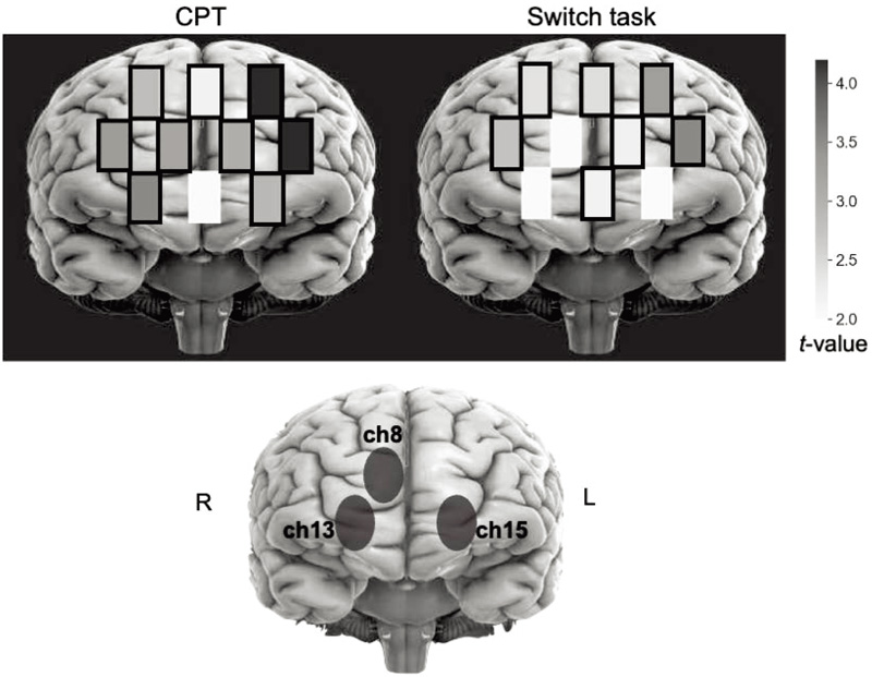
Estimation of ROI
- [1] S. Ishiai, “Learning of higher brain dysfunction,” 2nd Ed., Ishiyaku Publishers, Inc., 2017 (in Japanese).
- [2] Japan Society for Higher Brain Dysfunction, “Clinical assessment for attention and spontaneity,” Shinkoh Igaku Shuppansha Co., Ltd., 2008 (in Japanese).
- [3] M. G. Tana, E. Montin, S. Cerutti, and A. M. Bianchi, “Exploring cortical attentional system by using fMRI during a continuous performance test,” Computational Intelligence and Neuroscience, Article ID 329213, doi: 10.1155/2010/329213, 2010.
- [4] H. Yamasaki, K. S. LaBar, and G. McCarthy, “Dissociable prefrontal brain systems for attention and emotion,” PNAS, Vol.99, No.17, pp. 11447-11451, doi: 10.1073/pnas.182176499, 2002.
- [5] G. Bush, J. A. Frazier, S. L. Rauch, L. J. Seidman, P. J. Whalen, M. A. Jenike, B. R. Rosen, and J. Biederman, “Anterior cingulate cortex dysfunction in attention-deficit/hyperactivity disorder revealed by fMRI and the counting stroop,” Biological Psychiatry, Vol.45, No.12, pp. 1542-1552, 1999.
- [6] A. J. Fallgatter and W. K. Strik, “Reduced frontal functional asymmetry in schizophrenia during a cued continuous performance test assessed with near-infrared spectroscopy,” Schizophrenia Bulletin, Vol.26, No.4, pp. 913-919, doi: 10.1093/oxfordjournals.schbul.a033505, 2000.
- [7] A. Araki, M. Ikegami, A. Okayama, N. Matsumoto, S. Takahashi, H. Azuma, and M. Takahashi, “Improved prefrontal activity in AD/HD children treated with atomoxetine: A NIRS study,” Brain and Development, Vol.37, No.1, pp. 76-87, doi: 10.1016/j.braindev.2014.03.011, 2015.
- [8] B. D. Killory, X. Bai, M. Negishi, C. Vega, M. N. Spann, M. Vestal, J. Guo, R. Berman, N. Danielson, J. Trejo, D. Shisler, E. J. Novotny Jr., R. T. Constable, and H. Blumenfeld, “Impaired attention and network connectivity in childhood absence epilepsy,” NeuroImage, Vol.56, No.4, pp. 2209-2217, doi: 10.1016/j.neuroimage.2011.03.036, 2011.
- [9] G. Strangman, J. P. Culver, J. H. Thompson, and D. A. Boas, “A quantitative comparison of simultaneous BOLD fMRI and NIRS recordings during functional brain activation,” NeuroImage, Vol.17, No.2, pp. 719-731, doi: 10.1006/nimg.2002.1227, 2002.
- [10] T. J. Huppert, R. D. Hoge, S. G. Diamond, M. A. Franceschini, and D. A. Boas, “A temporal comparison of BOLD, ASL, and NIRS hemodynamic responses to motor stimuli in adult humans,” NeuroImage, Vol.29, No.2, pp. 368-382, doi: 10.1016/j.neuroimage.2005.08.065, 2006.
- [11] P. Pinti, I. Tachtsidis, A. Hamilton, J. Hirsch, C. Aichelburg, S. Gilbert, and P. W. Burgess, “The present and future use of functional near-infrared spectroscopy (fNIRS) for cognitive neuroscience,” Annals of the New York Academy of Sciences, Vol.1464, No.1, pp. 5-29, doi: 10.1111/nyas.13948, 2020.
- [12] R. E. Vanderwert and C. A. Nelson, “The use of near-infrared spectroscopy in the study of typical and atypical development,” NeuroImage, Vol.85, Part 1, pp. 264-271, doi: 10.1016/j.neuroimage.2013.10.009, 2014.
- [13] S. Shimizu, H. Inoue, H. Nara, T. Tsuruga, F. Miwakeichi, N. Hirai, S. Kikuchi, E. Watanabe, and S. Kato, “Basic study for new assistive technology based on brain activity during car driving,” J. Robot. Mechatron., Vol.26, No.2, pp. 253-260, doi: 10.20965/jrm.2014.p0253, 2014.
- [14] H. Sakaniwa, S. Sutoko, A. Obata, H. Atsumori, N. Fukuda, M. Kiguchi, and A. Kandori, “Effects of shape characteristics on tactile sensing recognition and brain activation,” J. Robot. Mechatron., Vol.23, No.6, pp. 1080-1088, doi: 10.20965/jaciii.2019.p1080, 2019.
- [15] T. Yamada, S. Umeyama, and K. Matsuda, “Separation of fNIRS signals into functional and systemic components based on differences in hemodynamic modalities,” PLOS ONE, Vol.7, No.11, Article No.e50271, doi: 10.1371/journal.pone.0050271, 2012.
- [16] Y. Hoshi, N. Kobayashi, and M. Tamura, “Interpretation of near-infrared spectroscopy signals: a study with a newly developed perfused rat brain model,” J. of Applied Physiology, Vol.90, Issue 5, pp. 1657-1662, doi: 10.1152/jappl.2001.90.5.1657, 2001.
- [17] T. Tsujimoto, H. Shimazu, Y. Isomura, and K. Sasaki, “Theta oscillations in primate prefrontal and anterior cingulate cortices in forewarned reaction time tasks,” J. of Neurophysiology, Vol.103, No.2, pp. 827-843, doi: 10.1152/jn.00358.2009, 2010.
- [18] H. D. Critchley, C. J. Mathias, O. Josephs, J. O’Doherty, S. Zanini, B.-K. Dewar, L. Cipolotti, T. Shallice, and R. J. Dolan, “Human cingulate cortex and autonomic control: converging neuroimaging and clinical evidence,” Brain, Vol.126, No.10, pp. 2139-2152, doi:10.1093/brain/awg216, 2003.
- [19] C. Nakagawa, “Special Issues No.3: Measurement technique for ergonomics, Section 4: Measurements and analyses of bioelectric phenomena and others (5) measurement and analysis of autonomic indices,” The Japanese J. of Ergonomics, Vol.52, pp. 6-12, doi: 10.5100/jje.52.6, 2016 (in Japanese).
- [20] C. A. Riccio, C. R. Reynolds, P. Lowe, and J. J. Moore, “The continuous performance test: a window on the neural substrates for attention?,” Archives of Clinical Neuropsychology, Vol.17, Issue 3, pp. 235-272, doi: 10.1093/arclin/17.3.235, 2002.
- [21] K. J. Friston, J. T. Ashburner, S. J. Kiebel, T. E. Nichols, and W. D. Penny, “Statistical Parametric Mapping – The Analysis of Functional Brain Images,” Academic Press, 2006.
- [22] J. C. Ye, S. Tak, K. E. Jang, J. Jung, and J. Jang, “NIRS-SPM: Statistical parametric mapping for near-infrared spectroscopy,” NeuroImage, Vol.44, No.2, pp. 428-447, doi: 10.1016/j.neuroimage.2008.08.036, 2009.
- [23] P. Sumner and M. Husain, “At the Edge of consciousness: Automatic motor activation and voluntary control,” The Neuroscientist, Vol.14, No.5, pp. 474-486, doi: 10.1177/1073858408314435, 2008.
- [24] C. E. Curtis and M. D’Esposito, “Persistent activity in the prefrontal cortex during working memory,” Trends in Cognitive Sciences, Vol.7, No.9, pp. 415-423, doi: 10.1016/S1364-6613(03)00197-9, 2003.
- [25] J. A. Richeson, A. A. Baird, H. L. Gordon, T. F. Heatherton, C. L. Wyland, S. Trawalter, and J. N. Shelton, “An fMRI investigation of the impact of interracial contact on executive function,” Nature Neuroscience, Vol.6, pp. 1323-1328, doi: 10.1038/nn1156, 2003.
This article is published under a Creative Commons Attribution-NoDerivatives 4.0 Internationa License.