Research Paper:
Fragility Fracture of Pelvis Prediction from Computed Tomography Using Boring Survey and Convolutional Neural Network
Rashedur Rahman*,†, Naomi Yagi**
 , Keigo Hayashi***, Akihiro Maruo***, Hirotsugu Muratsu***, and Syoji Kobashi*
*Graduate School of Engineering, University of Hyogo
2167 Shosha, Himeji, Hyogo 671-2280, Japan
†Corresponding author
**Advanced Medical Engineering Research Institute, University of Hyogo
3-264 Kamiya-cho, Himeji, Hyogo 670-0836, Japan
***Hyogo Prefectural Harima-Himeji General Medical Center
3-264 Kamiya-cho, Himeji, Hyogo 670-8560, Japan

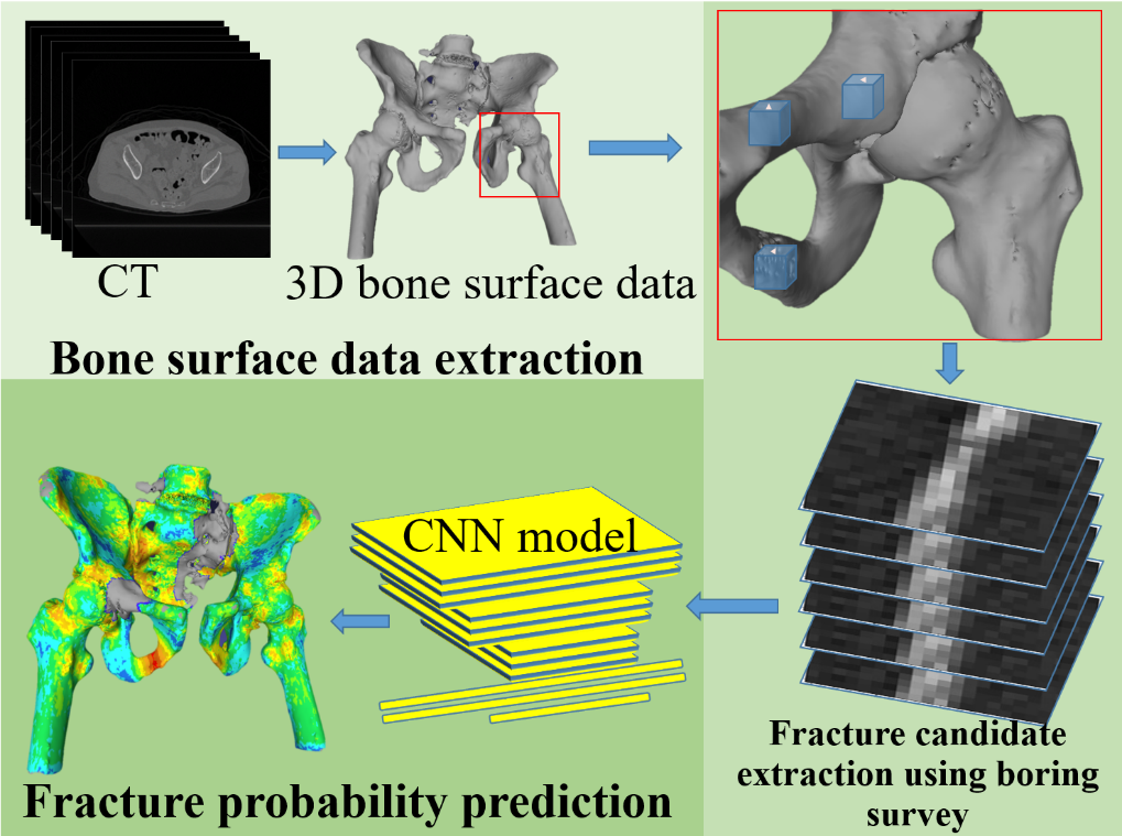
Fragility fracture of pelvis (FFP) is increasingly affecting elderly population. Although computed tomography (CT) imaging is considered superior to conventional radiographic image for diagnosing FFP, clinicians face challenges in recognizing pelvic fractures owing to imaging contrast or feature size. This study proposes a method that combines boring survey based FFP candidate extraction from CT images and a newly developed convolutional neural network model. In addition, the proposed method also visualizes the probability of fracture on 3D bone surface data. The accuracy, precision, and recall of the proposed method were found to be 79.7%, 60.0%, and 80.6%, respectively. Furthermore, the 3D view of fracture probability on the pelvic bone surface allows for qualitative assessment and can support physicians to diagnose FFP. The findings indicate that the proposed method has potential for predicting FFP.
FFP prediction using boring survey and CNN
- [1] M. P. Sullivan et al., “Geriatric fractures about the hip: Divergent patterns in the proximal femur, acetabulum, and pelvis,” Orthopedics, Vol.37, No.3, pp. 151-157, 2014. https://doi.org/10.3928/01477447-20140225-50
- [2] G. L. Nanninga et al., “Increasing rates of pelvic fractures among older adults: The Netherlands, 1986–2011,” Age Ageing, Vol.43, No.5, pp. 648-653, 2014. https://doi.org/10.1093/ageing/aft212
- [3] S. Andrich et al., “Epidemiology of pelvic fractures in Germany: Considerably high incidence rates among older people,” PLOS ONE, Vol.10, No.9, Article No.e0139078, 2015. https://doi.org/10.1371/journal.pone.0139078
- [4] P. M. Rommens et al., “Progress of instability in fragility fractures of the pelvis: An observational study,” Injury, Vol.50, No.11, pp. 1966-1973, 2019. https://doi.org/10.1016/j.injury.2019.08.038
- [5] N. Suhm et al., “Low acceptance of osteoanabolic therapy with parathyroid hormone in patients with fragility fracture of the pelvis in routine clinical practice: A retrospective observational cohort study,” Arch. Orthop. Trauma Surg., Vol.140, No.3, pp. 321-329, 2020. https://doi.org/10.1007/s00402-019-03241-4
- [6] H.-G. Palm et al., “Dual-energy CT as an innovative method for diagnosing fragility fractures of the pelvic ring: A retrospective comparison with MRI as the gold standard,” Arch. Orthop. Trauma. Surg., Vol.140, No.4, pp. 473-480, 2020. https://doi.org/10.1007/s00402-019-03283-8
- [7] I. D. Alexa et al., “Importance of CT scan in fragility fracture of the pelvis,” 2020 Int. Conf. e-Health Bioeng. (EHB), 2020. https://doi.org/10.1109/EHB50910.2020.9280254
- [8] A. Bar et al., “Compression fractures detection on CT,” Proc. SPIE Vol.10134, Article No.1013440, 2017. https://doi.org/10.1117/12.2249635
- [9] N. Tomita, Y. Y. Cheung, and S. Hassanpour, “Deep neural networks for automatic detection of osteoporotic vertebral fractures on CT scans,” Comput. Biol. Med., Vol.98, pp. 8-15, 2018. https://doi.org/10.1016/j.compbiomed.2018.05.011
- [10] X. H. Meng et al., “A fully automated rib fracture detection system on chest CT images and its impact on radiologist performance,” Skelet. Radiol., Vol.50, No.9, pp. 1821-1828, 2021. https://doi.org/10.1007/s00256-021-03709-8
- [11] K. Ukai et al., “Detecting pelvic fracture on 3D-CT using deep convolutional neural networks with multi-orientated slab images,” Sci. Rep., Vol.11, Article No.11716, 2021. https://doi.org/10.1038/s41598-021-91144-z
- [12] N. Yamamoto et al., “Boring survey based fracture detection (BSFD) for fragility fracture of the pelvis in CT images,” 2021 Int. Conf. Mach. Learn. Cybern. (ICMLC), 2021. https://doi.org/10.1109/ICMLC54886.2021.9737242
- [13] G. Huang et al., “Densely connected convolutional networks,” 2017 IEEE Conf. Comput. Vis. Pattern Recognit. (CVPR), pp. 2261-2269, 2017. https://doi.org/10.1109/CVPR.2017.243
- [14] M. Tan and Q. Le, “EfficientNet: Rethinking model scaling for convolutional neural networks,” Proc. 36th Int. Conf. Mach. Learn. (ICML), pp. 6105-6114, 2019.
- [15] N. Yamamoto et al., “An automated fracture detection from pelvic CT images with 3-D convolutional neural networks,” 2020 Int. Symp. Community-Centric Syst. (CcS), 2020. https://doi.org/10.1109/CcS49175.2020.9231453
- [16] N. Yamamoto et al., “Automatic detection of fragility fractures of the pelvic using convolutional neural network with 3D features,” Proc. 37th Fuzzy Syst. Symp., pp. 713-715, 2021 (in Japanese). https://doi.org/10.14864/fss.37.0_713
- [17] IXI dataset. https://brain-development.org/ixi-dataset/ [Accessed January 10, 2020]
- [18] M. Martinek, R. Grosso, and G. Greiner, “Fast and efficient 3D chamfer distance transform for polygonal meshes,” Proc. 16th Int. Workshop Vis. Model. Vis. (VMV 2011), pp. 121-128, 2011. https://doi.org/10.2312/PE/VMV/VMV11/121-128
- [19] D. P. Kingma and J. Ba, “Adam: A method for stochastic optimization,” Proc. 3rd Int. Conf. Learn. Represent. (ICLR), 2015.
This article is published under a Creative Commons Attribution-NoDerivatives 4.0 Internationa License.