Research Paper:
Effect of Alcohol Consumption on the Frequency of Microsaccades
Toumi Ohara and Fumiya Kinoshita†

Electrical and Computer Engineering, Graduate School of Engineering, Toyama Prefectural University
5180 Kurokawa, Imizu-shi, Toyama 939-0398, Japan
†Corresponding author

In recent years, as eye movement measurement devices have become relatively cheap, many attempts have been made to quantitatively evaluate covert attention by focusing on microsaccades. However, the measurement of microsaccades still has many unclear points, and a unified analysis method is still lacking. As such, the interpretation of results differs among different research groups. To solve this problem, it is important to conduct empirical studies on microsaccades to evaluate them using a unified method. In this study, we conducted an empirical experiment on the effects of alcohol consumption on microsaccades by temporarily suppressing cerebellar activity with alcohol consumption. The results showed that the frequency of microsaccades was significantly reduced after 30, 50, and 70 min of drinking compared to after drinking (p< 0.05). These results suggest that the decrease in brain function caused by alcohol consumption suppresses the frequency of microsaccades, and that this may be the cause of constriction in the peripheral visual field when drinking.
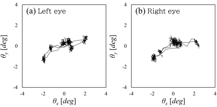
Eye movement over 2 s (angular coordinate)
- [1] M. I. Posner, J. A. Walker, F. A. Friedrich, and R. D. Rafal, “How do the parietal lobes direct covert attention?,” Neuropsychologia, Vol.25, No.1, pp. 135-145, 1987.
- [2] H. Kaneko, “Fixational Eye Movements,” J. of the Institute of Image Information and Television Engineers, Vol.63, No.11, pp. 1538-1539, 2009 (in Japanese).
- [3] D. Troxler, “Ueber das Verschwinden gegebener Gegenstande innerhalb unseres Gesichtskreises,” Ophthalmologische Bibliothek, Vol.2, pp. 1-119, 1804 (in German).
- [4] H. V. Helmholtz, “Handbuch der physiologischen Optik,” Leopold Voss, 1867 (in German).
- [5] R. Engbert and R. Kliegl, “Microsaccades uncover the orientation of covert attention,” Vision Research, Vol.43, No.9, pp. 1035-1045, 2003.
- [6] T. Tanaka, T. Kohama, and H. Yoshida, “Arousal evaluation based on the analysis of microsaccade rate and pupil fluctuation,” ITE Technical Report, Vol.36, No.13, pp. 51-54, 2012 (in Japanese).
- [7] M. Takahashi, H. Isogai, and J. L. V. Raalte, “Atempts to Detect Microsaccades from Eye Movements in Anticipatory Response Situation: Introducing Anticipatory Response Tasks Toward Tennis Serve,” J. of Japan Society of Sports Industry, Vol.28, No.1, pp. 13-29, 2018.
- [8] Z. Kapoula, Q. Yang, J. Otero-Millan, S. Xiao, S. L. Macknik, A. Lang, M. Verny, and S. Martinez-Conde, “Distinctive features of microsaccades in Alzheimer’s disease and in mild cognitive impairment,” Age, Vol.36, No.2, pp. 535-543, 2014.
- [9] F. R. Robinson and A. F. Fuchs, “The role of the cerebellum in voluntary eye movements,” Annu. Rev. Neurosci., Vol.24, pp. 981-1004, 2001.
- [10] A. Kheradmand and D. S. Zee, “Cerebellum and Ocular Motor Control,” Front Nuerol., Vol.2, Article No.53, 2011.
- [11] H. Takada, Y. Kitaoka, M. Ichikawa, and M. Miyao, “Physical Meaning on Geometrical Index for Stabilometry,” Equilibrium Res., Vol.62, No.3, pp. 168-180, 2003.
- [12] K. Hirayanagi, N. Yamaguchi, and T. Shiozawa, “Effects of fixed alcohol concentrations in expiration on task performance,” The Japanese J. of Ergonomics, Vol.42, No.5, pp. 337-442, 2006.
- [13] C.-Y. Chen and Z. M. Hafed, “Postmicrosaccadic enhancement of slow eye movements,” J. Neuroscience, Vol.33, No.12, pp. 5375-5386, 2013.
- [14] J. Otero-Millan, J. L. A. Castro, S. L. Macknik, and S. Martinez-Conde, “Unsupervised clustering method to detect microsaccades,” J. of Vision, Vol.14, No.2, Article No.18, 2014.
- [15] A. Mihali, B. V. Opheusden, and W. J. Ma, “Bayesian microsaccade detection,” J. of Vision, Vol.17, No.1, Article No.13, 2017.
- [16] S. Martinez-Conde, S. L. Macknik, and D. H. Hubel, “The role of fixational eye movements in visual perception,” Nature Reviews Neuroscience, Vol.5, No.3, pp. 229-240, 2004.
- [17] S. Martinez-Conde and S. L. Macknik, “Windows on the Mind,” Scientific American, Vol.297, No.2, pp. 56-63, 2007.
- [18] R. Engbert, “Microsaccades: a microcosm for research on oculomotor control, attention, and visual perception,” Progress in Brain Research, Vol.154, Part A, pp. 177-192, 2006.
- [19] M. Yoneya, L. Hsin-I, S. Furukawa, and M. Kashio, “Decoding Perceptual Performance for Sounds from Eye Movement and Pupil Size Change,” The 30th Annual Conf. of the Japanese Society for Artificial Intelligence, Article No.2I4-OS-07a-3, 2016 (in Japanese).
- [20] J. Emoto and Y. Hirata, “Realtime Microsaccade Detection with Convolutional Neural Network,” IEICE Trans. on Information and Systems (Japanese Edition), Vol.J101-D, No.2, pp. 456-467, 2018.
This article is published under a Creative Commons Attribution-NoDerivatives 4.0 Internationa License.